

Risk Factors for Mucosal Involvement in Bullous Pemphigoid and the Possible Mechanism: A Review
- PMID: 34095183
- PMCID: PMC8172594
- DOI: 10.3389/fmed.2021.680871
Risk Factors for Mucosal Involvement in Bullous Pemphigoid and the Possible Mechanism: A Review
Abstract
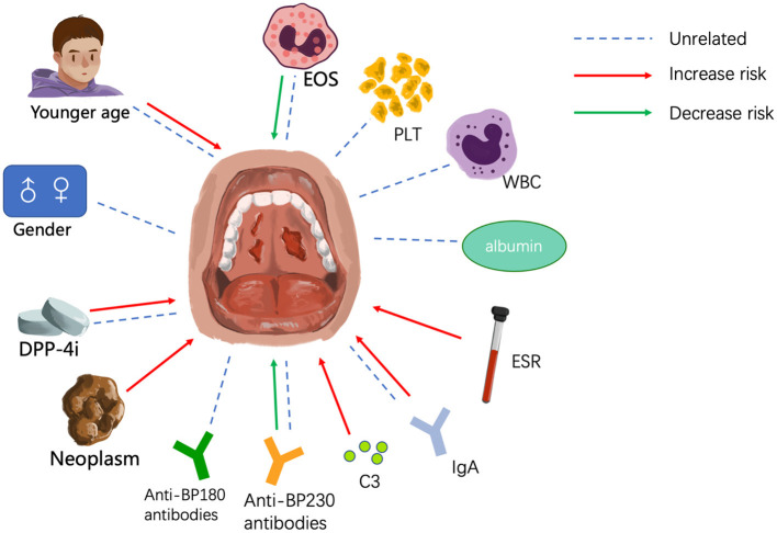
Bullous pemphigoid (BP) is the most common type of autoimmune bullous disease and is characterized by the presence of circulating anti-BP180 and/or anti-BP230 autoantibodies. Patients with BP often present with tense blisters and erythema, mainly on the trunk and limbs, but a few patients also have mucosal involvement. In this article, we discuss the fact that BP patients with mucosal involvement tend to have more serious conditions and their disease is more difficult to control. Potential risk factors for mucous involvement include earlier age at onset, drugs such as dipeptidyl peptidase-4 inhibitors, cancer, and blood/serum biomarkers, including lower eosinophil count, higher erythrocyte sedimentation rate, IgG autoantibodies against both the NH2- and COOH-termini of BP180, and the absence of anti-BP230 antibodies. IgA and C3 deposition at the dermo-epidermal junction may also be present. Understanding these risk factors may benefit earlier diagnosis of these patients and promote the development of novel treatments. What's more, it's helpful in deeper understanding of BP development and the relationship between BP and mucous membrane pemphigoid (MMP).
Keywords: autoantibody; bullous pemphigoid; mucous; risk factors; treatment.
Copyright © 2021 Chen, Zhao, Jin and Li.
Conflict of interest statement
The authors declare that the research was conducted in the absence of any commercial or financial relationships that could be construed as a potential conflict of interest.
Figures
References
-
- Baigrie D, Nookala V. Bullous Pemphigoid. StatPearls. Treasure Island, FL: StatPearls Publishing Copyright © 2020, StatPearls Publishing LLC; (2020). - PubMed
Publication types
LinkOut - more resources
Full Text Sources
Miscellaneous