

Case Report: A Rare Case of Nasal Forehead Mass in Kimura's Disease
- PMID: 34095210
- PMCID: PMC8176203
- DOI: 10.3389/fsurg.2021.672291
Case Report: A Rare Case of Nasal Forehead Mass in Kimura's Disease
Abstract
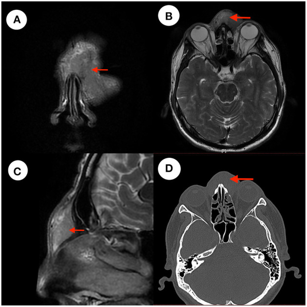
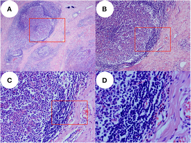
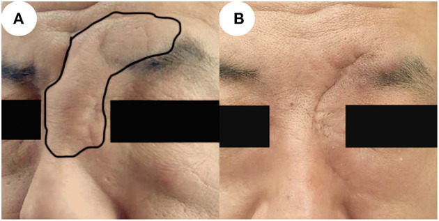
Background: Kimura's disease is a rheumatic immune disease and head and neck lymph nodes are often involved. A mass occurring in the nasal forehead is rare. Good prognosis after surgical resection by glucocorticoid therapy is more rare. Case Summary: We report the rare case of a nasal forehead mass in a 45-year-old male patient with Kimura's disease. The patient underwent resection of the mass in October 2018 in a local hospital and the postoperative pathology was unclear. He then underwent a second resection in our department in December 2019 mainly because growth of the mass was affecting his appearance. Postoperative pathology confirmed that the patient had Kimura's disease, and he accepted systemic treatment with prednisone. We followed the patient for 10 months after surgery. He is now recovering well and continues to be closely monitored during follow-up. Conclusion: It is rare that the painless mass in the nasal forehead is diagnosed as a Kimura's disease.After completely resection of the mass and systemic treatment with prednisone, the patient had a good outcome. We provide experience for the treatment of Kimura's disease in nasal forehead.
Keywords: Kimura disease; case report; nasal forehead mass; prednisone; surgery.
Copyright © 2021 Zhao, Cao and Gu.
Conflict of interest statement
The authors declare that the research was conducted in the absence of any commercial or financial relationships that could be construed as a potential conflict of interest.
Figures
Similar articles
-
Kimura's disease sequentially involving multiple sites in the head and neck: A case report with a 13-year follow-up and literature review.J Int Med Res. 2025 May;53(5):3000605251337422. doi: 10.1177/03000605251337422. Epub 2025 May 13. J Int Med Res. 2025. PMID: 40357910 Free PMC article. Review.
-
Mepolizumab as an effective treatment for Kimura's disease associated with ulcerative colitis: A case report.J Family Med Prim Care. 2019 Sep 30;8(9):3028-3031. doi: 10.4103/jfmpc.jfmpc_373_19. eCollection 2019 Sep. J Family Med Prim Care. 2019. PMID: 31681687 Free PMC article.
-
Kimura's Disease: A Rare Cause of Unilateral Tonsillar Enlargement.Case Rep Otolaryngol. 2021 Jan 7;2021:8815317. doi: 10.1155/2021/8815317. eCollection 2021. Case Rep Otolaryngol. 2021. PMID: 33505749 Free PMC article.
-
Kimura's Disease Diagnosed in the Department of Orthopedic Surgery Treated With Wide Excision: Report of Two Cases.In Vivo. 2023 May-Jun;37(3):1373-1378. doi: 10.21873/invivo.13219. In Vivo. 2023. PMID: 37103071 Free PMC article.
-
Kimura's disease successively affecting multiple body parts: a case-based literature review.BMC Ophthalmol. 2022 Apr 2;22(1):154. doi: 10.1186/s12886-022-02378-y. BMC Ophthalmol. 2022. PMID: 35366827 Free PMC article. Review.
Cited by
-
Multiomic landscape of immune pathogenesis in Kimura's disease.iScience. 2023 Apr 1;26(4):106559. doi: 10.1016/j.isci.2023.106559. eCollection 2023 Apr 21. iScience. 2023. PMID: 37123228 Free PMC article.
-
Case Report: Successful Treatment of Recurrent Candida Albicans Meningitis with Kimura's Disease Using Amphotericin B Colloidal Dispersion Combined with Fluconazole.Infect Drug Resist. 2023 Oct 27;16:6905-6909. doi: 10.2147/IDR.S416040. eCollection 2023. Infect Drug Resist. 2023. PMID: 37915503 Free PMC article.
References
-
- Dokania V, Patil D, Agarwal K, Thakur P, Prajapati P. Kimura's disease without peripheral eosinophilia: an unusual and challenging case simulating venous malformation on imaging studies-case report and review of literature. J Clin Diagn Res. (2017) 11:ME01–4. 10.7860/JCDR/2017/28603.10063 - DOI - PMC - PubMed
Publication types
LinkOut - more resources
Full Text Sources