. 2021 May 23;6(12):4707-4716.
doi: 10.1016/j.bioactmat.2021.05.010. eCollection 2021 Dec.

Platinum-crosslinking polymeric nanoparticle for synergetic chemoradiotherapy of nasopharyngeal carcinoma

Affiliations

Platinum-crosslinking polymeric nanoparticle for synergetic chemoradiotherapy of nasopharyngeal carcinoma

Yuxun Ding et al. Bioact Mater. .

Abstract

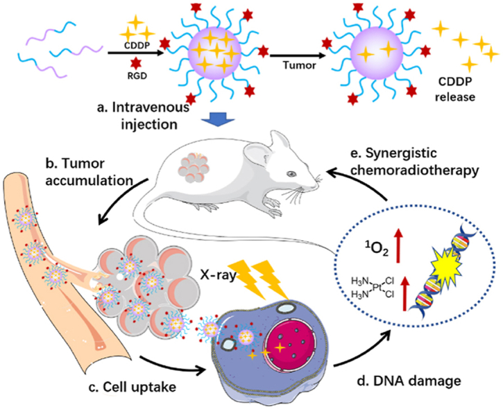
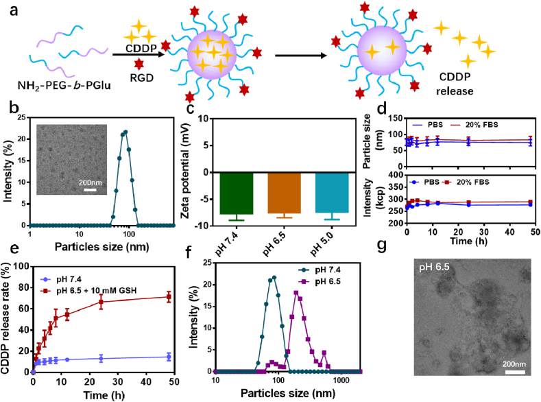
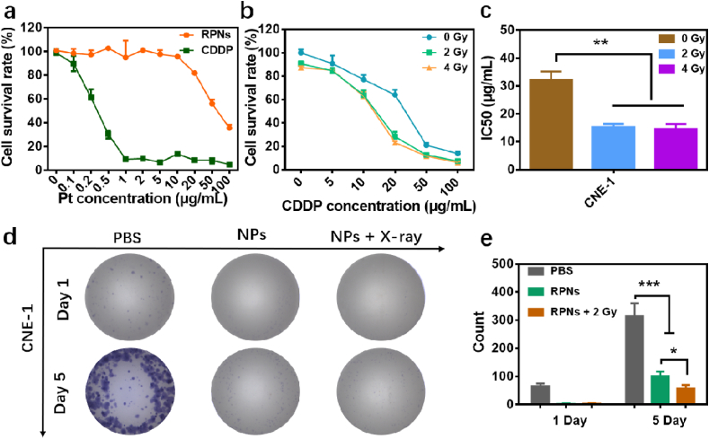
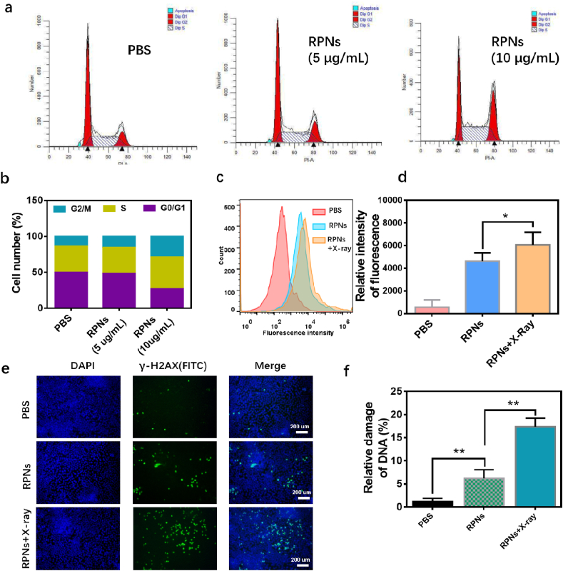
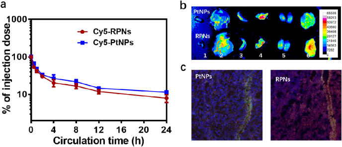
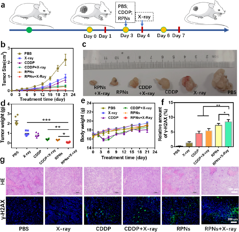
Despite extensive use of radiotherapy in nasopharyngeal carcinoma (NPC) treatment because of its high radiosensitivity, there have been huge challenges in further improving therapeutic effect, meanwhile obviously reducing radiation damage. To this end, synergistic chemoradiotherapy has emerged as a potential strategy for highly effective NPC therapy. Here, we developed RGD-targeted platinum-based nanoparticles (RGD-PtNPs, denoted as RPNs) to achieve targeted chemoradiotherapy for NPC. Such nanoparticles consist of an RGD-conjugated shell and a cis-platinum (CDDP) crosslinking core. Taking advantage of RGD, the RPNs may effectively accumulate in tumor, penetrate into tumor tissues and be taken by cancer cells, giving rise to a high delivery efficiency of CDDP. When they are fully enriched in tumor sites, the CDDP loaded RPNs can act as radiotherapy sensitizer and chemotherapy agents. By means of X-ray-promoted tumor cell uptake of nanoparticle and CDDP-induced cell cycle arrest in radiation-sensitive G2/M phases, RPNs may offer remarkable therapeutic outcome in the synergistic chemoradiotherapy for NPC.

Keywords: Chemoradiotherapy; Nasopharyngeal carcinoma (NPC); Polymeric nanoparticles; Precise treatment.


Conflict of interest statement

The authors declare no competing interests.

Figures

Image 1
Graphical abstract
Scheme 1
Scheme 1
Schematic illustration of the preparation and the application of RPNs in synergistic chemoradiotherapy.
Fig. 1
Fig. 1
Fabrication and characterization of RPNs. a. The preparation scheme of RPNs and the responsive release of CDDP from RPNs. b. The hydrodynamic diameters and TEM image of RPNs. c. Zeta potentials of RPNs. d. The stability of RPNs determined by particle size and intensity of RPNs over 48 hours. e. The in vitro pH-responsive CDDP release of RPNs. f. The particles size of RPNs in acidic condition at 48 h. g. The TEM image of RPNs in acidic condition at 48 h.
Fig. 2
Fig. 2
In vitro synergistic therapeutic effect of RPNs. a. The cytotoxicities of CDDP and RPNs monitored in 3T3 cells. b-c. The cytotoxicities (b) and IC50 (c) for CNE-1 cells treated with RPNs and X-ray (0, 2, and 4 Gy). d-e. The clone formation (d) and statistical analysis (e) in CNE-1 cells after treating with PBS, RPNs, and RPNs+X-ray (2 Gy). Statistical analysis was operated by one-way ANOVA for multiple groups and Student's t-test for two groups. *P < 0.05, **P < 0.01, ***P < 0.001.
Fig. 3
Fig. 3
In vitro synergistic mechanism of RPNs for CNE-1 cells. a. Flow cytometric analysis of cell cycles for CNE-1 cells, which treated with PBS, RPNs (5 μg/mL), and RPNs (10 μg/mL). b. Quantitative analysis of the G1, G2/M, S phases of CNE-1 cells treated with PBS, RPNs (5 μg/mL), and RPNs (10 μg/mL)). c-d. Flow cytometric (c) and quantitative analysis (d) of cell uptake in CNE-1 cells treated with PBS, Cy5-RPNs, and Cy5-RPNs+X-ray. e-f. γ-H2AX immunofluorescent staining (e) and statistical analysis (f) in CNE-1 cells, administrated with PBS, RPNs, and RPNs+X-ray. Statistical analysis was operated by Student's t-test for two groups. *P < 0.05, **P < 0.01, ***P < 0.001.
Fig. 4
Fig. 4
The in vivo delivery efficiency of RPNs. The in vivo pharmacokinetics (a), biodistribution (b) (1. Heart, 2. Liver, 3. Spleen, 4. Lung, 5. Kidney, 6. Tumor) and tumor penetration (c) of RPNs and PtNPs.
Fig. 5
Fig. 5
In vivo radiochemotherapy studies. a. Schematic illustration of the treatment for NPC-bearing nude mice. b-f. Tumor volume (b), photographs (c), weight (d), body weight (e), DNA damage levels (f) of mice with various treatments including PBS, X-ray, CDDP, CDDP+X-ray, RPNs, and RPNs+X-ray at the dose of 2 Gy and 2 mg CDDP/kg. g. The effect of radiochemotherapy on the histopathology and γ-H2AX immunofluorescent staining from mice in each group over 21 days. Data represent the mean ± SD (n = 6). Statistical analysis was operated by one-way ANOVA for multiple groups and Student's t-test for two groups. *P < 0.05, **P < 0.01, ***P < 0.001.
Fig. 6
Fig. 6
In vivo biocompatibility and biodegradability of RPNs. H&E staining of major organs (heart, liver, spleen, lung, and kidney) from NPC-bearing mice treated with RPNs+X-ray on the 21st day.

Similar articles

Cited by

References

    1. Chua M.L.K., Wee J.T.S., Hui E.P., Chan A.T.C. Lancet. 2016;387:1012–1024. - PubMed
    1. Wei W.I., Sham J.S.T. Lancet. 2005;365:2041–2054. - PubMed
    1. Chen Y.-P., Chan A.T.C., Le Q.-T., Blanchard P., Sun Y., Ma J. Lancet. 2019;394:64–80. - PubMed
    1. Blanchard P., Lee A., Marguet S., Leclercq J., Ng W.T., Ma J., Chan A.T.C., Huang P.-Y., Benhamou E., Zhu G., Chua D.T.T., Chen Y., Mai H.-Q., Kwong D.L.W., Cheah S.L., Moon J., Tung Y., Chi K.-H., Fountzilas G., Zhang L., Hui E.P., Lu T.-X., Bourhis J., Pignon J.P. Lancet Oncol. 2015;16:645–655. - PubMed
    1. Caudell J.J., Torres-Roca J.F., Gillies R.J., Enderling H., Kim S., Rishi A., Moros E.G., Harrison L.B. Lancet Oncol. 2017;18:e266–e273. - PMC - PubMed

LinkOut - more resources