. 2021 Mar 31;6(5):416-427.
doi: 10.1016/j.jacbts.2021.01.012. eCollection 2021 May.

Downstream Paclitaxel Released Following Drug-Coated Balloon Inflation and Distal Limb Wound Healing in Swine

Affiliations

Downstream Paclitaxel Released Following Drug-Coated Balloon Inflation and Distal Limb Wound Healing in Swine

Juan F Granada et al. JACC Basic Transl Sci. .

Abstract

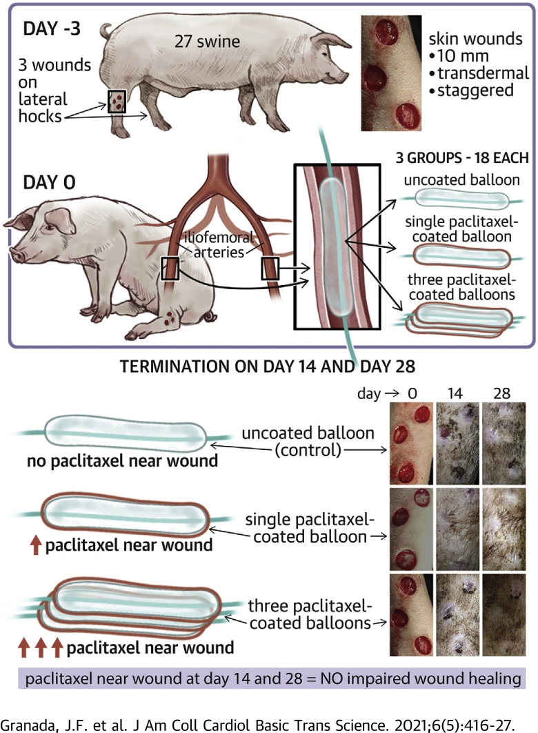
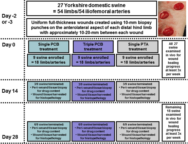
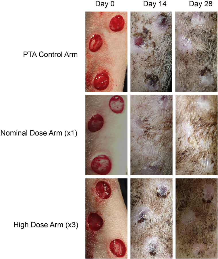
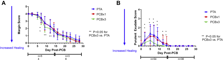
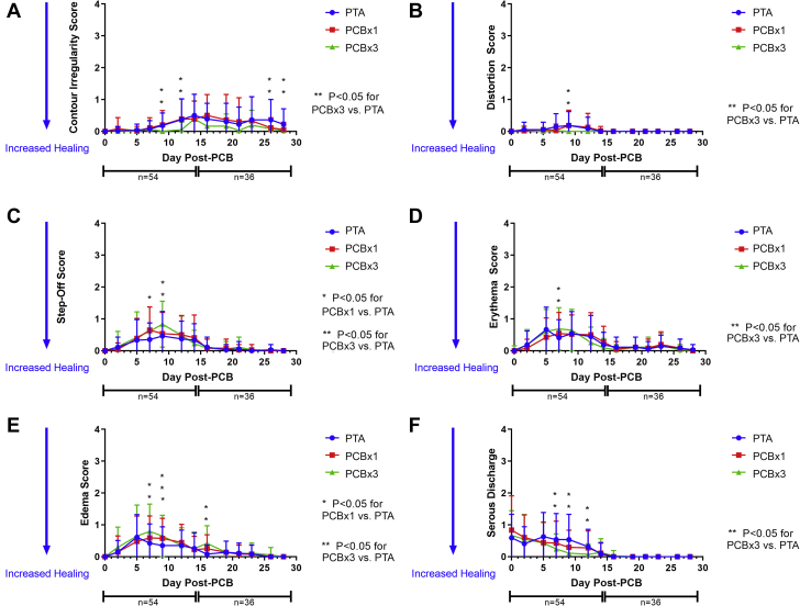
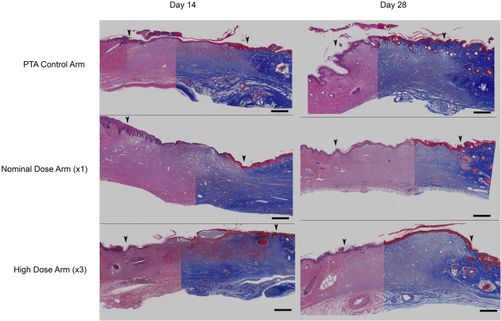
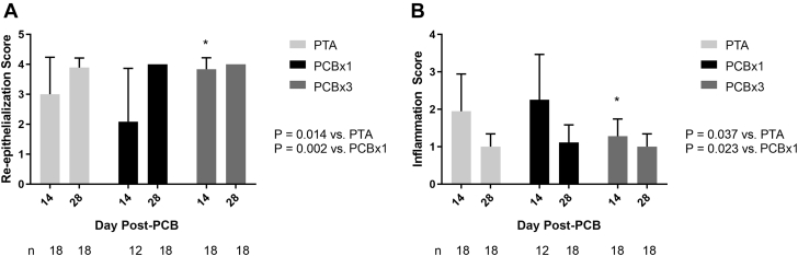
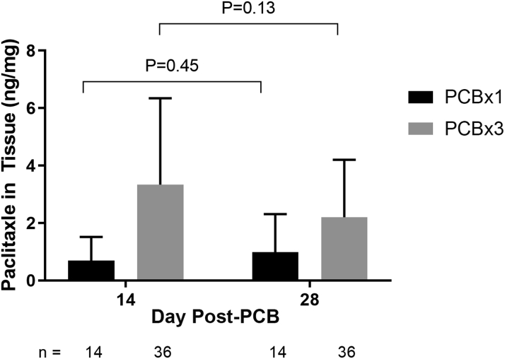
The authors evaluated the presence of paclitaxel and healing of distal hind limb wounds created in 27 swine using biopsy punches followed by paclitaxel-coated balloon (PCB) use in the iliofemoral arteries of healthy swine. After 14 and 28 days, no differences were seen in time course, appearance, and histopathology of wound healing between the single or triple PCB and uncoated balloon treatment despite clinically relevant paclitaxel concentrations in the skin adjacent to the healing wounds. Presence of paclitaxel downstream from the PCB treatment site does not impair the wound healing response of preexisting distal cutaneous lesions in healthy swine.

Keywords: BTK, below the knee; CLI, critical limb ischemia; PCB, paclitaxel coated balloon; PTA, percutaneous transluminal angioplasty; SFA, superficial femoral artery; histopathology; paclitaxel-coated balloon; swine; translational; wound healing.


Conflict of interest statement

Ms. Crookall, and Drs. Schulz-Jander, Tunev, and Melder are full-time employees of Medtronic PLC, the manufacturer of the IN.PACT Admiral product evaluated in the study. Dr. Granada is the CEO, and Dr. Kaluza is an employee of the Cardiovascular Research Foundation which receives research and educational grant support from Medtronic. All other authors have reported that they have no relationships relevant to the contents of this paper to disclose.

Figures

None
Graphical abstract
Figure 1
Figure 1
Study Flow Chart Chart shows key study design aspects, methods and milestones. PCB = paclitaxel-coated balloon; PTA = percutaneous transluminal angioplasty.
Figure 2
Figure 2
Visual Assessment of Wound Healing Progression Photographs showing representative examples of leg wounds from control and treatment arms over the course of the study. Granulation tissue was grossly well established in all arms after 14 days of healing with complete closure seen in most cases by day 28. Abbreviation as in Figure 1.
Figure 3
Figure 3
Margin Separation Score and Purulent Exudate Score (A) Margin separation score of all wounds by study arm. Mean scores ± SD are shown for 0 to 28 days post-treatment. The number (n) of monitored wounds decreased after 14 days due to termination of the first cohort of animals for histological and drug content sampling at the study midpoint. The observed rate of wound closure trended consistently across all 3 study arms, with scores decreasing with increased healing. (B) Purulent exudate score of all wounds by study arm. Mean scores ± SD are shown for 0 to 28 days post-treatment. The number (n) of scored wounds decreased after 14 days due to termination of the first cohort of animals for histological and drug content sampling. Scores for all arms trended in a similar manner with exudate largely resolved after 2 weeks, although scores for high-dose arm (PCB × 3) were consistently less than the other 2 arms. Abbreviations as in Figure 1.
Figure 4
Figure 4
Inflammation and Cosmesis Parameter Scores (A to F) Markers with maximum scores <1. Mean and SD shown for each timepoint. All arms followed the same wound resolution trends with regard to magnitude and time course. Distortion, step-off, erythema, edema and serous discharge were most apparent during the first 2 weeks after wounding, resolving during the following 2 weeks. Slight contour irregularity developed later during healing, but also showed similar trends across all arms. Abbreviations as in Figures 1 and 3.
Figure 5
Figure 5
Healing Response Histology Composite images (Masson’s trichrome, right; hematoxylin and eosin stain, left) of PTA control arm, nominal-dose (PCB × 1) arm and high-dose (PCB × 3) arm at 14 and 28 days post-treatment, respectively (bar scale = 1 mm). Arrows indicate the approximate demarcation of the normal (nonwounded) skins to the left and right of the healing wound. Histological appearance of the healing responses across the study arms is similar; at 14 days, the dermal collagen is not as dense, and the collagen bundles are not as thick appearing in the center of the wound as in the flanking nonwounded skin areas and there is gradation of the collagen density along the wound border and from the base to the surface representing normal healing and progressive production of dermal collagen. At 28 days the wound surfaces are covered by keratinized epidermis and the wound defect is filled with mature granulation tissue. Abbreviations as in Figure 1.
Figure 6
Figure 6
Re-Epithelialization Score and Dermal Inflammation Score (A) Re-epithelialization mean scores and SD by study arm. Numbers (n) of independent histological samples evaluated for epithelialization score are indicated. Significantly higher re-epithelialization score was found in the high-dose (PCB × 3) arm relative to both the PTA control and nominal-dose (PCB × 1) arms, although no significant differences were found between any arms at 28 days post-treatment (Mann-Whitney test). (B) Dermal inflammation mean scores and SD by study arm. Numbers (n) of independent histological samples evaluated for inflammation score indicated. Significantly lower inflammation score was found in the PCB × 3 arm relative to both the PTA control and PCB × 1 arms, although no significant differences were found between any arms at 28 days post-treatment (Mann-Whitney test). Abbreviations as in Figure 1.
Figure 7
Figure 7
Paclitaxel Tissue Concentration Mean amount of paclitaxel in full-thickness biopsy punch sample adjacent to wound. Mean drug concentration and SD are indicated for PCB × 1 and PCB × 3 study arms showing consistently greater drug concentrations with the high dose (PCB × 3) at both time points with no significant decline of drug concentrations from 14 to 28 days post-treatment in either arm. Abbreviations as in Figure 1.

Comment in

References

    1. Laird J.A., Schneider P.A., Jaff M.R. Long-term clinical effectiveness of a drug-coated balloon for the treatment of femoropopliteal lesions. Circ Cardiovasc Interv. 2019;12 - PMC - PubMed
    1. Rosenfield K., Jaff M.R., White C.J. Trial of a paclitaxel-coated balloon for femoropopliteal artery disease. N Engl J Med. 2015;373:145–153. - PubMed
    1. Brodmann M., Werner M., Meyer D.R. Sustainable antirestenosis effect with a low-dose drug-coated balloon: the ILLUMENATE European randomized clinical trial 2-year results. J Am Coll Cardiol Intv. 2018;11:2357–2364. - PubMed
    1. Cremers B., Speck U., Kaufels N. Drug-eluting balloon: very short-term exposure and overlapping. Thromb Haemost. 2009;101:201–206. - PubMed
    1. Gongora C.A., Shibuya M., Wessler J.D. Impact of paclitaxel dose on tissue pharmacokinetics and vascular healing: a comparative drug-coated balloon study in the familial hypercholesterolemic swine model of superficial femoral in-stent restenosis. J Am Coll Cardiol Intv. 2015;8:1115–1123. - PubMed