. 2021 Apr 7;6(5):431-443.
doi: 10.1016/j.jacbts.2021.01.013. eCollection 2021 May.

Selective Interleukin-6 Trans-Signaling Blockade Is More Effective Than Panantagonism in Reperfused Myocardial Infarction

Affiliations

Selective Interleukin-6 Trans-Signaling Blockade Is More Effective Than Panantagonism in Reperfused Myocardial Infarction

Marc Jonathan George et al. JACC Basic Transl Sci. .

Abstract

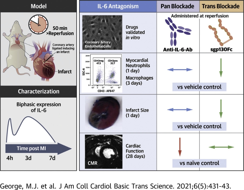
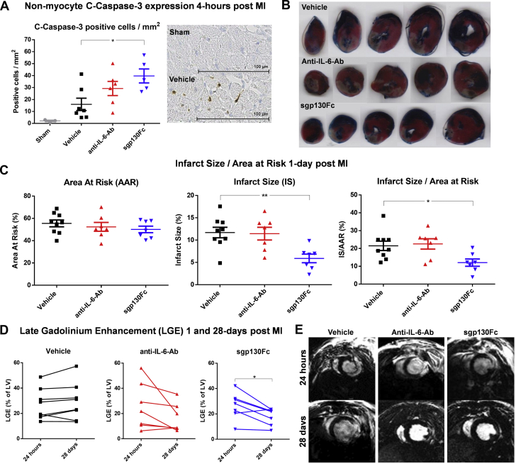
Interleukin (IL)-6 is an emerging therapeutic target in myocardial infarction (MI). IL-6 has 2 distinct signaling pathways: trans-signaling, which mediates inflammation, and classic signaling, which also has anti-inflammatory effects. The novel recombinant fusion protein sgp130Fc achieves exclusive trans-signaling blockade, whereas anti-IL-6 antibodies (Abs) result in panantagonism. In a rat model of reperfused MI, sgp130Fc, but not anti-IL-6-Ab, attenuated neutrophil and macrophage infiltration into the myocardium, reduced infarct size, and preserved cardiac function 28 days after MI. These data demonstrate the efficacy of exclusive IL-6 trans-signaling blockade and support further investigation of sgp130Fc as a potential novel therapy in MI.

Keywords: AAR, area at risk; Ab, antibody; CCL, C-C motif chemokine ligand; CMR, cardiac magnetic resonance; CXCL, C-X-C motif ligand; ICAM-1, intercellular adhesion molecule 1; IL, interleukin; IS, infarct size; LGE, late-gadolinium enhancement; LVEF, left ventricular ejection fraction; MHC, major histocompatibility complex; MI, myocardial infarction; NSTEMI, non–ST-segment-elevation MI; RCAEC, rat coronary artery endothelial cell; STEMI, ST-segment-elevation MI; TCZ, tocilizumab; Trop-T, troponin T; c-caspase-3, cleaved caspase-3; inflammation; interleukin-6; myocardial infarction; reperfusion; sIL-6R, soluble IL-6 receptor.


Conflict of interest statement

This work was supported by the Wellcome Trust (108735/Z/15/Z to Dr. George and 212937/Z/18/Z to Dr. Stuckey), the British Heart Foundation (FS/15/33/31608 and RM/17/1/33377 to Dr. Stuckey), the Medical Research Council (MR/R026416/1 to Dr. Stuckey), the National Institute for Health Research (Senior Investigator: Dr, Hingorani), and a King Scholarship of Malaysia (Ms. Jasmin). Dr. Woollard is an employee of AstraZeneca. No funding or support was received from AstraZeneca. All other authors have reported that they have no relationships relevant to the contents of this paper to disclose.

Figures

None
Graphical abstract
Figure 1
Figure 1
IL-6 Stimulation of RCAECs and In Vitro Antagonism of IL-6 Signaling With Anti–IL-6-Ab and sgp130Fc (A) RCAECs were seeded in 24-well plates at P8 and grown until confluent in serum-containing medium. Confluent cells were imaged with a light microscope to confirm endothelial cell morphology. (B) The cells were stimulated with 1, 10, or 100 ng/ml IL-6 ± 500 ng/ml sIL-6R in serum-free medium for 16 h. CCL2 was measured in the supernates by means of enzyme-linked immunosorbent assay (ELISA), and surface ICAM-1 expression was quantified by means of flow cytometry. (C) Shed sIL-6R was measured by means of ELISA in the supernate of RCAECs incubated for 16 h with and without 100 ng/ml IL-6. RCAECs were stimulated with 100 ng/ml IL-6 for 16 h with increasing concentrations of (D) anti–IL-6-Ab (AF506) and (E) sgp130Fc alone and (F) with sIL-6R. n = 3 (technical repeats), data shown as mean + SEM. Statistical significance was tested with the use of analysis of variance with multiple comparisons (comparing each condition with [B, C] unstimulated or [D to F], 100 ng/ml IL-6; black asterisks) or unpaired Student's t-test (D to F, unstimulated vs. 10 ng/ml IL-6; gray asterisks). Multiplicity-adjusted p < 0.05 was considered to be significant. p < 0.05; ∗∗p < 0.01; ∗∗∗p < 0.001. ICAM = intercellular adhesion molecule; IL-6 = interleukin; RCAEC = rat coronary artery endothelial cell; sIL-6R = soluble IL-6 receptor.
Figure 2
Figure 2
Temporal Profile of IL-6, sIL-6R, sgp130Fc, and Leukocytes After MI With Reperfusion Rats were subjected to surgical MI with 50 min of ischemia before reperfusion. (A) IL-6, (B) sIL-6R, and (C) sgp130Fc were measured by means of enzyme-linked immunosorbent assay in plasma (blue) ± supernates of heart digests (red) at 7 time points (0 h to 7 days after MI) (n = 3 to 4/group). (D) Leukocyte counts in the myocardium and (E) their CD11b expression were measured by means of flow cytometry at 6 time points (0 h to 7 days after MI) (n = 3 to 4/group). Statistical significance was tested by means of analysis of variance with multiple comparisons (comparing each time point with naive rats). Data presented as mean ± SEM. Multiplicity-adjusted p < 0.05 was considered to be significant. p < 0.05; ∗∗p < 0.01; ∗∗∗p < 0.001. MI = myocardial infarction; other abbreviations as in Figure 1.
Figure 3
Figure 3
Effect of Anti–IL-6-Ab and sgp130Fc on Soluble Inflammatory Mediators and Leukocytes After MI With Reperfusion Rats were subjected to surgical myocardial infarction with 50 min of ischemia before reperfusion. One minute before reperfusion, vehicle (phosphate-buffered saline solution [PBS]), anti–IL-6-Ab (0.1 μg/mg, second dose intraperitoneally 3 days after MI), or sgp130Fc (0.5 μg/mg) was administered intravenously in 1 ml PBS (n = 5–9/group). (A to D) Cytokines in plasma and supernate of myocardial digests were measured by means of enzyme-linked immunosorbent assay and enzyme immunoassay. (E to K) Cell concentrations and CD11b expression were measured by means of flow cytometry. Data presented as mean ± SEM. Statistical significance was tested by means of analysis of variance (ANOVA) with multiple comparisons (comparing drug groups with vehicle; black asterisks and daggers) or unpaired Student’s t-tests (comparing sham with vehicle controls; gray asterisks and daggers). Where ANOVA was significant but differences between vehicle and the drug groups were not, a post hoc unpaired Student’s t-test was performed between the two drug groups (red asterisk). Multiplicity-adjusted (asterisks) and ANOVA (daggers) p < 0.05 was considered to be significant. †p<0.05; ∗∗††p<0.01; ∗∗∗†††p<0.001. Ab = antibody; MHC = major histocompatibility complex; Mo = monocytes; other abbreviations as in Figures 1 and 2.
Figure 4
Figure 4
The Effect of Anti–IL-6-Ab and sgp130Fc on Infarct Size, c-Caspase-3 Expression, and LGE After MI With Reperfusion Rats were subjected to surgical myocardial infarction with 50 min ischemia prior to reperfusion. 1-min prior to reperfusion, vehicle (phosphate-buffered saline solution [PBS]), anti–IL-6-Ab (0.1 μg/mg, second dose intraperitoneally 3 days after MI) or sgp130Fc (0.5 μg/mg) were administered intravenously in 1 mL PBS (n = 7–9/group). (A) Hearts were excised 4 h after MI and stained with an anti–c-caspase-3 antibody. Nonmyocytes positive for c-caspase-3 were manually counted in a 4-mm2 transmural grid sited over the infarct area and expressed as positive cells/mm2 (n = 5–7/group). (B, C) Area at risk and infarct size were measured histologically by staining with 2,3,5-triphenyl-tetrazolium-chloride and Evans Blue and analyzed with the use of ImageJ 1 day after MI. (D, E) In further groups, electrocardiography-gated cardiac magnetic resonance–cine and gadolinium scans were performed 1 and 28 days after MI. LGE as percentage of total LV mass was calculated. Data presented as mean ± SEM. (A, B) Statistical significance tested with the use of analysis of variance with multiple comparisons (comparing the drug groups with vehicle) or unpaired Student's t-test (sham vs. vehicle). (C) Paired Student’s t-tests were performed to compare means of the 2 time points within each group. Multiplicity-adjusted p < 0.05 was considered to be significant. p < 0.05; ∗∗p < 0.01; ∗∗∗p < 0.001. c-Caspase-3, cleaved caspase-3; LGE = late gadolinium enhancement; LV = left ventricular; other abbreviations as in Figures 1 and 2.

Comment in

References

    1. Ziaeian B., Fonarow G.C. Epidemiology and aetiology of heart failure. Nat Rev Cardiol. 2016;13:368–378. - PMC - PubMed
    1. Fanola C.L., Morrow D.A., Cannon C.P. Interleukin-6 and the risk of adverse outcomes in patients after an acute coronary syndrome: observations from the SOLID-TIMI 52 (Stabilization of Plaque Using Darapladib-Thrombolysis in Myocardial Infarction 52) trial. J Am Heart Assoc. 2017;6 - PMC - PubMed
    1. Chimen M., Yates C.M., McGettrick H.M. Monocyte subsets coregulate inflammatory responses by integrated signaling through TNF and IL-6 at the endothelial cell interface. J Immunol. 2017;198:2834–2843. - PMC - PubMed
    1. Romano M., Sironi M., Toniatti C. Role of IL-6 and its soluble receptor in induction of chemokines and leukocyte recruitment. Immunity. 1997;6:315–325. - PubMed
    1. Modur V., Li Y., Zimmerman G.A., Prescott S.M., McIntyre T.M. Retrograde inflammatory signaling from neutrophils to endothelial cells by soluble interleukin-6 receptor alpha. J Clin Invest. 1997;100:2752–2756. - PMC - PubMed