

3-Dimensional Bioprinting of Cardiovascular Tissues: Emerging Technology
- PMID: 34095635
- PMCID: PMC8165127
- DOI: 10.1016/j.jacbts.2020.12.006
3-Dimensional Bioprinting of Cardiovascular Tissues: Emerging Technology
Abstract
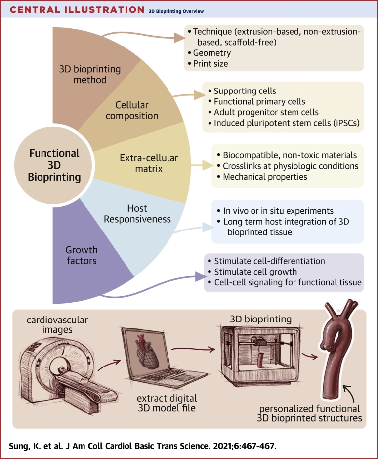
Three-dimensional (3D) bioprinting may overcome challenges in tissue engineering. Unlike conventional tissue engineering approaches, 3D bioprinting has a proven ability to support vascularization of larger scale constructs and has been used for several cardiovascular applications. An overview of 3D bioprinting techniques, in vivo translation, and challenges are described.
Keywords: 3-dimensional; 3D, 3-dimensional; ECM, extracellular matrix; HUVEC, human umbilical vein endothelial cell(s); MSC, mesenchymal stem cell(s); UV, ultraviolet; bioink; bioprinting; cardiovascular disease; hCPC, human cardiac-derived progenitor cell(s); hiPSC, human induced-pluripotent stem cell(s); regenerative medicine; tissue engineering.
Conflict of interest statement
Dr. Ashammakhi has received grant support from the National Institutes of Health (UG3TR003148) and the American Heart Association (18TPA34230036, 442611-NU-80922). Dr. Nguyen has received grant support from the American Heart Association (18TPA34170049); the National Heart, Lung, and Blood Institute (R01HL148182); and the Veterans Health Administration (VA-MERIT, I01-CX001901). All other authors have reported that they have no relationships relevant to the contents of this paper to disclose.
Figures

Similar articles
-
Mechanically robust cryogels with injectability and bioprinting supportability for adipose tissue engineering.Acta Biomater. 2018 Jul 1;74:131-142. doi: 10.1016/j.actbio.2018.05.044. Epub 2018 May 26. Acta Biomater. 2018. PMID: 29842971
-
Progress in 3D bioprinting technology for tissue/organ regenerative engineering.Biomaterials. 2020 Jan;226:119536. doi: 10.1016/j.biomaterials.2019.119536. Epub 2019 Oct 11. Biomaterials. 2020. PMID: 31648135 Review.
-
Bioink with cartilage-derived extracellular matrix microfibers enables spatial control of vascular capillary formation in bioprinted constructs.Biofabrication. 2022 Apr 20;14(3). doi: 10.1088/1758-5090/ac6282. Biofabrication. 2022. PMID: 35354130
-
Advances in three-dimensional bioprinted stem cell-based tissue engineering for cardiovascular regeneration.J Mol Cell Cardiol. 2022 Aug;169:13-27. doi: 10.1016/j.yjmcc.2022.04.017. Epub 2022 May 12. J Mol Cell Cardiol. 2022. PMID: 35569213 Free PMC article. Review.
-
ECM Based Bioink for Tissue Mimetic 3D Bioprinting.Adv Exp Med Biol. 2018;1064:335-353. doi: 10.1007/978-981-13-0445-3_20. Adv Exp Med Biol. 2018. PMID: 30471042 Review.
Cited by
-
Alternative Therapies in Transplantology as a Promising Perspective in Medicine.Ann Transplant. 2024 Jun 4;29:e943387. doi: 10.12659/AOT.943387. Ann Transplant. 2024. PMID: 38831572 Free PMC article. Review.
-
Hydrogels for Cardiac Tissue Regeneration: Current and Future Developments.Int J Mol Sci. 2025 Mar 5;26(5):2309. doi: 10.3390/ijms26052309. Int J Mol Sci. 2025. PMID: 40076929 Free PMC article. Review.
-
Emerging 3D bioprinting applications in plastic surgery.Biomater Res. 2023 Jan 3;27(1):1. doi: 10.1186/s40824-022-00338-7. Biomater Res. 2023. PMID: 36597149 Free PMC article. Review.
-
A deep dive into the darning effects of biomaterials in infarct myocardium: current advances and future perspectives.Heart Fail Rev. 2022 Jul;27(4):1443-1467. doi: 10.1007/s10741-021-10144-3. Epub 2021 Aug 3. Heart Fail Rev. 2022. PMID: 34342769 Review.
-
Development and Prospective Applications of 3D Membranes as a Sensor for Monitoring and Inducing Tissue Regeneration.Membranes (Basel). 2023 Sep 18;13(9):802. doi: 10.3390/membranes13090802. Membranes (Basel). 2023. PMID: 37755224 Free PMC article. Review.
References
-
- Weinand C., Jian W.X., Peretti G.M., Bonassar L.J., Gill T.J. Conditions affecting cell seeding onto three-dimensional scaffolds for cellular-based biodegradable implants. J Biomed Mater Res B Appl Biomater. 2009;91:80–87. - PubMed
-
- Radisic M., Euloth M., Yang L., Langer R., Freed L.E., Vunjak-Novakovic G. High-density seeding of myocyte cells for cardiac tissue engineering. Biotechnol Bioeng. 2003;82:403–414. - PubMed
Publication types
LinkOut - more resources
Full Text Sources
