. 2021 May 7;2(5):100262.
doi: 10.1016/j.xcrm.2021.100262. eCollection 2021 May 18.

Vaccine-induced ICOS+CD38+ circulating Tfh are sensitive biosensors of age-related changes in inflammatory pathways

Affiliations

Vaccine-induced ICOS+CD38+ circulating Tfh are sensitive biosensors of age-related changes in inflammatory pathways

Ramin Sedaghat Herati et al. Cell Rep Med. .

Abstract

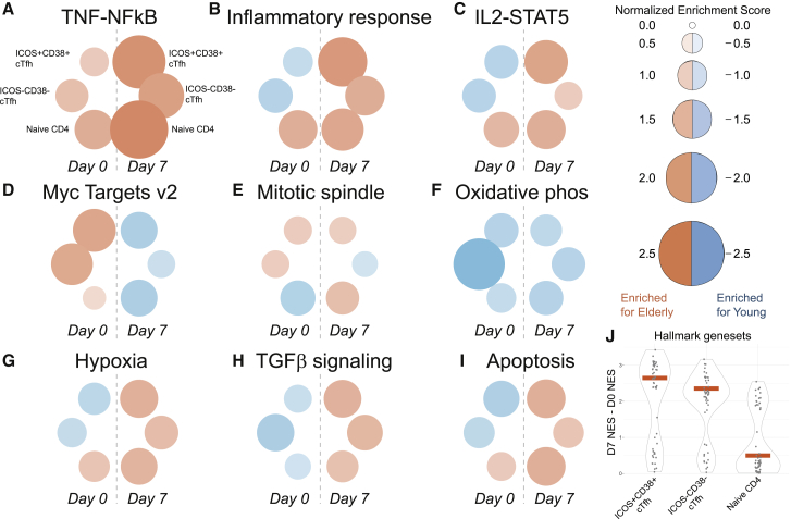
Humoral immune responses are dysregulated with aging, but the cellular and molecular pathways involved remain incompletely understood. In particular, little is known about the effects of aging on T follicular helper (Tfh) CD4 cells, the key cells that provide help to B cells for effective humoral immunity. We performed transcriptional profiling and cellular analysis on circulating Tfh before and after influenza vaccination in young and elderly adults. First, whole-blood transcriptional profiling shows that ICOS+CD38+ cTfh following vaccination preferentially enriches in gene sets associated with youth versus aging compared to other circulating T cell types. Second, vaccine-induced ICOS+CD38+ cTfh from the elderly had increased the expression of genes associated with inflammation, including tumor necrosis factor-nuclear factor κB (TNF-NF-κB) pathway activation. Finally, vaccine-induced ICOS+CD38+ cTfh display strong enrichment for signatures of underlying age-associated biological changes. These data highlight the ability to use vaccine-induced cTfh as cellular "biosensors" of underlying inflammatory and/or overall immune health.

Keywords: CD4; NF-kB; T follicular helper; aging; cellular biosensors; influenza; network analysis; vaccine.


Conflict of interest statement

The authors declare no competing interests. E.J.W. consults or is an advisor for Merck, Elstar, Janssen, Related Sciences, Synthekine, and Surface Oncology, unrelated to the present study. E.J.W. is a founder of Surface Oncology and Arsenal Biosciences, unrelated to the present study. E.J.W. is an inventor on a patent (US patent no. 10,370,446) submitted by Emory University that covers the use of PD-1 blockade to treat infections and cancer.

Figures

None
Graphical abstract
Figure 1
Figure 1
ICOS+CD38+ cTfh responses appear similar with aging A cohort of young adults (n = 28) and elderly adults (n = 35) received influenza vaccination in Fall 2014. (A and B) Flow cytometry for days 0 and 7 after vaccination in the frequency of cTfh co-expressing ICOS and CD38 for young (orange, n = 27) and elderly (purple, n = 35) adults. (C and D) Fold change between days 0 and 7 for the change in ICOS+CD38+ cTfh frequency (C) or geometric MFI of PD-1 (D) in ICOS+CD38+ cTfh for young (orange, n = 27) and elderly (purple, n = 35) adults. (E) Correlation between ICOS+CD38+ cTfh frequency fold change at day 7 compared to day 0, and the plasmablast fold change at day 7 compared to day 0 for young (orange, n = 25) and elderly (purple, n = 26) adults. (F) Peripheral blood mononuclear cells (PBMCs) from young (n = 6) and elderly (n = 8) adults were sorted for ICOS+CD38+ cTfh, ICOSCD38 cTfh, and naive CD4 at days 0 and 7 after vaccination. Log-transformed transcriptional profiling data were queried for selected genes for young (orange) and elderly (purple) adults at day 7 after vaccination. The heatmap is row normalized.
Figure 2
Figure 2
TNF-NF-κB pathway is enriched with aging in ICOS+CD38+ cTfh (A) Schematic for RNA-seq analyses. (B and C) Weighted gene correlation network analysis on ICOS+CD38+ cTfh at day 7 from young (B) and elderly (C) adults. (D) Module preservation analysis was performed using Fisher’s exact test. Heatmap color and value indicate the −log10(p value) for the overlap in genes in modules. (E) Genes ranked based on module membership for elderly module EM4. Then, GSEA was performed using the MSigDB HALLMARK collection. Positive enrichment scores indicate enrichment for genes in EM4. (F) Transcription factors with module membership >0.80 in EM4 displayed as a multiple association network (GeneMANIA without gene prediction). (G) Ingenuity Pathway Analysis for predicted upstream regulators for ICOS+CD38+ cTfh at day 7 for young and elderly adults. (H) Differentially expressed NF-κB target genes comparing ICOS+CD38+ cTfh at day 7 from elderly (purple) and young (orange) adults. (I) GSEA for NF-κB target genes to compare ICOS+CD38+ cTfh from elderly and young adults. (J) Example plot from 1 young adult and 1 elderly adult for total NF-κB p50 protein from an independent cohort of young and elderly adults. Geometric mean fluorescence intensity (MFI) shown. (K) Total NF-κB p50 for ICOS+CD38+ cTfh at baseline in young (orange) and elderly (purple) adults (n = 16 in each group), as taken from an independent cohort of young and elderly adults. (L and M) Gene expression for TNFRSF1A (L) (n = 6 for young; n = 8 for elderly) and TNFRSF1B (M) (n = 8 for elderly) shown at day 7 after vaccine from log2-transformed counts data for young (orange) and elderly (purple) adults.
Figure 3
Figure 3
TNF signaling promotes Tfh-B cell interactions (A and B) PBMC from young adults were freshly isolated and sorted for coculture of T cell subsets (naive, gated as CD4+CD45RA+CD27+; CXCR5 memory, non-naive CD4+CXCR5; cTfh, non-naive CD4+CXCR5+PD-1+) with autologous naive B cells (CD3CD19+CD27loIgD+). Supernatant IgM (A) and IgG1 (B) were measured after 7 days, as shown for the following conditions: unstimulated (gray), SEB alone (0.5 μg/mL, green), SEB with recombinant human TNF (125 ng/mL, tan), or SEB with α-TNF antibodies (2 μg/mL, sienna) (1-way repeated-measures ANOVA with Holm-Sidak’s test; n = 12 per group). (C and D) Correlation shown for the TNFRSF1A in ICOS+CD38+ cTfh at day 7 compared with the H1N1-specific (C) hemagglutinin inhibitory (HAI) titer (n = 14) or H3N2-specific (D) HAI titer (n = 14) in young (orange) and elderly (purple) adults. (E) Correlation of apoptosis and NF-κB gene set GSVA scores for ICOS+CD38+ cTfh at day 7 for young (orange, n = 6) and elderly (purple, n = 8) adults. (F) Pearson coefficients for BCL family member genes compared to GSVA scores for TNF-NF-κB signaling in ICOS+CD38+ cTfh at day 7 for young (x axis) and elderly (y axis) adults. Genes classified as either pro-survival (dark blue) or apoptotic (sea green).
Figure 4
Figure 4
Age-dependent differences in transcriptional profiles of ICOS+CD38+ cTfh (A) t-Distributed stochastic neighbor embedding (t-SNE) analysis was performed for all CD4 T cell subsets. ICOS+CD38+ cTfh shown before and after vaccination for young (n = 6) and elderly (n = 8) adults. (B) Aggregated GSEA results for Hallmark gene sets comparing day 7 versus day 0 for ICOS+CD38+ cTfh in young (orange) and elderly (purple) adults. (C–F) GSEA shown for day 7 versus day 0 for ICOS+CD38+ cTfh from young (orange) and elderly (purple) adults for TGF-β signaling (C), E2F targets (D), IL-2 signaling (E), and TNF-NF-κB signaling (F) gene sets.
Figure 5
Figure 5
Age-related differences in signaling pathways are preferentially revealed by subset and time point (A–I) GSEA performed to compare young versus elderly for each CD4 T cell subset for each time point. Normalized enrichment score shown as circle size and color. (J) “Delta-NES” score was calculated by subtracting the day 0 NES score from the day 7 NES score with aging for each gene set for each subset. Each dot represents 1 gene set. The red bar indicates median.
Figure 6
Figure 6
ICOS+CD38+ cTfh transcriptional profiles reveal signatures of aging (A) Signatures for youth and aging were constructed from the differential expression of young versus elderly whole-blood transcriptional profiling from Immport study SDY739. These signatures were tested by pre-ranked GSEA for other vaccine years in this study (SDY622, SDY648, and SDY819). (B) The signatures of youth and aging were validated against publicly available whole-blood transcriptional profiles (GSE accessions GEO: GSE123696, GEO: GSE123697, GEO: GSE123698, and GEO: GSE79396) and Nanostring target profiling (EGAS00001002460, Milieu Intérieur) for young (ages 20–40) and elderly (ages 60 and older) adults. Normalized enrichment score (NES) shown for youth (left) and aging (right) signatures. (C) CD4 T cell subsets were tested for signatures of youth and aging. Naive (yellow), ICOSCD38 cTfh (green), and ICOS+CD38+ cTfh (orange) are shown, with days 0 and 7 connected by dotted lines. The yellow region indicates FDR < 0.05.

Similar articles

Cited by

References

    1. Lang P.-O., Mendes A., Socquet J., Assir N., Govind S., Aspinall R. Effectiveness of influenza vaccine in aging and older adults: comprehensive analysis of the evidence. Clin. Interv. Aging. 2012;7:55–64. - PMC - PubMed
    1. Goronzy J.J., Weyand C.M. Understanding immunosenescence to improve responses to vaccines. Nat. Immunol. 2013;14:428–436. - PMC - PubMed
    1. Gustafson C.E., Weyand C.M., Goronzy J.J. T follicular helper cell development and functionality in immune ageing. Clin. Sci. (Lond.) 2018;132:1925–1935. - PMC - PubMed
    1. Henry C., Zheng N.-Y., Huang M., Cabanov A., Rojas K.T., Kaur K., Andrews S.F., Palm A.E., Chen Y.-Q., Li Y. Influenza Virus Vaccination Elicits Poorly Adapted B Cell Responses in Elderly Individuals. Cell Host Microbe. 2019;25:357–366.e6. - PMC - PubMed
    1. Xu W., Larbi A. Markers of T Cell Senescence in Humans. Int. J. Mol. Sci. 2017;18:E1742. - PMC - PubMed

Publication types

MeSH terms