. 2021 Jun 16;143(23):8925-8934.
doi: 10.1021/jacs.1c04191. Epub 2021 Jun 5.

Redefining Protein Interfaces within Protein Single Crystals with DNA

Affiliations

Redefining Protein Interfaces within Protein Single Crystals with DNA

Benjamin E Partridge et al. J Am Chem Soc. .

Abstract

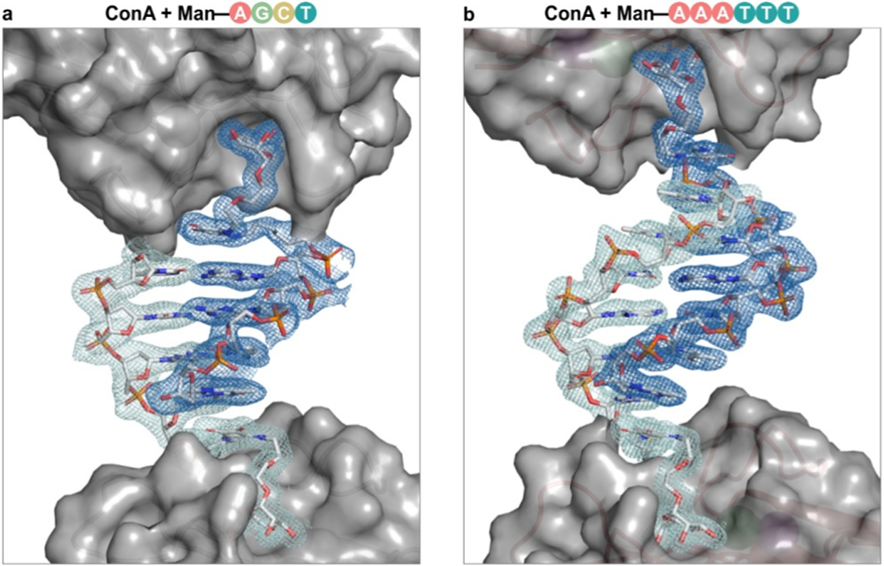
Proteins are exquisite nanoscale building blocks: molecularly pure, chemically addressable, and inherently selective for their evolved function. The organization of proteins into single crystals with high positional, orientational, and translational order results in materials where the location of every atom can be known. However, controlling the organization of proteins is challenging due to the myriad interactions that define protein interfaces within native single crystals. Recently, we discovered that introducing a single DNA-DNA interaction between protein surfaces leads to changes in the packing of proteins within single crystals and the protein-protein interactions (PPIs) that arise. However, modifying specific PPIs to effect deliberate changes to protein packing is an unmet challenge. In this work, we hypothesized that disrupting and replacing a highly conserved PPI with a DNA-DNA interaction would enable protein packing to be modulated by exploiting the programmability of the introduced oligonucleotides. Using concanavalin A (ConA) as a model protein, we circumvent potentially deleterious mutagenesis and exploit the selective binding of ConA toward mannose to noncovalently attach DNA to the protein surface. We show that DNA association eliminates the major PPI responsible for crystallization of native ConA, thereby allowing subtle changes to DNA design (length, complementarity, and attachment position) to program distinct changes to ConA packing, including the realization of three novel crystal structures and the deliberate expansion of ConA packing along a single crystallographic axis. These findings significantly enhance our understanding of how DNA can supersede native PPIs to program protein packing within ordered materials.


Conflict of interest statement

The authors declare no competing financial interest.

Figures

Figure 1.
Figure 1.
Crystal structures of ConA with 5′-Man–DNA glycoconjugates. (a) Native ConA crystallizes into structure I, in which (left) the major interaction between tetramers occurs via a vertex–face interface, denoted by an arrow. Proteins are arranged in (center) densely packed sheets that give a (right) porous structure. (b, c) When mixed with complementary or self-complementary Man–DNA glycoconjugates, ConA crystallizes into two novel packings defined by DNA length: (b) structure II, 4-bp DNA, and (c) structure III, 6-bp DNA. In these structures, (left) DNA defines the primary interaction between two tetramers (denoted by an arrow). Proteins assemble into (center) staggered sheets that stack via (right) DNA interactions. Distances noted in b, c (right) are measured between the Cα atoms of two D78 residues.
Figure 2.
Figure 2.
Conformation of DNA in ConA-DNA single crystals. DNA glycoconjugates in crystal structures of ConA with (a) Man-AGCT and (b) Man-AAATTT adopt nearly identical conformations and protrude from the mannose-binding site in the same direction. Difference maps comparing electron density observed experimentally and calculated from a protein-only model (FoFc) show substantial unmodeled electron density. This density correlates closely with a double helix of B-form DNA. FoFc maps are depicted at 1.0 σ in light and dark blue.
Figure 3.
Figure 3.
Crystal structures of ConA with internally modified DNA glycoconjugates. (a) ConA crystallizes with self-complementary internally modified 4-bp DNA (A(Man-T)AT, G(Man-T)AC) into a novel packing, structure IV. In this crystal, regions of solvent space are surrounded by the mannose-binding sites of four ConA tetramers. (b) Crystals of ConA with a noncomplementary analogue (T(Man-T)TT) were solved into a distinct structure, structure V, in which regions of solvent space are surrounded by the mannose-binding sites of two ConA tetramers. Within structure V, proteins pack into staggered sheets identical to those observed in structures II and III (Figure 1b,c, center).
Figure 4.
Figure 4.
Interface analysis of ConA-DNA single crystals. (a) In native ConA crystals (structure I), the major interface between tetramers exists between the vertex of one tetramer (surface residues in dark blue) and the face of another tetramer (light blue). (b) Upon binding, Man-DNA (dark blue) sterically blocks the surface residues surrounding the mannose-binding site (red), thus eliminating the predominant interaction in structure I. (c) ConA packs into identical, staggered layers in structures II, III, and V. ConA tetramers interact via orthogonal interfaces along the cand a-directions (surface residues in green and orange, respectively). The structures differ only in their interactions along the b-direction (blue arrows). (d) DNA design leads to specific changes in the interaction between ConA tetramers along the b-direction. In structure V, there are no complementary DNA interactions, and thus proteins interact via PPIs (surface residues in blue). In structures II and III, interactions between proteins along the b-direction are defined by self-complementary DNA–DNA interactions, with a corresponding increase in the unit cell parameter b with increasing DNA length.
Scheme 1.
Scheme 1.. Crystallization of Concanavalin A (ConA) with DNAa
a(a) ConA is a homotetramer with an approximately tetrahedral topology. (Left) Tetrahedral ConA binds mannose through four binding sites at its vertices (red and blue). For ease of visualization, two amino acid chains are colored in red and two are colored in blue. (Right) In its native crystal packing (PDB: 1JBC), the major interface between tetramers exists between the vertex of one tetramer (dark blue) and the face of another tetramer (light blue). (b) Mixtures of ConA and a mannose-containing DNA glycoconjugate (Man-DNA) were crystallized in high-throughput screens. (Bottom) Crystals are represented as (left to right) molecular structures, schematic depictions, and optical microscope images.

References

    1. Sleytr UB; Schuster B; Egelseer EM; Pum D S-Layers: Principles and Applications. FEMS Microbiol. Rev 2014, 38, 823–864. - PMC - PubMed
    1. Gotti C; Sensini A; Zucchelli A; Carloni R; Focarete ML Hierarchical Fibrous Structures for Muscle-inspired Soft-actuators: A Review. Appl. Mater. Today 2020, 20, 100772.
    1. Bachand GD; Spoerke ED; Stevens MJ Microtubule-Based Nanomaterials: Exploiting Nature’s Dynamic Biopolymers. Biotechnol. Bioeng 2015, 112, 1065–1073. - PubMed
    1. Luo Q; Hou C; Bai Y; Wang R; Liu J Protein Assembly: Versatile Approaches to Construct Highly Ordered Nanostructures. Chem. Rev 2016, 116, 13571–13632. - PubMed
    1. Wilson CJ; Bommarius AS; Champion JA; Chernoff YO; Lynn DG; Paravastu AK; Liang C; Hsieh MC; Heemstra JM Biomolecular Assemblies: Moving from Observation to Predictive Design. Chem. Rev 2018, 118, 11519–11574. - PMC - PubMed

Publication types