Review
. 2021 Aug 10;65(3):587-602.
doi: 10.1042/EBC20200158.

Bioprinting of kidney in vitro models: cells, biomaterials, and manufacturing techniques

Affiliations
Review

Bioprinting of kidney in vitro models: cells, biomaterials, and manufacturing techniques

Maaike F J Fransen et al. Essays Biochem. .

Abstract

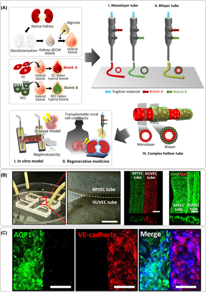
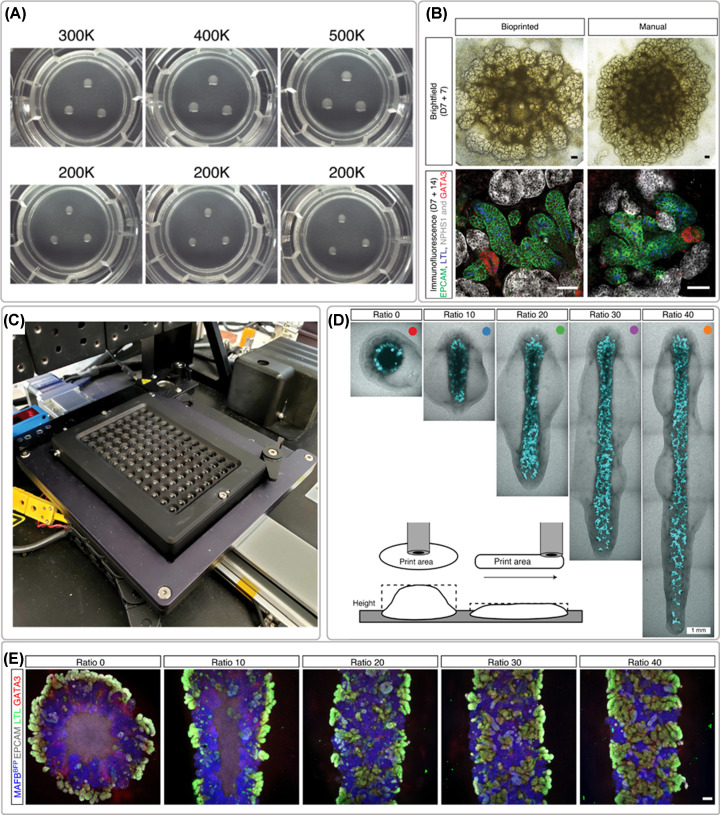
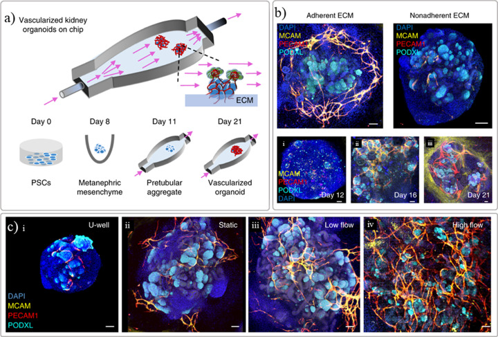
The number of patients with end-stage renal disease is continuously increasing worldwide. The only therapies for these patients are dialysis and organ transplantation, but the latter is limited due to the insufficient number of donor kidneys available. Research in kidney disease and alternative therapies are therefore of outmost importance. In vitro models that mimic human kidney functions are essential to provide better insights in disease and ultimately novel therapies. Bioprinting techniques have been increasingly used to create models with some degree of function, but their true potential is yet to be achieved. Bioprinted renal tissues and kidney-like constructs presents challenges, for example, choosing suitable renal cells and biomaterials for the formulation of bioinks. In addition, the fabrication of complex renal biological structures is still a major bottleneck. Advances in pluripotent stem cell-derived renal progenitors has contributed to in vivo-like rudiment structures with multiple renal cells, and these started to make a great impact on the achieved models. Natural- or synthetic-based biomaterial inks, such as kidney-derived extracellular matrix and gelatin-fibrin hydrogels, which show the potential to partially replicate in vivo-like microenvironments, have been largely investigated for bioprinting. As the field progresses, technological, biological and biomaterial developments will be required to yield fully functional in vitro tissues that can contribute to a better understanding of renal disease, to improve predictability in vitro of novel therapeutics, and to facilitate the development of alternative regenerative or replacement treatments. In this review, we resume the main advances on kidney in vitro models reported so far.

Keywords: Bioprinting; biomaterials; in vitro models; kidney.


Conflict of interest statement

The authors declare that there are no competing interests associated with the manuscript.

Figures

Figure 1
Figure 1. Bioprinting techniques used to create kidney in vitro models
(A) extrusion-based (pressure-assisted), (B) microfluidics, and (C) inkjet or droplet on demand bioprinting with (D) thermal, (E) piezo, (F) electromagnetic, or (G) acoustic actuation. Adapted with permission from Mota et al. [6].
Figure 2
Figure 2. Anatomy of the kidney
Schematic representation of the anatomy of the kidney with details of the nephrons and glomerulus compartment where the filtration occurs. Created with Biorender.com.
Figure 3
Figure 3. Renal proximal tubule in vitro model within perfusable chip
( A) manufacturing strategy using sacrificial 3D printing to create hollow channel after post-encapsulation with a gelatin-fibrin extracellular matrix hydrogel, (B) proximal tubule-like fully epithelialized after post-seeding and cultured with proximal tubule epithelial cells (PTECs), (C) control group of tubule model used to screen the toxicity effect of cyclosporine A and (D) disrupted epithelial barrier observed when model was exposed with a high concentration (100 μM) of the chemical compound. Adapted with permission from ref. [23].
Figure 4
Figure 4. Renal proximal tubule model with parallel vascular channel
( A) manufacturing strategy using sacrificial 3D printing to create two parallel hollow channels after post encapsulation with a gelatin-fibrin extracellular matrix hydrogel, (B) tubules can be manufactured in different configurations, including out of the plane, (C) full endothelialized and epithelialized tubules stained with cell-specific markers obtained after post-seeding with respective cells and cultured under perfusion up to 5 days, and (E) functionality test performed on the model to evaluate the influence of hyperglycaemic conditions on filtration and reabsorption response with and without the presence of a glucose transport inhibitor. Adapted with permission from ref. [25].
Figure 5
Figure 5. Bioprinted renal tubule model
( A) Schematics of the manufacturing approach combining kidney decellularized extracellular matrix with and alginate with endothelial or epithelial cells to form bioinks dispensed with core–shell bioprinting approach with individual cells to form tubular constructs or multi-shell arrangements, and the envisioned applications, (B) perfusable model manufactured with parallel epithelial and endothelial tubules in close contact showing high cellular viability post printing and hollow channels with a cellular lumen, and (C) immunostaining of cell specific markers. The barrier function of cell layers was tested in vitro and implanted in vivo under the kidney capsule envisioning future regenerative medicine application. Adapted with permission from ref. [20].
Figure 6
Figure 6. Bioprinting of iPSCs-derived renal cells for the formation of organoids
( A) example of organoids formed after printing different cell numbers (300,000–500,000 cells) and the reproducibility test with 200,000 cells, (B) brightfield and whole-mount immunofluorescence morphological comparison between bioprinted and manually formed organoids, (C) 96-well plate platform for compound testing, (D) organoid conformation studies, and (E) immunofluorescence staining of organoids created with different length while maintaining the same number of cells showing thinner cell mass with improved morphology. Adapted with permission from ref. [50].
Figure 7
Figure 7. Maturation of renal organoids in vitro with a perfusable chip
(A) schematic representation of the manufactured chip where PSC-derived kidney organoids are seeded onto a prevascularized ECM bed, (B) results demonstrating that the selection of ECM is important for the success of the vascularization, and (C) the importance of fluid-flow on the vascularization and growth of the organoids as demonstrated by endothelial and podocyte markers (MCAM, PECAM1, and PODXL). Adapted with permission from ref. [70].

References

    1. Bikbov B., Purcell C.A., Levey A.S., Smith M., Abdoli A., Abebe M.et al. . (2020) Global, regional, and national burden of chronic kidney disease, 1990–2017: a systematic analysis for the Global Burden of Disease Study 2017. Lancet North Am. Ed. 395, 709–733 10.1016/S0140-6736(20)30045-3 - DOI - PMC - PubMed
    1. Bulow R.D. and Boor P. (2019) Extracellular matrix in kidney fibrosis: more than just a scaffold. J. Histochem. Cytochem. 67, 643–661 10.1369/0022155419849388 - DOI - PMC - PubMed
    1. Peired A.J., Mazzinghi B., De Chiara L., Guzzi F., Lasagni L., Romagnani P.et al. . (2020) Bioengineering strategies for nephrologists: kidney was not built in a day. Expert Opin. Biol. Ther. 20, 467–480 10.1080/14712598.2020.1709439 - DOI - PubMed
    1. Peloso A., Katari R., Murphy S.V., Zambon J.P., DeFrancesco A., Farney A.C.et al. . (2015) Prospect for kidney bioengineering: shortcomings of the status quo. Expert Opin. Biol. Ther. 15, 547–558 10.1517/14712598.2015.993376 - DOI - PubMed
    1. Wilm B., Tamburrini R., Orlando G. and Murray P. (2016) Autologous cells for kidney bioengineering. Curr. Transplant. Rep. 3, 207–220 10.1007/s40472-016-0107-8 - DOI - PMC - PubMed

Publication types

Substances