. 2021 Jun 21;49(11):6437-6455.
doi: 10.1093/nar/gkab452.

An essential role of the autophagy activating kinase ULK1 in snRNP biogenesis

Affiliations

An essential role of the autophagy activating kinase ULK1 in snRNP biogenesis

Katharina Schmitz et al. Nucleic Acids Res. .

Abstract

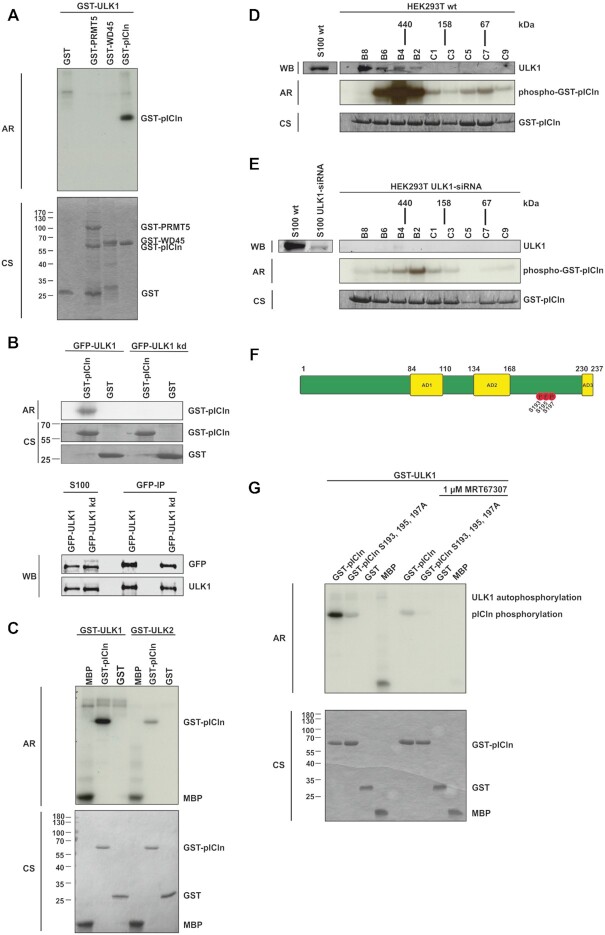
The biogenesis of small uridine-rich nuclear ribonucleoproteins (UsnRNPs) depends on the methylation of Sm proteins catalyzed by the methylosome and the subsequent action of the SMN complex, which assembles the heptameric Sm protein ring onto small nuclear RNAs (snRNAs). In this sophisticated process, the methylosome subunit pICln (chloride conductance regulatory protein) is attributed to an exceptional key position as an 'assembly chaperone' by building up a stable precursor Sm protein ring structure. Here, we show that-apart from its autophagic role-the Ser/Thr kinase ULK1 (Uncoordinated [unc-51] Like Kinase 1) functions as a novel key regulator in UsnRNP biogenesis by phosphorylation of the C-terminus of pICln. As a consequence, phosphorylated pICln is no longer capable to hold up the precursor Sm ring structure. Consequently, inhibition of ULK1 results in a reduction of efficient UsnRNP core assembly. Thus ULK1, depending on its complex formation, exerts different functions in autophagy or snRNP biosynthesis.


Figures

Figure 1.
Figure 1.
ULK1 interacts directly with the PRMT5 complex independent of its role in autophagy. (A) Flp-In T-REx 293-GFP-ULK1 and Flp-In T-REx 293-GFP cells were stimulated with 0.1 μg/ml doxycycline for 18 h, followed by 1 h of starvation treatment with EBSS. After cytoplasm extraction (S100) GFP-IP was performed and analyzed by Tris/Glycine-SDS-PAGE and western blotting using antibodies against GFP, PRMT5, WD45, and pICln. (B) Flp-In T-REx 293-GFP-ULK1 and Flp-In T-REx 293-GFP cells were stimulated with 0.1 μg/ml doxycycline for 18 h, followed by treatment with full or starvation medium (EBSS) in the absence or presence of bafilomycin A1 (BafA1; 10 nM) for 1 h. Afterward, cells were harvested, lysed, and cleared cellular lysates were subjected to Tris/Glycine-SDS-PAGE and immunoblotting for GFP, ULK1 pS757, ATG14, ATG14 pS29, p62, LC3, GAPDH, and Actin. (C) Flp-In T-REx 293-GFP-ULK1, -GFP-ULK1/ΔCTD and -GFP cells were stimulated with 0.1 μg/ml doxycycline for 18 h. After cytoplasm extraction (S100) GFP-IP was performed and analyzed by Tris/Glycine-SDS-PAGE and western blotting using antibodies against FIP200, GFP, PRMT5, ATG13, WD45, pICln and ATG101. (D) HEK293T cells were stably transfected with pMSCVbsd-GFP-ULK1 kinase domain (k. dom.) and pMSCVbsd-GFP constructs. Additionally, HEK293T cells were transiently transfected with pcDNA5-FRT-TO-GFP-ULK1 GABARAP domain (GABARAP dom.) and -ULK1 C-terminal domain (CTD) constructs. After cytoplasm extraction (S100) GFP-IP was performed and analyzed by Tris/Glycine-SDS-PAGE and western blotting using antibodies against GFP, FIP200, ATG13, ATG101, PRMT5, WD45, and pICln. (E) S100 extract was generated of HEK293T cells and applied to a Superdex 200 increase column. Fractions were analyzed by Tris/Glycine-SDS-PAGE and immunoblotting using antibodies against ULK1, ATG13, ATG101, PRMT5, WD45 and pICln. (F) S100 extract of HEK293T cells was applied to a Superdex 200 increase column and subsequent immunoprecipitation of endogenous was performed with antibody against ULK1. Immunoprecipitation was analyzed by Tris/Glycine-SDS-PAGE using antibodies against ULK1 and pICln.
Figure 2.
Figure 2.
ULK1 phosphorylates pICln in the C-terminal region on residues S193, S195 and S197. (A) In vitro kinase assay using recombinant active GST-ULK1 expressed in Sf9 insect cells and GST-PRMT5, -WD45, and -pICln purified from E. coli as substrate proteins were incubated with 10 μCi [32P]-ATP for 45 min. at 30°C. Samples were separated by Tris/Glycine-SDS-PAGE and analyzed by autoradiography. (B) In vitro kinase assay with purified GFP-ULK1 or GFP-ULK1 kinase-dead mutant (GFP-ULK1kd) overexpressed in Flp-In T-REx 293 cells and GST-pICln was executed as described in (A). Amounts of the GFP precipitation from GFP-ULK1 and GFP-ULK1 kinase-dead were directly compared by Tris/Glycine-SDS-PAGE and Western-Blot analysis using antibodies against ULK1 and GFP. (C) In vitro kinase assay using recombinant active GST-ULK1 or -ULK2 expressed in Sf9 cells and GST-pICln was executed as described in (A). (D, E) Gel filtration was performed with HEK293T wild type (D) and HEK293T ULK1-siRNA knockdown (E) S100 extracts fractionated by a Superdex 200 column and evaluated by western blotting. Appropriate fractions were used for in vitro kinase assay using exogenous GST-pICln substrate protein and 10 μCi [32P]-ATP for 45 min. at 30°C and analyzed by autoradiography (Exposure time of 30 minutes for (D) and (E)). (F) Schematic view of pICln protein with its three acidic domains (AD1-3) and the ULK1-dependent phosphorylation sites. In vitro kinase assay was performed with recombinant active GST-ULK1 from Sf9 cells and GST-pICln as described in (A). After Tris/Glycine-SDS-PAGE and coomassie blue staining, the pICln band was excised and phosphorylation status was analyzed by mass spectrometry (LC-MS/MS). Three phosphosites were detected: S193 (P = 3.05E-05, 94.5%), S195 (P = 2.3E–07, 100%), S197 (P = 2.8E–06, 100%). (G) Recombinant active GST-ULK1 from Sf9 insect cells was incubated with 1 μM ULK inhibitor MRT67307 for 30 min at 30°C. In vitro kinase assay with inhibitor-treated and non-treated GST-ULK1 was performed using substrate proteins GST-pICln wild type and alanine mutant purified from E. coli as described in (A). AR: autoradiography, CS: coomassie blue staining, WB: western blotting. See also Supplementary Figure SD2.
Figure 3.
Figure 3.
ULK1 dependent phosphorylation of pICln regulated binding of pICln towards SmG. (A–D) In vitro translated L-[35S]-Methionine labelled Sm Proteins D1, D3, B, and G were applied to an interaction assay with GST-pICln wt and phosphomutants purified from E. coli. After incubation for 1.5 h at 4°C and two times washing, purified proteins were separated by Tris/Glycine-SDS-PAGE and analyzed by autoradiography. (E-H) In vitro translated L-[35S]-Methionine labelled Sm Proteins D1, D3, B, and G were applied to an interaction assay with GST-pICln wt and GST-pICln pre-phosphorylated by ULK1. Pre-phosphorylation of pICln was performed for 1.5 h with 100 ng of active ULK1. After incubation for 1.5 h at 4°C with translated Sm Proteins and two times washing, purified proteins were separated by Tris/Glycine-SDS-PAGE and analyzed by autoradiography. (I, J) SmG (analyte), and GST-pICln (ligand) wild type, and aspartate mutant were purified from E. coli. Affinity dissociation constants were analyzed by surface plasmon resonance by steady-state analysis (SmG – GST-pICln wt, Kd 11.5 ± 1.5 μM; SmG – GST-pICln S193,195,197D, Kd 84.7 ± 14.7 μM; n = 3). AR: autoradiography, CS: coomassie blue staining.
Figure 4.
Figure 4.
Phosphorylation of pICln by ULK1 alters the structure of the 6S complex. (A) Sedimentation velocity (SV) analysis of GFP-pICln wild type and aspartate mutant in S100 extracts. The c(s) distributions for wild type pICln complex (magenta curve) and pICln complex with S193, S195, S197D mutations (blue curve) obtained from SV analysis at 40 000 rpm at 20°C for 5 h are shown. For better comparability normalization according to the area under the curve was done. (B–E), Size exclusion chromatography of Flp-In T-REx 293-GFP-pICln S100 extract using a Superdex 200 increase column was performed following a sedimentation velocity analysis (B) for GFP-pICln wild type of the 6S fractions B13 and B12. (C) Sedimentation coefficient distribution for wild-type pICln complex incubated with recombinant active GST-ULK1. (D) Sedimentation coefficient distribution for wild-type pICln complex incubated with recombinant active GST-ULK1 and ATP. (E) Overlay of c(s) distributions for all three samples acquired under the same conditions. All measurements were performed at 40 000 rpm at 20°C for 5 h. See also Supplementary Figure SD3.
Figure 5.
Figure 5.
Phosphorylated pICln is not able to build the 6S complex and to promote the subsequent Sm protein transfer onto the SMN complex. (A) Pulldown experiments using GST-pICln wild type and phosphomutants were executed in HEK293T S100 extract overnight at 4°C and co-purified Sm proteins were quantified by mass spectrometry and normalized to pICln wild type (LC-MS/MS; *P < 0.05; **P < 0.01). (B) Sm proteins D1, D2, E, F and G were in vitro translated and labelled with l-[35S]-Methionine. D1/D2 and E/F/G were pre-incubated for 1 h at 30°C and applied to an interaction assay with GST-pICln wt and phosphomutants purified from E. coli. Following incubation for 1.5 h at 4°C and three times washing purified proteins were separated by Tris/Glycine-SDS-PAGE and analyzed by autoradiography. (C) Sm proteins D1, D2, E, F and G were in vitro translated and labelled with L-[35S]-Methionine. To form the 6S structure D1/D2 and E/F/G were pre-incubated for 1 h at 30°C. To assess the influence of pICln as an ‘assembly chaperone’ Sm protein complex D1/D2 was first incubated (1 h at 4°C) using GST-pICln wt or phosphomutants purified from E. coli. After three times washing of the resulting pICln–SmD1/D2 complex, SmE/F/G complex was added to the mixture for a further 1 h at 4°C. After three times washing purified proteins were separated by Tris/Glycine-SDS-PAGE and analyzed by autoradiography. AR: autoradiography, CS: coomassie blue staining.
Figure 6.
Figure 6.
Decrease of endogenous ULK1 results in a decreased number of Cajal bodies. (AB) HEK293T cells were treated with 50 nM ULK1,2 siRNA or non-targeting control for 48 h. (A, C) The cells were fixed and Cajal bodies were visualized with antibody staining against Coilin (green) and SMN (red). The DNA was stained with DAPI (blue). (B) Cell lysates of siRNA-treated cells were analyzed by Tris/Glycine-SDS-PAGE using antibodies against ULK1, ULK2, and Tubulin. The downregulation of ULK1 causes a reduction in the snRNP storage pool. In the boxplot diagram, the ‘box’ represents 25–75% of all values and the mean (red), standard deviation, and out layers are visualized. HEK293T cells show an average of 1.54 (n = 500) Cajal bodies. Treatment of cells with siRNA caused a 1. significant decrease in the number of Cajal bodies. (C, D) The phosphorylation status of pICln influences the number of Cajal bodies per cell. (D) Overexpression of pICln in Flp-In T-Rex cells causes an increase in the number of Cajal bodies, mean 1.69 (n = 502). Phosphomutants of pICln (S193, 195, 197A, or D), cause a decrease in the number of Cajal bodies per cell. The P-value was calculated with Origin using the Mann–Whitney U test. ***P < 0.005; scale bars: 10 μm (A and C).
Figure 7.
Figure 7.
ULK1 phosphorylation of pICln regulates UsnRNP biogenesis. (A, B) GFP-pICln IP contains all proteins necessary for U1 snRNP core assembly. In vitro transcribed U1 snRNA labeled with 10 μCi [32P]-UTP was incubated with GFP-pICln IP. After incubation samples were directly analyzed by native gel electrophoresis (2, 4, 6, 8), or the same samples were subjected to supershift analysis with the Y12 antibody to show the specific formation of snRNPs (3, 5, 7, 9). The formation of snRNPs was quantified with Image Studio to compare the efficiency of the snRNP biogenesis. Adding ATP to the GFP-pICln IP increases efficiency (1.4) while adding ATP and ULK1 (2.6) leads to the highest efficiency in snRNP formation compared to the GFP-pICln IP alone. Adding ULK2 and ATP to the GFP-pICln IP caused no effect at all (1.3). (C) In vitro transcribed U1 snRNA labelled with 10 μCi [32P]-UTP was incubated with S100 extract from HEK293T cells treated with siRNA against ULK1, ULK 1+2, or a non-targeting control. After a native gel electrophoresis supershift analysis was performed using the Y12 antibody. (D) S100 extract from Mouse Embryonic Fibroblasts (MEFs) lacking ULK1/2, or reconstituted with vector control, ULK1 or ULK2 were incubated with [32P] labelled U1 snRNA, after a native gel electrophoresis supershift analysis occurred.
Figure 8.
Figure 8.
Schematic illustration summarizing the new role of ULK1 in the UsnRNP assembly as well as its well-known function in autophagy induction.

Similar articles

Cited by

References

    1. Matera A.G., Wang Z.. A day in the life of the spliceosome. Nat. Rev. Mol. Cell Biol. 2014; 15:108–121. - PMC - PubMed
    1. Kambach C., Walke S., Young R., Avis J.M., de la Fortelle E., Raker V.A., Luhrmann R., Li J., Nagai K.. Crystal structures of two Sm protein complexes and their implications for the assembly of the spliceosomal snRNPs. Cell. 1999; 96:375–387. - PubMed
    1. Newman A.J., Nagai K.. Structural studies of the spliceosome: blind men and an elephant. Curr. Opin. Struct. Biol. 2010; 20:82–89. - PubMed
    1. Friesen W.J., Wyce A., Paushkin S., Abel L., Rappsilber J., Mann M., Dreyfuss G.. A novel WD repeat protein component of the methylosome binds Sm proteins. J. Biol. Chem. 2002; 277:8243–8247. - PubMed
    1. Friesen W.J., Paushkin S., Wyce A., Massenet S., Pesiridis G.S., Van Duyne G., Rappsilber J., Mann M., Dreyfuss G.. The methylosome, a 20S complex containing JBP1 and pICln, produces dimethylarginine-modified Sm proteins. Mol. Cell. Biol. 2001; 21:8289–8300. - PMC - PubMed

Publication types

MeSH terms

Substances