. 2021 Jul 8;162(2):149-160.
doi: 10.1530/REP-21-0053.

CRISPR/Cas13d-mediated efficient KDM5B mRNA knockdown in porcine somatic cells and parthenogenetic embryos

Affiliations

CRISPR/Cas13d-mediated efficient KDM5B mRNA knockdown in porcine somatic cells and parthenogenetic embryos

Dengfeng Bi et al. Reproduction. .

Abstract

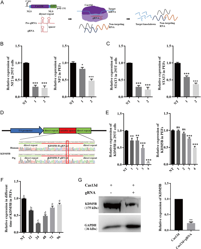
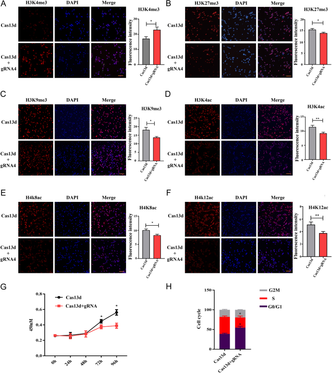
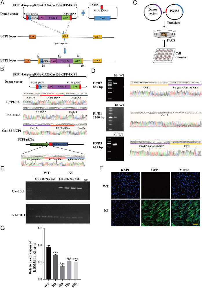
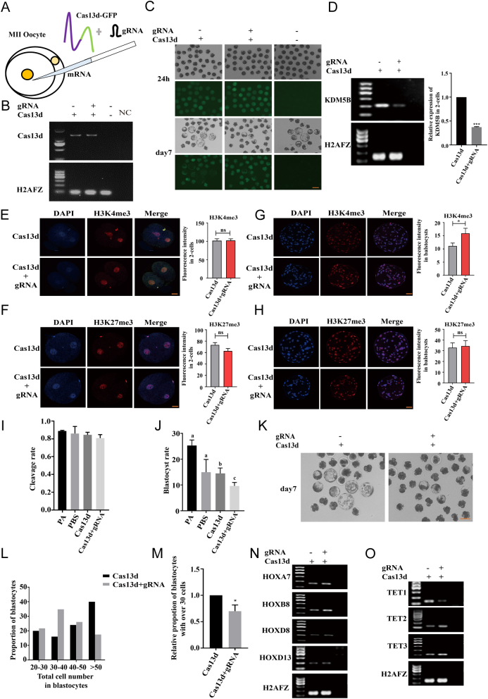
An efficient mRNA knockdown strategy is needed to explore gene function in cells and embryos, especially to understand the process of maternal mRNA decay during early embryo development. Cas13, a novel RNA-targeting CRISPR effector protein, could bind and cleave complementary single-strand RNA, which has been employed for mRNA knockdown in mouse and human cells and RNA-virus interference in plants. Cas13 has not yet been reported to be used in pigs. In the current study, we explored the feasibility of CRISPR/Cas13d-mediated endogenous RNA knockdown in pigs. KDM5B, a histone demethylase of H3K4me3, was downregulated at the transcriptional level by 50% with CRISPR/Cas13d in porcine fibroblast cells. Knockdown of KDM5B-induced H3K4me3 expression and decreased the abundance of H3K27me3, H3K9me3, H3K4ac, H4K8ac, and H4K12ac. These changes affected cell proliferation and cell cycle. Furthermore, stable integration of the CRISPR/Cas13d system into the porcine genome resulted in the continuous expression of Cas13d and persistent knockdown of KDM5B. Finally, the RNA-targeting potential of Cas13d was further validated in porcine parthenogenetic embryos. By microinjection of Cas13d mRNA and gRNA targeting KDM5B into porcine oocytes, the expression of KDM5B was downregulated, the abundance of H3K4me3 increased as expected, and the expression of embryonic development-related genes was changed accordingly. These results indicate that CRISPR/Cas13d provides an easily programmable platform for spatiotemporal transcriptional manipulation in pigs.


Conflict of interest statement

The authors declare that there is no conflict of interest that could be perceived as prejudicing the impartiality of the research reported.

Figures

Figure 1
Figure 1
Cas13d-mediated RNA knockdown in human cells and pig cells. (A) Schematic of constructs encoding Cas13d and gRNA. NLS, nuclear localization signal. Pre-gRNA, unprocessed guide RNA containing a single 30nt spacer sequence flanked by two 36nt direct repeats. gRNA, matured guide RNA with 22nt spacer sequence. The purple box, Cas13d protein. (B and C) Cas13d-mediated knockdown of NF2 (B) and STAT3 (C) in 293T and PEF cells (left and right) as determined by quantitative PCR. 1 and 2, gRNA1 and gRNA2; NT, non-targeting gRNA. The transcript levels were normalized against GAPDH. Data are presented as mean ± s.e.m., n  = 3. *P < 0.05, **P < 0.01, ***P < 0.001. (D) Sequence of gRNA4 targeting human and pig KDM5B sequence. There is one base difference in gRNA4 targeting the human and pig KDM5B sequences, as indicated in red rectangle. (E) The knockdown of KDM5B mediated by Cas13d via transient transfection in 293T (left) and PEF cells (right) with different gRNAs (1–4) and non-targeting gRNA (NT). The transcript levels were normalized against GAPDH. Data are presented as mean ± s.e.m., n  = 3. *P < 0.05, **P < 0.01, ***P < 0.001. (F) Cas13d-mediated knockdown of KDM5B with gRNA4 at different time points post-transfection. The transcript levels were normalized against GAPDH. Data are presented as mean ± s.e.m., n  = 3. Values with different letter are significantly different, P < 0.05. (G) Western blot analysis of KDM5B expression. The quantitative result is shown on the right. Data are presented as mean ± s.e.m., n  = 3. **P < 0.01.
Figure 2
Figure 2
Knockdown of KDM5B resulted in the abundance change of histone modifications and the arrest of PEF cell proliferation and cell cycle. (A, B, C, D, E and F) Immunofluorescence staining of H3K4me3 (A), H3K27me3 (B), H3K9me3 (C), H3K4ac (D), H4K8ac (E), and H4K12ac (F)in PEF cells transfected with Cas13d and gRNA4 targeting KDM5B or Cas13d only. The quantitative analysis of fluorescence intensity is shown on the right. Data are presented as the mean ± s.e.m.; *P < 0.05. Scale bar, 40 μm. At least 10 randomly selected fields were counted for each group. (G) Cell proliferation analysis of PEF cells transfected with Cas13d and gRNA4 (Cas13d only as control). *P < 0.05. 450 nm, the optical density (OD) absorbance at the wavelength of 450nm. (H) Cell cycle analysis of PEF cells transfected with Cas13d and gRNA4 (Cas13d only as control.) *P < 0.05.
Figure 3
Figure 3
Cas13d-mediated continuous knockdown of KDM5B in PEF cells. (A) Schematic representation of the design of CRISPR/Cas9-mediated, HR-independent integration of Cas13d and gRNA. Exons of UCP1 are shown in yellow rectangle. The blue lightning bolt in exon2 represents the Cas9-UCP1-gRNA target site, and the red boxes represent UCP1-gRNA. The U6-pre-gRNA-CAG-Cas13d-GFP expression plasmid is shown on the upper left, and the Cas9-UCP1-gRNA expression plasmid is shown on the upper right. The bottom showed forward integration of Cas13d and gRNA in UCP1. Three pairs of primers were designed for genotyping. F2/R2 and F3/R3 were designed for genotyping the 5’ and 3’ junctions in the transgenic colonies, respectively. F1/R1 were designed for detecting the integration of the UCP1-U6-pre-gRNA-CAG-Cas13d-GFP-UCP1 expression plasmid. (B) Confirmation of the successful construction of UCP1-U6-pre-gRNA-CAG-Cas13d-GFP-UCP1 and Cas9-UCP1-gRNA vector by Sanger sequencing. (C) Schematic diagram of the process to generate Cas13d-gRNA knockin cell colonies. (D) Genotyping results of the positive knockin colony by PCR (left) and Sanger sequencing (right). The primers used for PCR is indicated in A. (E) Semi-quantitative PCR analysis of the expression of Cas13d from the WT and KI cell at different time points post-transfection. (F) The GFP expression in the positive KI colony was detected by immunofluorescence imaging. Scale bar, 40 μm. (G) Quantitative PCR analysis of the expression of KDM5B in the KI colony against WT in different times.
Figure 4
Figure 4
Cas13d-mediated KDM5B knockdown in porcine oocytes. (A) Schematic diagram of microinjection in MII oocytes. (B) Expression of Cas13d transcript in blastocysts after injection of Cas13d mRNA with or without gRNA was determined by semi-quantitative PCR. (C) GFP expression in two-cell embryos and blastocysts after injection of Cas13d mRNA with or without gRNA. Scale bar, 40 μm. (D) Semi-quantitative PCR analysis of expression of KDM5B after injection Cas13d with or without gRNA in two-cell embryos is shown on the left. The quantitative result is shown on the right. (E, F, G and H) Immunofluorescence staining of H3K4me3 and H3K27me3 in two-cell embryos (E and F) and blastocysts (G and H) after injection of Cas13d RNA with or without gRNA. The quantitative results are shown on the right. At least 10 embryos were counted for each group. Scale bar, 40 μm. (I) The cleavage rate in both groups with or without injection. (J and K) The blastocyst rate in both groups with or without injection. A representative picture of embryos developed to the blastocyst stage in both groups is shown in K. (L) Proportion of blastocysts with different cell number was counted after injection of Cas13d with or without gRNA. (M) The proportion of blastocysts with over 30 cells was compared after injection Cas13d and gRNA vs Cas13d only. (N and O) Semi-quantitative PCR analysis of the expression of HOX genes and TET genes in two-cell embryos.

Similar articles

Cited by

References

    1. Abudayyeh OO, Gootenberg JS, Konermann S, Joung J, Slaymaker IM, Cox DB, Shmakov S, Makarova KS, Semenova E, Minakhin Let al. 2016. C2c2 is a single-component programmable RNA-guided RNA-targeting CRISPR effector. Science 353 aaf5573. (10.1126/science.aaf5573) - DOI - PMC - PubMed
    1. Abudayyeh OO, Gootenberg JS, Essletzbichler P, Han S, Joung J, Belanto JJ, Verdine V, Cox DBT, Kellner MJ, Regev Aet al. 2017. RNA targeting with CRISPR-Cas13. Nature 550 280–284. (10.1038/nature24049) - DOI - PMC - PubMed
    1. Agger K, Cloos PA, Christensen J, Pasini D, Rose S, Rappsilber J, Issaeva I, Canaani E, Salcini AE, Helin K. 2007. UTX and JMJD3 are histone H3K27 demethylases involved in HOX gene regulation and development. Nature 449 731–734. (10.1038/nature06145) - DOI - PubMed
    1. Albert M, Schmitz SU, Kooistra SM, Malatesta M, Morales Torres C, Rekling JC, Johansen JV, Abarrategui I, Helin K. 2013. The histone demethylase Jarid1b ensures faithful mouse development by protecting developmental genes from aberrant H3K4me3. PLoS Genetics 9 e1003461. (10.1371/journal.pgen.1003461) - DOI - PMC - PubMed
    1. Ali Z, Mahas A, Mahfouz M. 2018. CRISPR/Cas13 as a tool for RNA interference. Trends in Plant Science 23 374–378. (10.1016/j.tplants.2018.03.003) - DOI - PubMed

Publication types

MeSH terms