. 2022 Feb;25(1):57-70.
doi: 10.1007/s10456-021-09800-x. Epub 2021 Jun 7.

The HGR motif is the antiangiogenic determinant of vasoinhibin: implications for a therapeutic orally active oligopeptide

Affiliations

The HGR motif is the antiangiogenic determinant of vasoinhibin: implications for a therapeutic orally active oligopeptide

Juan Pablo Robles et al. Angiogenesis. 2022 Feb.

Abstract

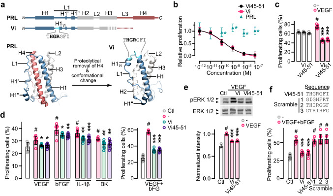
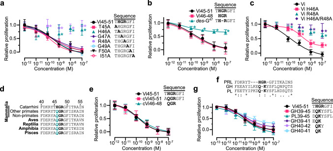
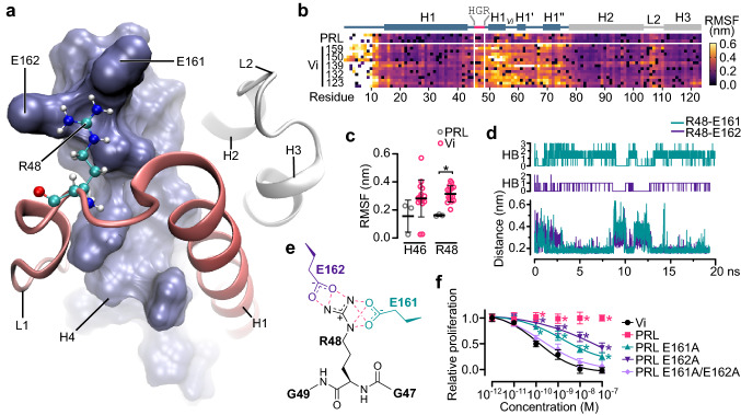
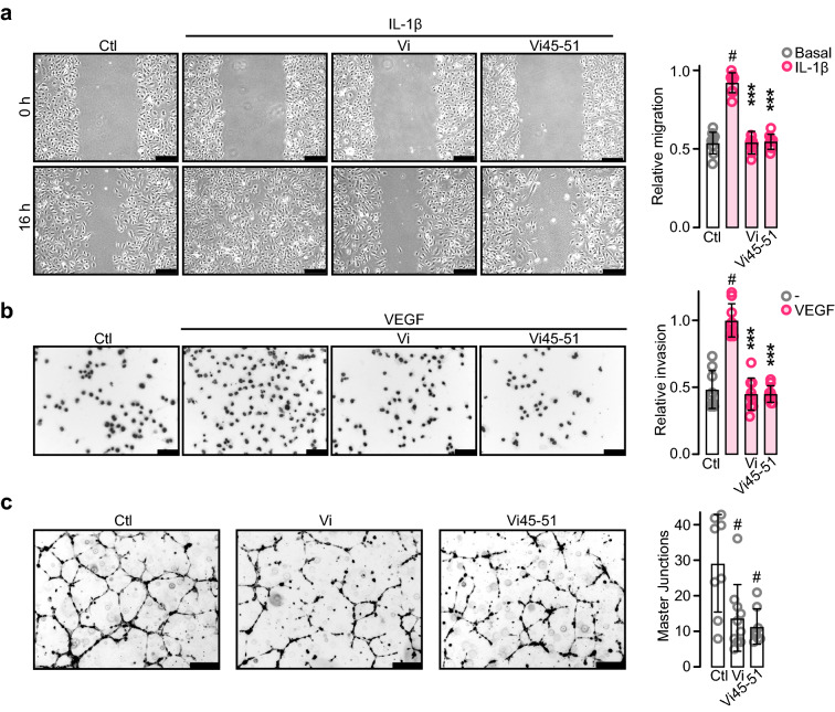
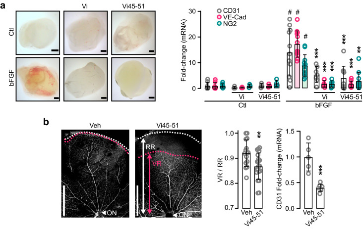
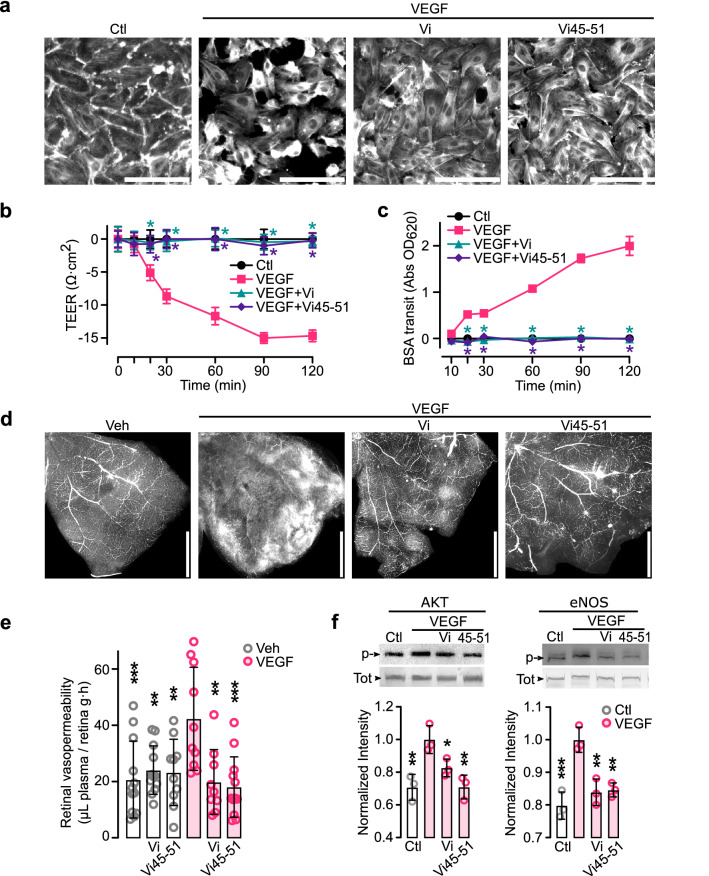
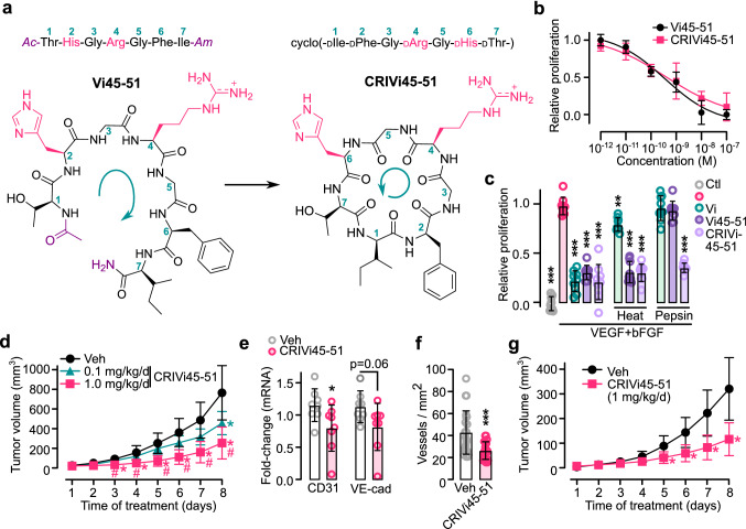
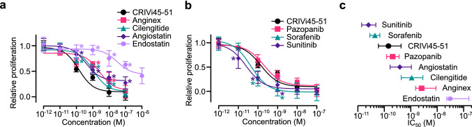
The hormone prolactin acquires antiangiogenic and antivasopermeability properties after undergoing proteolytic cleavage to vasoinhibin, an endogenous prolactin fragment of 123 or more amino acids that inhibits the action of multiple proangiogenic factors. Preclinical and clinical evidence supports the therapeutic potential of vasoinhibin against angiogenesis-related diseases including diabetic retinopathy, peripartum cardiomyopathy, rheumatoid arthritis, and cancer. However, the use of vasoinhibin in the clinic has been limited by difficulties in its production. Here, we removed this barrier to using vasoinhibin as a therapeutic agent by showing that a short linear motif of just three residues (His46-Gly47-Arg48) (HGR) is the functional determinant of vasoinhibin. The HGR motif is conserved throughout evolution, its mutation led to vasoinhibin loss of function, and oligopeptides containing this sequence inhibited angiogenesis and vasopermeability with the same potency as whole vasoinhibin. Furthermore, the oral administration of an optimized cyclic retro-inverse vasoinhibin heptapeptide containing HGR inhibited melanoma tumor growth and vascularization in mice and exhibited equal or higher antiangiogenic potency than other antiangiogenic molecules currently used as anti-cancer drugs in the clinic. Finally, by unveiling the mechanism that obscures the HGR motif in prolactin, we anticipate the development of vasoinhibin-specific antibodies to solve the on-going challenge of measuring endogenous vasoinhibin levels for diagnostic and interventional purposes, the design of vasoinhibin antagonists for managing insufficient angiogenesis, and the identification of putative therapeutic proteins containing HGR.

Keywords: 16K prolactin; Angiogenesis; Melanoma; Oligopeptide; Retina; Vasoinhibin; Vasopermeability.


Conflict of interest statement

The authors declare the following competing interests: C.C., J.P.R., M.Z., G.M.E., J.T., and T.B. are inventors of submitted Mexican (MX/E/2019/079075) and multinational (PCT/EP2020/069154) patent applications. The Universidad Nacional Autónoma de México (UNAM) and the authors J.T. and T.B. are owners of the pending patents.

Figures

Fig. 1
Fig. 1
THGRGFI heptapeptide mimics vasoinhibin. a Diagrams of secondary and tertiary structures of prolactin (PRL) and vasoinhibin (Vi) indicating α-helixes (H1-4), loops (L1-3), and residues 45–51 corresponding to the THGRGFI. Vi originates when H4 is removed by specific proteolysis causing conformational changes in L1 including a new α-helix (H1Vi). b Dose–response inhibition of HUVEC proliferation by the THGRGFI heptapeptide comprising residues 45–51 of Vi (Vi45–51), Vi of 123 residues (Vi), and PRL. Inhibition was against VEGF/bFGF-induced HUVEC proliferation. Proliferation relative to those of the lowest and highest dose of Vi positive control, n = 9, *P < 0.001 versus Vi (Two-way ANOVA, Dunnett). Dose–response curves fitted by least square regression analysis (r2 > 0.9). Effect of 100 nM Vi or Vi45–51 on BUVEC-E6E7 basal and VEGF-stimulated proliferation (c), or on human umbilical vein endothelial cell (HUVEC) proliferation stimulated by the proangiogenic factors VEGF, bFGF, IL-1β, or bradykinin (BK), or the VEGF + bFGF combination (d). e Representative Western blot showing the effect of Vi or Vi45–51 on VEGF-induced phosphorylation (p) and total levels of ERK 1/2 in HUVEC and the densitometric values of phosphorylated ERK 1/2 after normalization for total ERK 1/2. f Effect of 100 nM Vi, Vi45–51 and three different heptapeptides with Vi residues 45–51 in random order (scramble) on the VEGF + bFGF-induced proliferation of HUVEC. Proliferating cells are expressed relative to total cells. *P < 0.033, **P < 0.002, ***P < 0.001 versus stimulated control; # P < 0.001 versus basal proliferation (Ctl) (Two-way ANOVA, Tukey). In all cases, values are means ± SD, n = 9
Fig. 2
Fig. 2
HGR motif is the antiangiogenic determinant of vasoinhibin. Dose–response inhibition of VEGF + bFGF-induced HUVEC proliferation by: Vi45–51 subjected to alanine scanning mutagenesis (a); by Vi45–51, HGR tripeptide (Vi46–48), and Vi45–51 with G47 deleted (des-G47) (b); and by vasoinhibin of 123 amino acids (Vi) or Vi alanine mutants H46A, R48A, or H46A/R48A (c). d Alignment of vertebrate PRL sequences. Conserved HGR residues in bold and substitution of H by Q in green. e Dose–response inhibition of VEGF + bFGF-induced HUVEC proliferation by Vi45–51, its Q-substituted conserved version (cVi45–51), and the QGR tripeptide (cVi46–48). f Alignment of human PRL, GH, and PL sequences indicating same (*), similar (:), less similar (.), and dissimilar (blank) residues. The HGR motif (residues 46–48) in PRL and the putative Q–K motif in GH and PL (residues 40–41) are in bold. g Dose–response inhibition of HUVEC proliferation by Vi45–51, heptapeptides comprising residues 39–45 of GH (GH39–45) and PL (PL39–45), EQK and QKY tripeptides (GH39–41 and GH40–42, respectively) and the QK dipeptide (GH40–41). Inhibition was against VEGF/bFGF-induced HUVEC proliferation. Values are means ± SD relative to those of the lowest and highest dose of Vi or Vi45–51 positive controls, n = 9, *P < 0.001 versus Vi45–51 or Vi (Two-way ANOVA, Dunnett). Dose–response curves fitted by least square regression analysis (r2 > 0.8)
Fig. 3
Fig. 3
E161 and E162 in PRL obscure the HGR motif by salt bridges restraining R48. a Detail of the interaction between R48 (balls and sticks) and E161 and E162 in the N-terminal region of α-helix 4 (H4) (blue surface). R48 is located in loop 1 (L1) near α-helix 1 (H1) (red ribbon). b Root mean square fluctuation (RMSF) of the first 123 residues of PRL and vasoinhibin (Vi) of different lengths (159, 150, 139, 132, and 123 residues) over 20 ns molecular dynamic (MD) simulation. Location of HGR, α-helixes (H1-3), and loops (L1-2) are indicated in the diagram above. c RMSF of H46 and R48 in PRL and Vi isoforms. Values are means ± SD, n = 3–9, *P = 0.042 (Two-way ANOVA, Bonferroni). d Formation of hydrogen bonds (HB) between R48 and E161 and R48 and E162 in PRL and their correlation with the minimum distance between the residue pairs revealed by MD simulation. e Molecular scheme showing the three and four possible hydrogen bonds (red dashed lines) formed between the side chains of R48 and E162 and E161, respectively. e Dose-dependent inhibition by Vi of 123 residues (Vi), PRL, or PRL mutants where E161, E162, or E161 and E162 were replaced by alanine on VEGF + bFGF-induced proliferation of HUVEC. Values are means ± SD relative to those of the lowest and highest dose of the Vi positive control, n = 9, *P < 0.001 versus Vi (Two-way ANOVA, Dunnett). Dose–response curves fitted by least square regression analysis (r2 > 0.9)
Fig. 4
Fig. 4
Vasoinhibin heptapeptide (Vi45–51) containing the HGR motif inhibits angiogenesis in vitro. a HUVEC monolayers at 0 and 16 h after wound scratch incubated in the absence (Ctl) or presence of IL-1β (10 ng mL−1) alone or together with 100 nM vasoinhibin of 123 residues (Vi) or Vi45–51. Values represent the area occupied by migrating cells relative to the initial wound area. b Inhibition of HUVEC invasion across a Matrigel barrier by 100 nM Vi or Vi45–51. VEGF (50 ng mL−1) was used as chemoattractant. Values are number of invading cells relative to those with VEGF alone. c Inhibition of HUVEC tube-network formation by 200 nM Vi or Vi45–51 quantified by number of master junctions. **P < 0.01, ***P < 0.001 versus stimulated control, # P < 0.01 versus Ctl. Values are means ± SD, n = 9, compared by Two-way ANOVA-Dunnett. Scale bar: 300 μm
Fig. 5
Fig. 5
Vasoinhibin heptapeptide (Vi45–51) containing the HGR motif inhibits angiogenesis in vivo. a Matrigel plugs 6 d after mice implants without (Ctl) or with bFGF containing or not Vi or Vi45–51. mRNA expression of endothelial cell (CD31 and VE-Cad) and pericyte (NG2) markers in plugs. **P < 0.01, ***P < 0.001 versus bFGF; # P < 0.001 versus Ctl. b CD31-immunostained flat-mounted retinas from postnatal d 8 neonate mice injected with vehicle (Veh) or Vi45–51. Radial vascular expansion from the optic nerve (ON) evaluated by the index between vascular (VR) and retinal (RR) ratios and by retinal CD31 mRNA levels. **P < 0.01, ***P < 0.001 versus Veh. (Unpaired t-test). Values are means ± SD, n = 9. Scale: 1 mm (a), 500 μm (b)
Fig. 6
Fig. 6
Vasoinhibin heptapeptide (Vi45–51) containing the HGR motif inhibits vasopermeability in vitro and in vivo. Effect of VEGF, or VEGF and 100 nM Vi, or VEGF and 100 nM Vi45–51 on: actin cytoskeleton distribution (200 ng mL−1 VEGF) (a), transendothelial-electrical resistance (TEER) (50 ng mL−1 VEGF) (b), and flux of Evans blue-linked albumin (BSA) (50 ng mL−1 VEGF) (c) in HUVEC monolayers. *P < 0.001 versus VEGF. Extravasation of fluorescein-labeled dextran in flat-mounted retinas (d) and of Evans blue-linked albumin in retinal extracts (e) from rats injected intravitreally with vehicle (Veh), 200 ng VEGF, or VEGF and 20 μM Vi, or VEGF and 20 μM Vi45–51. **P < 0.01, ***P < 0.001 versus VEGF. Values are means ± SD, n = 9, compared by Two-way ANOVA-Dunnett Scale bar: 300 μm (a), 1 mm (d)
Fig. 7
Fig. 7
Oral administration of the cyclic retro-inverse vasoinhibin heptapeptide (CRIVi45–51) reduces tumor growth and vascularization. a Vi45–51 is composed of L-amino acids and acetylated (Ac) and amidated (Am) at the N- and C-termini, respectively. CRIVi45–51 is composed of D-amino acid in reverse order. Numbers indicate the ⍺-carbons and green arrows the synthesis sense. Note the conserved configuration of HGR side chains (magenta) in the two heptapeptides. b Dose-dependent inhibition of VEGF + bFGF-induced HUVEC proliferation by Vi45–51 or CRIVi45–51. c Inhibition of VEGF + bFGF-induced HUVEC proliferation by 10 nM vasoinhibin of 123 residues (Vi), Vi45–51, or CRIVi45–51 before or after heat inactivation or pepsin incubation. **P < 0.01, ***P < 0.001 versus VEGF + bFGF alone. d Growth curves of B16–F10 tumors in mice intravenously injected with vehicle (Veh) or with different doses of CRIVi45–51 after tumor appearance. *P < 0.002 versus Veh, #P < 0.002 versus 0.1 mg kg−1 d−1. mRNA expression of endothelial cell markers (CD31 and VE-Cad) (e) and vascular density (f) in tumors. *P < 0.05, ***P < 0.001 versus Veh. g Growth curves of B16–F10 tumors in mice after oral administration of vehicle (Veh) or CRIVi45–51 after tumor appearance. *P < 0.001 versus Veh. All values are mean ± SD, n = 9
Fig. 8
Fig. 8
Potency of the cyclic retro-inverse vasoinhibin heptapeptide (CRIVi45–51) compared to that of other anti-cancer and antiangiogenic drugs. Dose–response inhibition of VEGF + bFGF-induced HUVEC proliferation (a, b) and respective IC50 values (c) of CRIVi45–51, antiangiogenic proteins (angiostatin and endostatin), peptides (anginex, cilengitide), and tyrosine-kinase inhibitors (pazopanib, sorafenib, sunitinib). *P < 0.001 versus CRIVi45–51. HUVEC proliferation relative to that of the lowest and highest dose of the CRIVi45–51 positive control. Dose–response curves fitted by least square regression analysis (r2 < 0.7). All values are mean ± SD, n = 9

Similar articles

Cited by

References

    1. Carmeliet P. Angiogenesis in health and disease. Nat Med. 2003;9:653–660. doi: 10.1038/nm0603-653. - DOI - PubMed
    1. Jayson GC, Kerbel R, Ellis LM, Harris AL. Antiangiogenic therapy in oncology: current status and future directions. Lancet. 2016;388:518–529. doi: 10.1016/S0140-6736(15)01088-0. - DOI - PubMed
    1. Apte RS, Chen DS, Ferrara N. VEGF in signaling and disease: beyond discovery and development. Cell. 2019;176:1248–1264. doi: 10.1016/j.cell.2019.01.021. - DOI - PMC - PubMed
    1. Fallah A, Sadeghinia A, Kahroba H, et al. Therapeutic targeting of angiogenesis molecular pathways in angiogenesis-dependent diseases. Biomed Pharmacother. 2019;110:775–785. doi: 10.1016/j.biopha.2018.12.022. - DOI - PubMed
    1. Qin S, Li A, Yi M, et al. Recent advances on anti-angiogenesis receptor tyrosine kinase inhibitors in cancer therapy. J Hematol Oncol. 2019;12:27. doi: 10.1186/s13045-019-0718-5. - DOI - PMC - PubMed

Publication types