. 2021 Jul 15;89(8):e0012121.
doi: 10.1128/IAI.00121-21. Epub 2021 Jul 15.

Salmonella Typhimurium Adhesin OmpV Activates Host Immunity To Confer Protection against Systemic and Gastrointestinal Infection in Mice

Affiliations

Salmonella Typhimurium Adhesin OmpV Activates Host Immunity To Confer Protection against Systemic and Gastrointestinal Infection in Mice

Deepinder Kaur et al. Infect Immun. .

Abstract

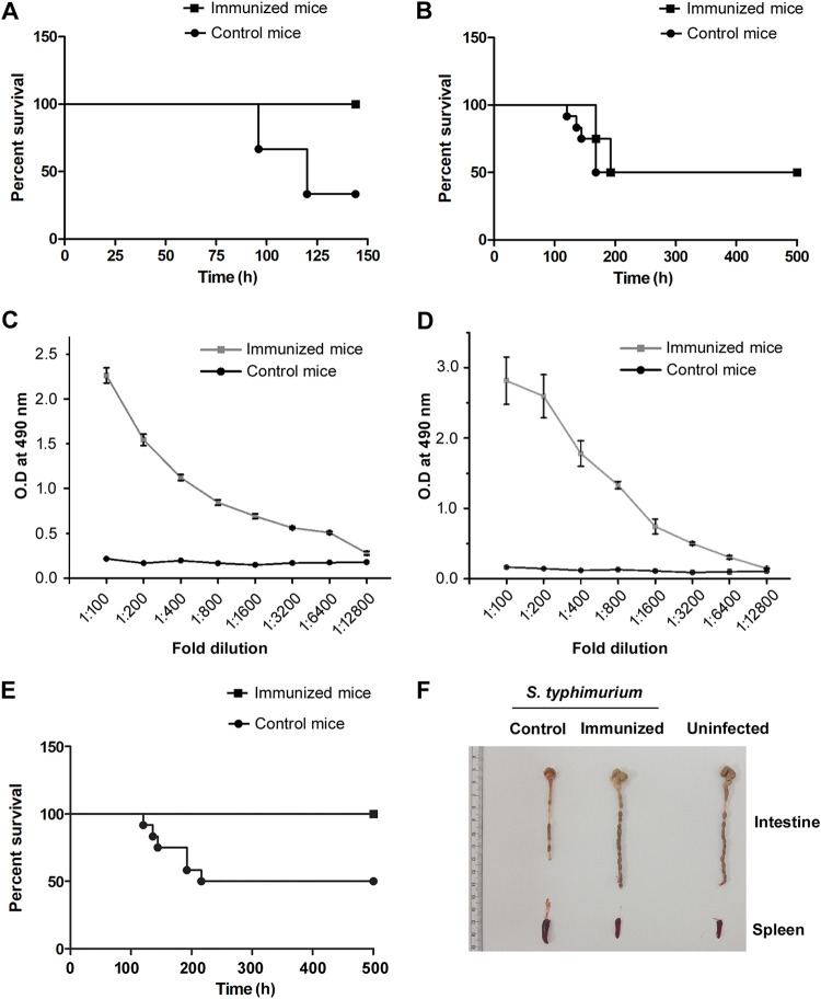
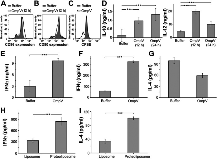
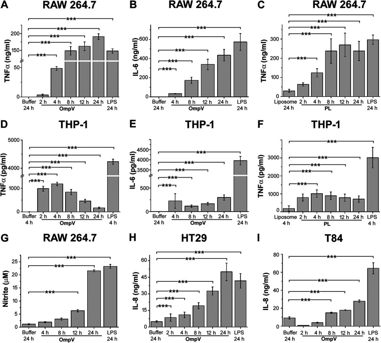
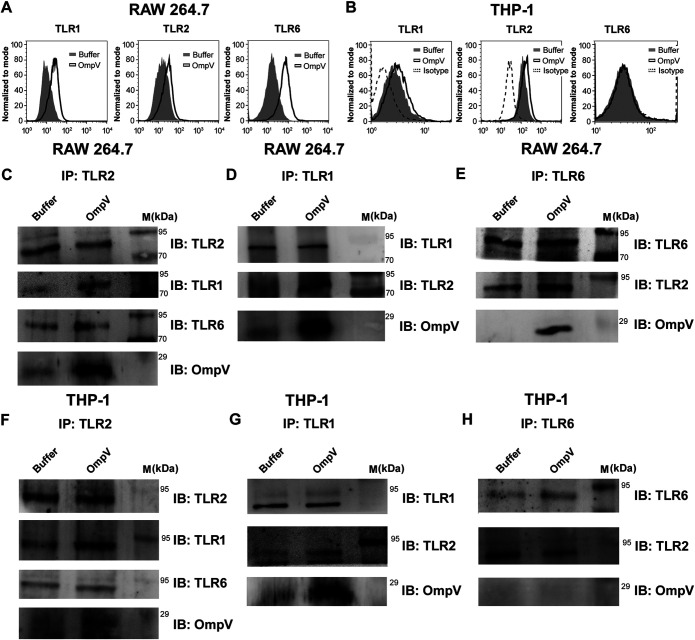
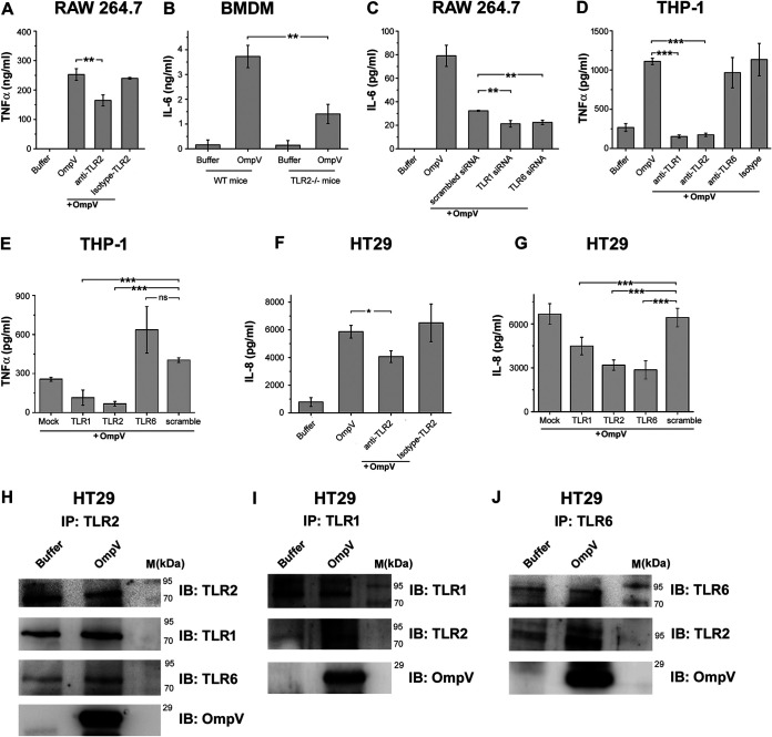
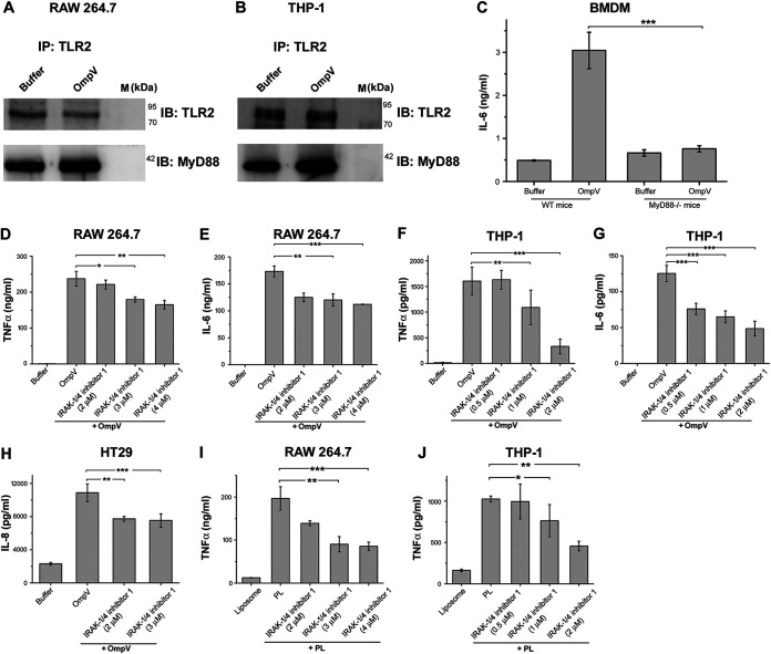
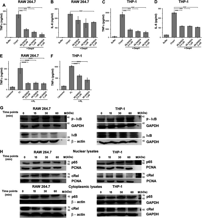
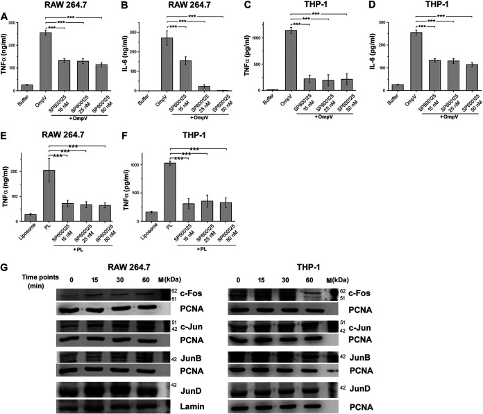
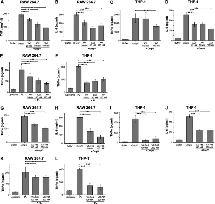
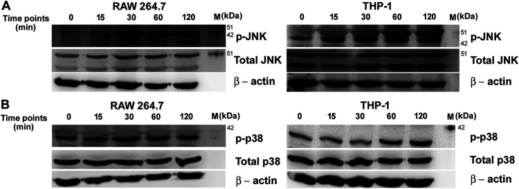
Salmonella enterica Typhimurium is a rod-shaped Gram-negative bacterium that mostly enters the human body through contaminated food. It causes a gastrointestinal disorder called salmonellosis in humans and typhoid-like systemic disease in mice. OmpV, an outer membrane protein of S. Typhimurium, helps in adhesion and invasion of bacteria to intestinal epithelial cells and thus plays a vital role in the pathogenesis of S. Typhimurium. In this study, we have shown that intraperitoneal immunization with OmpV is able to induce high IgG production and protection against systemic disease. Further, oral immunization with OmpV-incorporated proteoliposome (OmpV-proteoliposome [PL]) induces production of high IgA antibody levels and protection against gastrointestinal infection. Furthermore, we have shown that OmpV induces Th1 bias in systemic immunization with purified OmpV, but both Th1 and Th2 polarization in oral immunization with OmpV-proteoliposome (PL). Additionally, we have shown that OmpV activates innate immune cells, such as monocytes, macrophages, and intestinal epithelial cells, in a Toll-like receptor 2 (TLR2)-dependent manner. Interestingly, OmpV is recognized by the TLR1/2 heterodimer in monocytes, but by both TLR1/2 and TLR2/6 heterodimers in macrophages and intestinal epithelial cells. Further, downstream signaling involves MyD88, interleukin-1 receptor-associated kinase (IRAK)-1, mitogen-activated protein kinase (MAPK) (both p38 and Jun N-terminal protein kinase (JNK)), and transcription factors NF-κB and AP-1. Due to its ability to efficiently activate both the innate and adaptive immune systems and protective efficacy, OmpV can be a potential vaccine candidate against S. Typhimurium infection. Further, the fact that OmpV can be recognized by both TLR1/2 and TLR2/6 heterodimers increases its potential to act as good adjuvant in other vaccine formulations.

Keywords: OmpV; Salmonella; immune mechanisms.


Figures

FIG 1
FIG 1
OmpV induces protective immunity against S. Typhimurium infection. (A, B) Intraperitoneal immunization of mice with purified OmpV protects against systemic infection (A) but does not protect against gastrointestinal infection (B) of S. Typhimurium. Mice were immunized intraperitoneally at an interval of 7 days. Buffer was used for the control mice. Following 14 days after the last dose, mice were challenged with S. Typhimurium SL1344 intraperitoneally (A) or through the oral route (B), and survival was monitored until day 7 (A) or day 21 (B). (C, D) A high titer of IgG (C) and IgA (D) was observed in the serum of mice immunized intraperitoneally with OmpV (C) and in the stools of mice immunized orally with OmpV-proteoliposome (PL) (D). Mice were immunized intraperitoneally with four doses of OmpV, and buffer was used for the control mice (C). Mice were immunized with four doses of OmpV-proteoliposome (PL), and liposomes were used for control mice (D). Following 14 days after the last dose of immunization, serum and stool samples were obtained from the immunized and control mice (n = 3 mice/group, 3 biological replicates). (E) Oral immunization of mice with OmpV-proteoliposome (PL) protects against gastrointestinal infection of S. Typhimurium. Mice were immunized with OmpV-proteoliposome (PL). Following the last dose, mice were given gastrointestinal infection with oral challenge of S. Typhimurium SL1344, and survival was monitored. Mice immunized with liposomes were taken as controls. For panels A, B, and E, n = 12 mice/group (4 mice per group per experiment were taken, and 3 biological replicates were done). A Kaplan-Meier plot of cumulative mortality was prepared to compare the survival rate. (F) Similar intestinal length was observed in the naive noninfected mice as well as the infected mice immunized with OmpV-proteoliposome (PL). Mice were immunized with OmpV-proteoliposome (PL), and, after the last dose, mice were given gastrointestinal infection with S. Typhimurium SL1344. After 5 days of infection, the intestines and spleens of these mice were removed, observed, and matched with noninfected naive mice (n = 3 mice/group).
FIG 2
FIG 2
OmpV induces dendritic cell maturation and Th cell differentiation. (A) Increased expression of CD86 in BMDCs activated with OmpV compared to the control. (B) No increase in CD80 expression in OmpV-treated BMDCs compared to the control. (A, B) BMDCs were pretreated with polymyxin B (PmB) followed by treatment with OmpV. Following incubations, the surface expression of costimulatory molecules CD86 and CD80 were analyzed by flow cytometry. (C) OmpV induces proliferation of Th cells as indicated by a decrease in CFSE fluorescence. CFSE-labeled CD4+ T cells were cocultured with splenic DCs targeted with OmpV. At day 3, CFSE fluorescence was detected using flow cytometry. CFSE-labeled CD4+ T cells treated with buffer were used as controls. (A to C) Histograms are representative of three independent experiments. (D) OmpV leads to the production of a high IL-12/IL-10 ratio. BMDCs were treated with PmB followed by OmpV. Following incubations, the supernatants were collected and estimated for IL-10 and IL-12 cytokines using an ELISA. (E) OmpV leads to the production of high levels of IFN-γ. CD4+ T cells were cocultured with OmpV-targeted splenic DCs for 3 days. The collected supernatant was probed for the presence of IFN-γ using an ELISA. (D, E) Bar graphs are expressed as mean ± standard error of the mean (SEM) from three independent experiments (*, P < 0.05; **, P < 0.01; ***, P < 0.001; ns, P > 0.05 versus the buffer-treated cells). (F, G) Treatment with purified OmpV leads to the production of high levels of IFN-γ in vivo. (H, I) OmpV-proteoliposome (PL) treatment leads to the production of both IFN-γ and IL-4 in vivo. OmpV or buffer was administered to mice intraperitoneally (4 doses a week apart) (F, G). Mice were immunized orally with four doses of OmpV-proteoliposome (PL)/liposome (H, I). Following 14 days after the last dose, cells were isolated from peripheral lymph nodes (F, G) or mesenteric lymph nodes (H, I) and activated with PMA and ionomycin. The supernatants were collected, and IFN-γ and IL-4 were quantified by ELISA. Bar graphs are expressed as mean ± SEM from three independent experiments; *, P < 0.05; **, P < 0.01; ***, P < 0.001; ns, P > 0.05 versus the buffer-treated mice (F, G) or mice immunized with liposome (H, I).
FIG 3
FIG 3
Time-dependent analysis for the production of proinflammatory mediators in RAW 264.7 macrophages, THP-1 monocytes, and IECs. (A to C) Time-dependent increase in TNF-α and IL-6 production was observed in RAW 264.7 macrophages. (D to F) In THP-1 monocytes, TNF-α was maximally produced at 4 h and IL-6 at 24 h. (G) A time-dependent increase in nitric oxide production was observed in RAW 264.7 macrophages. (H, I) A time-dependent increase in IL-8 production was observed in HT29 (H) and T84 cells (I). (A to I) Cells were treated with PmB followed by OmpV or OmpV-proteoliposome (PL) and incubated for different time periods as indicated. Following respective incubations, supernatants were collected and analyzed for cytokines (A to F and H and I) or nitric oxide (G). LPS was used as a positive control. Bar graphs are expressed as mean ± SEM from three independent experiments (*, P < 0.05; **, P < 0.01; ***, P < 0.001; ns, P > 0.05 versus the buffer-treated or liposome-treated cells).
FIG 4
FIG 4
OmpV induces increases in surface expression of TLRs and coimmunoprecipitates with TLR2, TLR1, and TLR6 in macrophages and TLR1 and TLR2 in monocytes. (A, B) OmpV induces increased surface expression of TLR1, TLR2, and TLR6 on RAW 264.7 macrophages (A) and TLR1 and TLR2 on THP-1 monocytes (B). Cells were treated with PmB followed by OmpV. Following incubations, the surface expression of TLRs was analyzed using flow cytometry. Histograms are representatives of three independent experiments. (C to H) OmpV coimmunoprecipitates with TLR2, TLR1, and TLR6 in RAW 264.7 macrophages (C to E), whereas it only coimmunoprecipitates with TLR1 and TLR2 but not with TLR6 in THP-1 monocytes (F to H). Cells were treated with PmB followed by OmpV or buffer, and whole-cell lysates were prepared following incubation. Further, lysates were immunoprecipitated with anti-TLR2/anti-TLR-1/anti-TLR6 antibody and checked for the presence of TLR1, TLR2, TLR6, and OmpV. Buffer-treated cells were used as controls for coimmunoprecipitation experiments. IP indicates the antibody used for immunoprecipitation, whereas IB indicates the antibody used for immunoblotting. Western blots are representative of three independent experiments.
FIG 5
FIG 5
In macrophages and IECs, OmpV is recognized by both TLR1/TLR2 and TLR2/TLR6 heterodimers, whereas in monocytes, OmpV is recognized only by TLR1/TLR2 heterodimers. (A) Neutralization of TLR2 indicated its involvement in recognition of OmpV in RAW 264.7 macrophages. Cells were pretreated with anti-TLR2 neutralizing antibody followed by PmB and OmpV. Following incubation, supernatants were analyzed for TNF-α production. (B) BMDMs from mice deficient in TLR2 (TLR2−/− mice) confirmed involvement of TLR2 in recognition of OmpV in macrophages. BMDMs from wild-type mice and TLR2−/− mice were treated with PmB followed by OmpV for 24 h. Supernatants were collected and analyzed for IL-6 production. (C) A significant decrease in production of IL-6 was observed upon knockdown of TLR1 and TLR6 in macrophages. RAW 264.7 cells were transfected with siRNA followed by treatment with PmB and OmpV. Following incubation, supernatants were collected and quantified for IL-6 by ELISA. Nontargeted (scrambled) siRNA was used as a negative control. (D) A neutralization experiment indicated the involvement of TLR1 and TLR2 in recognition of OmpV in monocytes. THP-1 cells were pretreated with neutralizing antibodies followed by PmB and OmpV. Supernatants were collected following incubation and analyzed for TNF-α production. (E) A significant decrease in production of TNF-α was observed upon knockdown of TLR1 and TLR2 but not upon knockdown of TLR6 in monocytes. THP-1 cells were transfected with shRNA followed by treatment with PmB and OmpV, and supernatants were collected after incubation and checked for the presence of TNF-α. Nontargeted (scrambled) shRNA was used as a negative control. (F) Neutralization of TLR2 indicates its involvement in recognition of OmpV in IECs. HT29 cells were pretreated with neutralizing antibody for 1 h followed by PmB and OmpV. The supernatants were collected and analyzed for IL-8 production. (G) A significant decrease in IL-8 production was observed upon knockdown of TLR1, TLR2, and TLR6 in IECs. HT29 cells were transfected with shRNA and were then treated with PmB followed by OmpV. Following incubation, supernatants were collected, and IL-8 was quantified by ELISA. Nontargeted (scrambled) shRNA was used as a negative control. (A to G) Bar graphs are expressed as mean ± SEM from three independent experiments; *, P < 0.05; **, P < 0.01; ***, P < 0.001; ns, P > 0.05 versus only OmpV-treated cells (A, D, F), versus the OmpV-treated BMDMs from wild-type mice (B), versus scrambled siRNA-transfected OmpV-treated cells (C), or versus scrambled shRNA-transfected OmpV-treated cells (E, G). (H to J) OmpV coimmunoprecipitates with TLR1, TLR2, and TLR6 in IECs. HT29 cells were treated with PmB followed by OmpV. Following incubation, whole-cell lysates were prepared. These lysates were immunoprecipitated with anti-TLR2 (H), anti-TLR1 (I), or anti-TLR6 (J) antibody and checked for the presence of TLR1, TLR2, TLR6, and OmpV by immunoblotting. Buffer-treated cells were used as controls for coimmunoprecipitation experiments. IP indicates the antibody used for immunoprecipitation, whereas IB indicates the antibody used for immunoblotting. Western blots are representatives of three independent experiments.
FIG 6
FIG 6
MyD88 and IRAK are involved in OmpV-mediated signaling in macrophages, monocytes, and IECs. (A, B) Increased association of MyD88 with TLR2 in OmpV-activated macrophages and monocytes. Cell lysates of OmpV-treated RAW 264.7 macrophages (A) and THP-1 monocytes (B) were immunoprecipitated with anti-TLR2 antibody and checked for the presence of MyD88. Buffer-treated cells were used as controls. IP indicates the antibody used for immunoprecipitation, whereas IB indicates the antibody used for immunoblotting. Western blots are representatives of three independent experiments. (C) A significant decrease in proinflammatory cytokine production was observed in macrophages under MyD88-deficient conditions. BMDMs from wild-type and MyD88−/− mice were treated with PmB followed by OmpV. Following incubation, supernatants were collected and analyzed for IL-6 production. (D to G) A significant decrease in proinflammatory cytokine production was observed with inhibition of IRAK-1/4 in macrophages and monocytes. (H) A significant decrease in IL-8 was observed upon inhibition of IRAK-1/4 in IECs. (I, J) IRAK is involved in OmpV-proteoliposome (PL)-mediated signaling. (D to J) RAW 264.7 macrophages, THP-1 monocytes, or HT29 cells were pretreated with IRAK-1/4 inhibitor followed by treatment with PmB and OmpV or OmpV-proteoliposome (PL). Following incubations, supernatants were collected and analyzed for cytokine production by ELISA. Bar graphs are expressed as mean ± SEM from three independent experiments; *, P < 0.05; **, P < 0.01; ***, P < 0.001; ns, P > 0.05 versus the OmpV-activated BMDMs from wild-type mice (C), versus only OmpV-treated cells (D to H), or versus only OmpV-proteoliposome (PL)-treated cells (I, J).
FIG 7
FIG 7
NF-κB is involved in OmpV-mediated proinflammatory responses. (A to D) A significant decrease in TNF-α and IL-6 production was observed upon pretreatment with NF-κB inhibitor in OmpV-activated RAW 264.7 macrophages (A, B) and THP-1 cells (C, D). (E, F) OmpV-proteoliposome (PL)-mediated proinflammatory signaling involves NF-κB. (A to F) Cells were pretreated with NF-κB inhibitor followed by treatment with PmB and OmpV or OmpV-proteoliposome (PL). Following incubations, supernatants were collected and analyzed for cytokine production. Bar graphs are expressed as mean ± SEM from three independent experiments; *, P < 0.05; **, P < 0.01; ***, P < 0.001; ns, P > 0.05 versus only OmpV-treated cells (A to D) or versus only OmpV-proteoliposome (PL)-treated cells (E, F). (G) Phosphorylation of IκB was observed in OmpV-treated macrophages and monocytes. (H) Translocation of NF-κB subunits p65 and cRel from the cytoplasm to the nucleus was observed in OmpV-treated macrophages and monocytes. (G, H) RAW 264.7 and THP-1 cells were treated with PmB followed by OmpV. Following incubation, whole-cell lysates were prepared at different time points, and levels of p-IκB (phosphorylated IκB) and IκB were assessed by Western blotting (G), or nuclear and cytoplasmic fractions were prepared, and levels of p65 and cRel were checked by Western blotting (H). GAPDH and β-actin were used as loading controls for whole-cell and cytoplasmic lysates (G, H) and PCNA was used as a loading control for nuclear lysates (H). Western blots are representatives of three independent experiments.
FIG 8
FIG 8
OmpV-mediated proinflammatory signaling involves transcription factor AP-1. (A to D) A significant decrease in TNF-α and IL-6 was observed upon pretreatment with AP-1 inhibitor in OmpV-activated macrophages (A, B) and monocytes (C, D). (E, F) AP-1 is involved in OmpV-proteoliposome (PL)-mediated proinflammatory signaling. (A to F) RAW 264.7 and THP-1 cells were pretreated with AP-1 inhibitor followed by treatment with PmB and OmpV or OmpV-proteoliposome (PL). Following respective incubations, supernatants were collected and analyzed for cytokines by ELISA. Bar graphs are expressed as mean ± SEM from three independent experiments; *, P < 0.05; **, P < 0.01; ***, P < 0.001; ns, P > 0.05 versus only OmpV-treated cells or OmpV-proteoliposome (PL)-treated cells. (G) Translocation of AP-1 subunits to the nucleus was observed in OmpV-targeted cells. RAW 264.7 and THP-1 cells were treated with PmB followed by OmpV and incubated for different time points as indicated. Following incubations, nuclear fractions were extracted and probed for AP-1 subunits. PCNA and lamin were used as loading controls for nuclear lysates. Western blots are representatives of three independent experiments.
FIG 9
FIG 9
p38 and JNK are involved OmpV-mediated signaling. (A to D) A significant decrease in TNF-α and IL-6 production was observed upon pretreatment with JNK inhibitor in OmpV-activated macrophages (A, B) and monocytes (C, D). (E, F) JNK is involved in OmpV-proteoliposome (PL)-mediated proinflammatory signaling. (A to F) RAW 264.7 macrophages (A, B, E) and THP-1 monocytes (C, D, F) were pretreated with JNK inhibitor followed by treatment with PmB and OmpV (A to D) or OmpV-proteoliposome (PL) (E, F). Following incubations, supernatants were analyzed for proinflammatory cytokines by ELISA. Bar graphs are expressed as mean ± SEM from three independent experiments; *, P < 0.05; **, P < 0.01; ***, P < 0.001; ns, P > 0.05 versus only OmpV-treated cells (A to D) or OmpV-proteoliposome (PL)-treated cells (E, F). (G to J) A significant decrease in TNF-α and IL-6 was observed upon pretreatment with p38 inhibitor in OmpV-activated macrophages (G, H) and monocytes (I, J). (K, L) p38 is involved in OmpV-proteoliposome (PL)-mediated signaling in macrophages and monocytes. RAW 264.7 (G, H, K) and THP-1 cells (I, J, L) were pretreated with p38 inhibitor followed by treatment with PmB and OmpV or OmpV-proteoliposome (PL). Following incubations, supernatants were collected and analyzed for proinflammatory cytokines by ELISA. Bar graphs are expressed as mean ± SEM from three independent experiments; *, P < 0.05; **, P < 0.01; ***, P < 0.001; ns, P > 0.05 versus only OmpV-treated cells (G to J) or OmpV-proteoliposome (PL)-treated cells (K, L).
FIG 10
FIG 10
OmpV induces phosphorylation of JNK and p38 in macrophages, monocytes. (A, B) OmpV treatment leads to the phosphorylation of JNK and p38 in macrophages and monocytes. RAW 264.7 macrophages and THP-1 monocytes were treated with PmB followed by OmpV (2 μg/ml) for different time points. Following incubations, whole-cell lysates were prepared and probed for phosphorylated and total JNK (A) and p38 (B). β-Actin was used as a loading control. Western blots are representatives of three independent experiments.

Similar articles

Cited by

References

    1. Graham SM. 2010. Nontyphoidal salmonellosis in Africa. Curr Opin Infect Dis 23:409–414. 10.1097/QCO.0b013e32833dd25d. - DOI - PubMed
    1. Alghoribi MF, Doumith M, Alrodayyan M, Al Zayer M, Koster WL, Muhanna A, Aljohani SM, Balkhy HH, Desin TS. 2019. S. Enteritidis and S. Typhimurium harboring SPI-1 and SPI-2 are the predominant serotypes associated with human salmonellosis in Saudi Arabia. Front Cell Infect Microbiol 9:187. 10.3389/fcimb.2019.00187. - DOI - PMC - PubMed
    1. MacLennan CA, Martin LB, Micoli F. 2014. Vaccines against invasive Salmonella disease: current status and future directions. Hum Vaccin Immunother 10:1478–1493. 10.4161/hv.29054. - DOI - PMC - PubMed
    1. Sparham SJ, Kwong JC, Valcanis M, Easton M, Trott DJ, Seemann T, Stinear TP, Howden BP. 2017. Emergence of multidrug resistance in locally-acquired human infections with Salmonella Typhimurium in Australia owing to a new clade harbouring blaCTX-M-9. Int J Antimicrob Agents 50:101–105. 10.1016/j.ijantimicag.2017.02.014. - DOI - PubMed
    1. GBD 2017 Non-Typhoidal Salmonella Invasive Disease Collaborators. 2019. The global burden of non-typhoidal salmonella invasive disease: a systematic analysis for the Global Burden of Disease Study 2017. Lancet Infect Dis 19:1312–1324. 10.1016/S1473-3099(19)30418-9. - DOI - PMC - PubMed

Publication types