. 2021 Aug;116(2):707-722.
doi: 10.1111/mmi.14765. Epub 2021 Jun 25.

DNA damage checkpoint activation affects peptidoglycan synthesis and late divisome components in Bacillus subtilis

Affiliations

DNA damage checkpoint activation affects peptidoglycan synthesis and late divisome components in Bacillus subtilis

Emily A Masser et al. Mol Microbiol. 2021 Aug.

Abstract

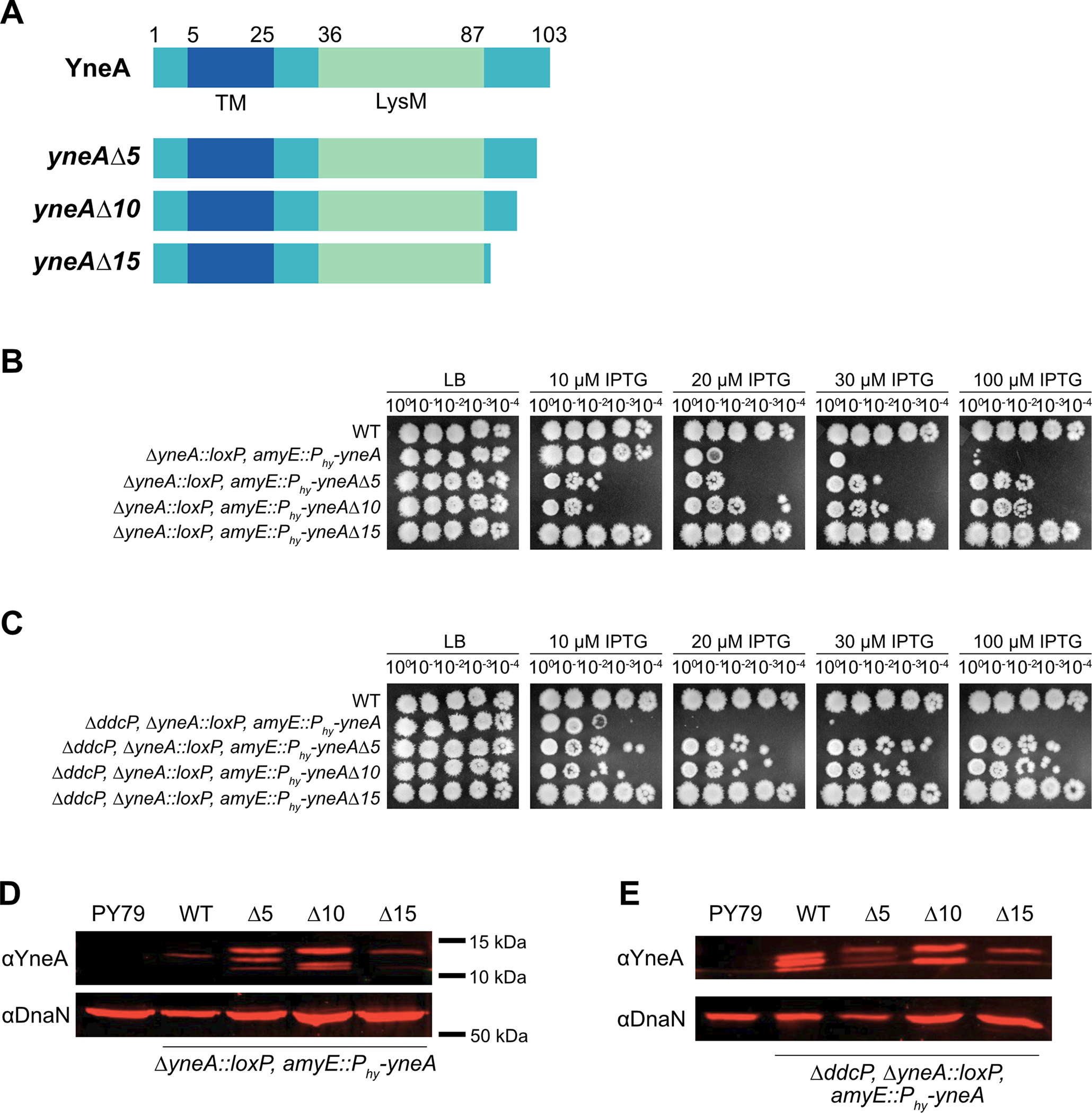
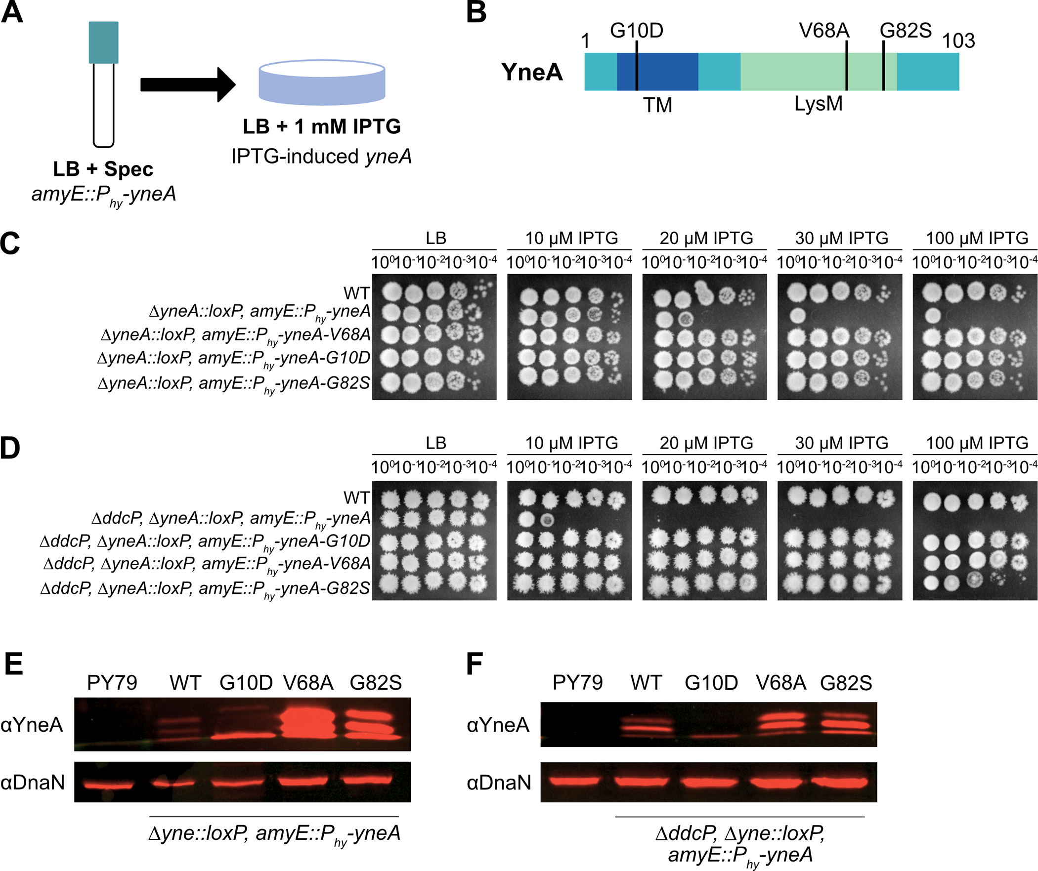
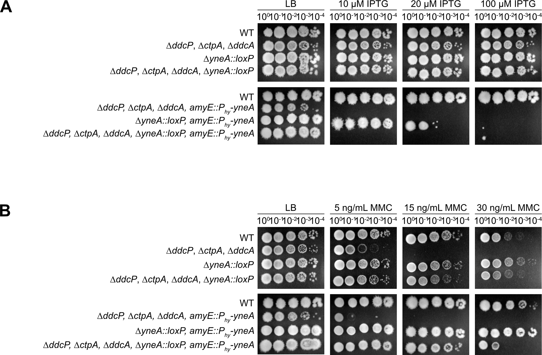
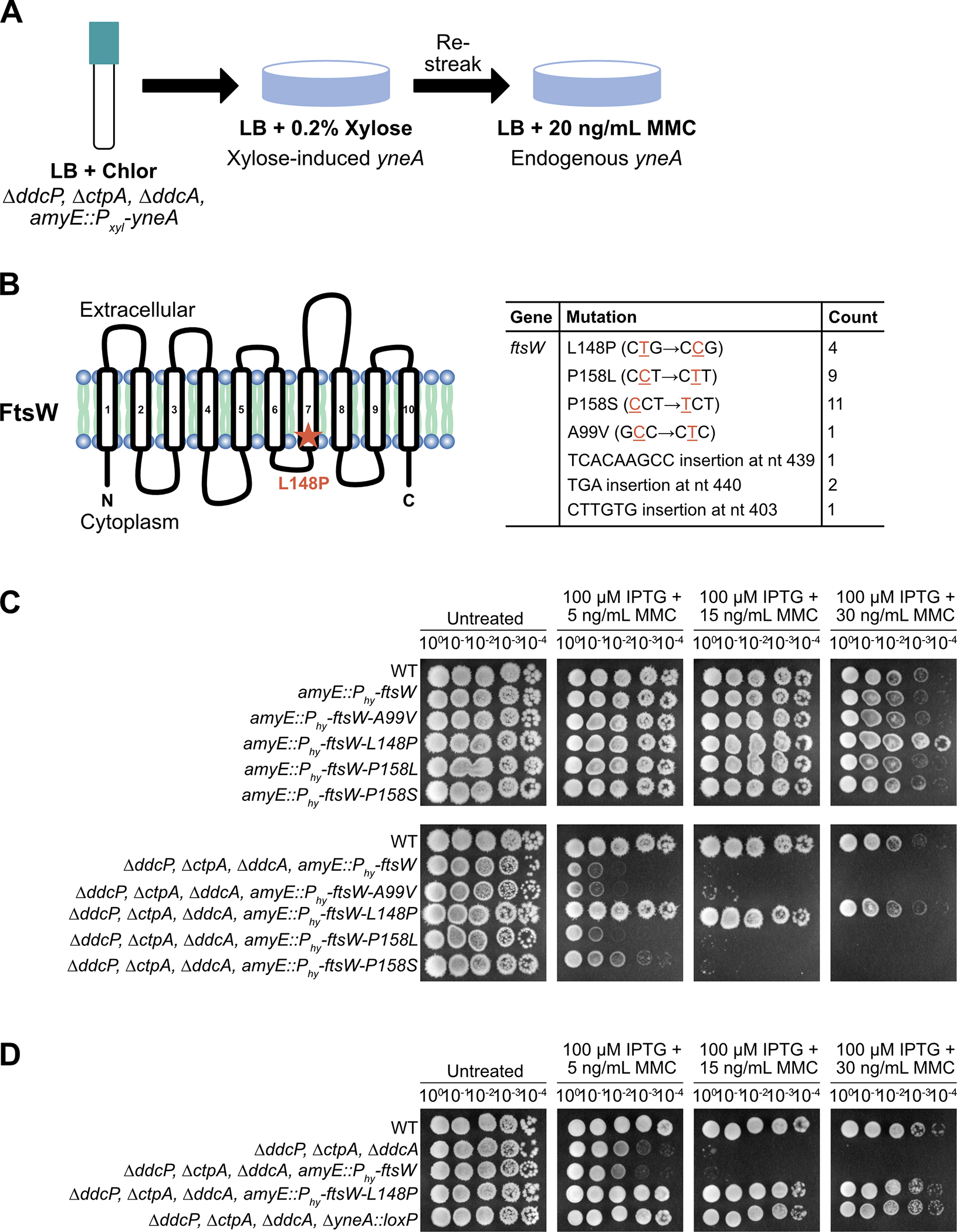
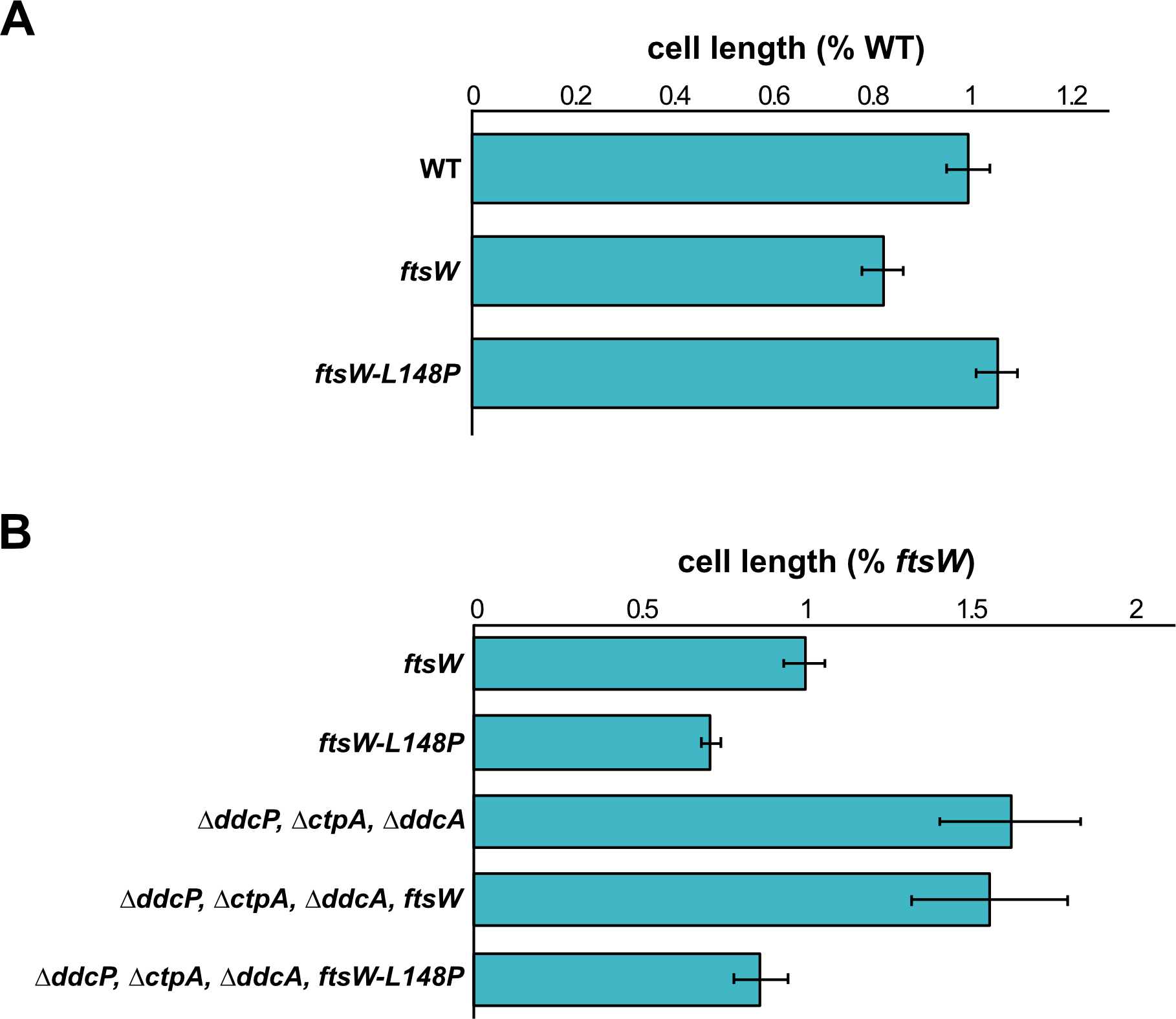
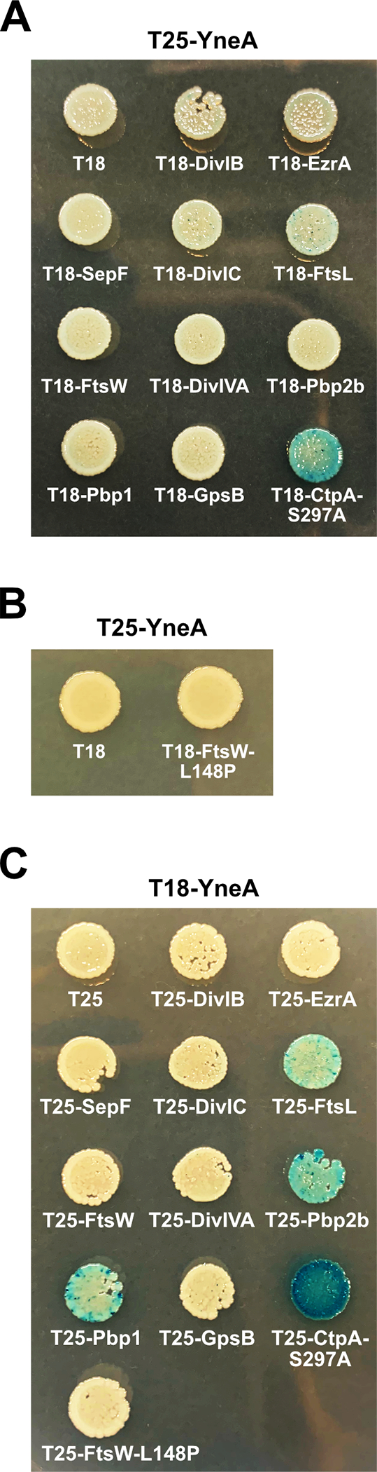
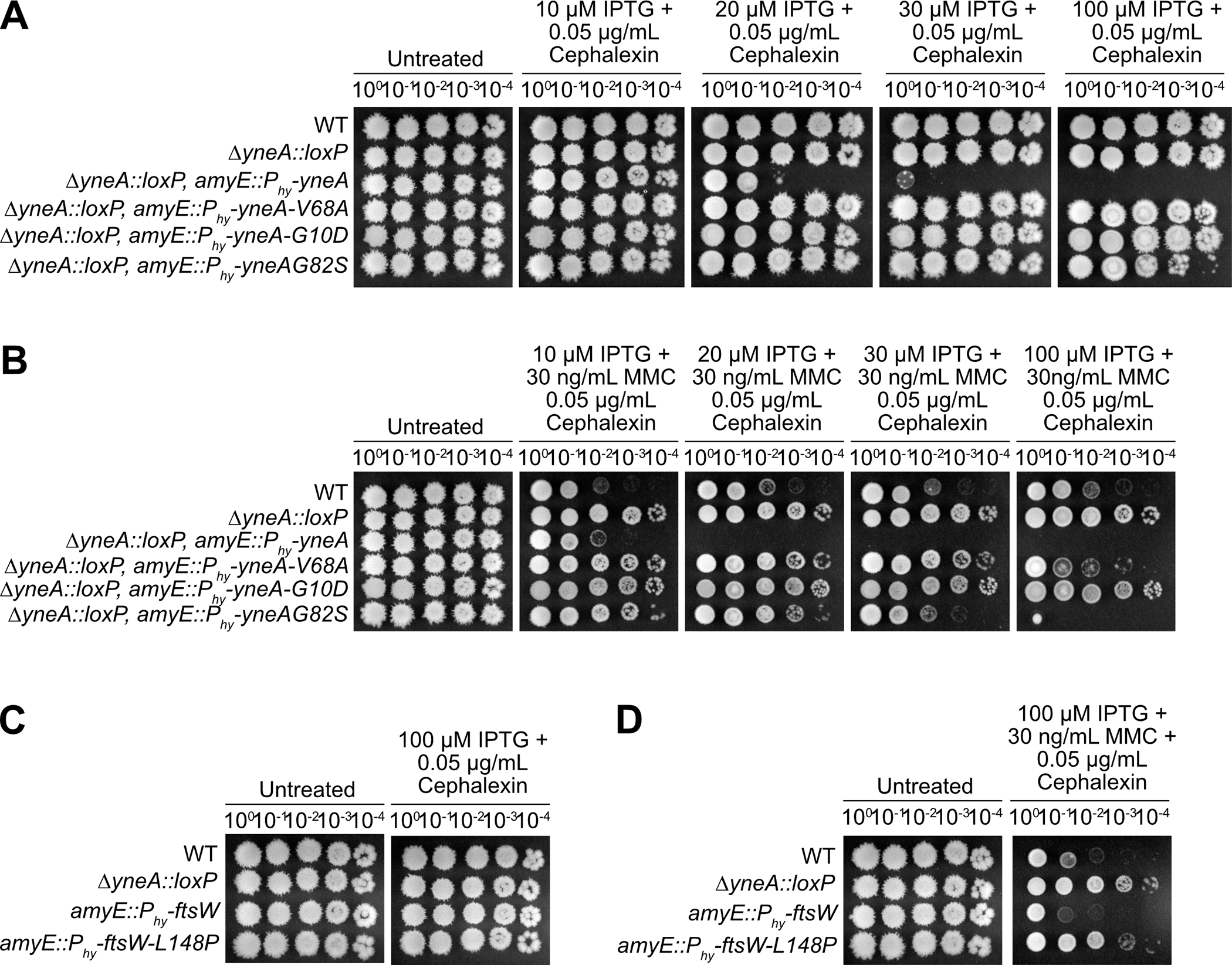
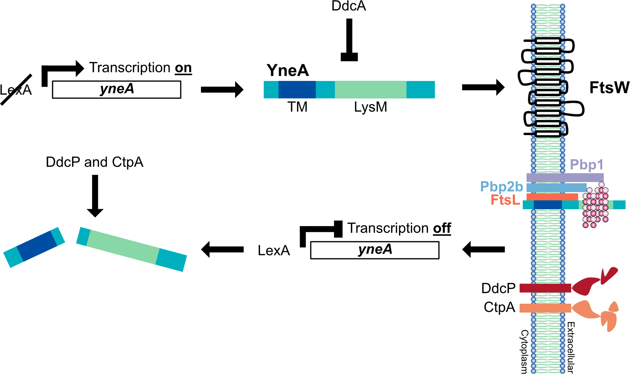
During normal DNA replication, all cells encounter damage to their genetic material. As a result, organisms have developed response pathways that provide time for the cell to complete DNA repair before cell division occurs. In Bacillus subtilis, it is well established that the SOS-induced cell division inhibitor YneA blocks cell division after genotoxic stress; however, it remains unclear how YneA enforces the checkpoint. Here, we identify mutations that disrupt YneA activity and mutations that are refractory to the YneA-induced checkpoint. We find that YneA C-terminal truncation mutants and point mutants in or near the LysM peptidoglycan binding domain render YneA incapable of checkpoint enforcement. In addition, we develop a genetic method which isolated mutations in the ftsW gene that completely bypassed checkpoint enforcement while also finding that YneA interacts with late divisome components FtsL, Pbp2b, and Pbp1. Characterization of an FtsW variant resulted in considerably shorter cells during the DNA damage response indicative of hyperactive initiation of cell division and bypass of the YneA-enforced DNA damage checkpoint. With our results, we present a model where YneA inhibits septal cell wall synthesis by binding peptidoglycan and interfering with interaction between late arriving divisome components causing DNA damage checkpoint activation.

Keywords: DNA damage checkpoint; FtsW; YneA; cell division.


Figures

Figure 1.
Figure 1.. yneA C-terminal truncations impair checkpoint activation.
(A) Schematic of the full-length YneA protein and the C-terminal truncation mutants lacking the last five (yneAΔ5), ten (yneAΔ10) and fifteen (yneAΔ15) amino acid residues. YneA is predicted to have a transmembrane domain (TM) and a LysM binding domain (LysM). (B) Spot titer assay using B. subtilis strains WT (PY79), ΔyneA::loxP amyE::Phy-yneA (EAM46), ΔyneA::loxP amyE::Phy-yneAΔ5 (EAM53), ΔyneA::loxP amyE::Phy-yneAΔ10 (EAM54) and ΔyneA::loxP amyE::Phy-yneAΔ15 (EAM55) spotted on the indicated media. (C) Spot titer assay using B. subtilis strains WT (PY79), ΔddcP ΔyneA::loxP amyE::Phy-yneA (EAM48), ΔddcP ΔyneA::loxP amyE::Phy-yneAΔ5 (EAM83), ΔddcP ΔyneA::loxP amyE::Phy-yneAΔ10 (EAM84) and ΔddcP ΔyneA::loxP amyE::Phy-yneAΔ15 (EAM85) spotted on the indicated media. (D) Western blot using antisera against YneA (upper panel) or DnaN (lower panel) using B. subtilis strains WT (PY79), ΔyneA::loxP amyE::Phy-yneA (EAM46), ΔyneA::loxP amyE::Phy-yneAΔ5 (EAM53), ΔyneA::loxP amyE::Phy-yneAΔ10 (EAM54) and ΔyneA::loxP amyE::Phy-yneAΔ15 (EAM55) after growing in the presence of IPTG until an OD600 = 1. (E) Western blot using antisera against YneA (upper panel) or DnaN (lower panel) using B. subtilis strains WT (PY79), ΔddcP ΔyneA::loxP amyE::Phy-yneA (EAM48), ΔddcP ΔyneA::loxP amyE::Phy-yneAΔ5 (EAM83), ΔddcP ΔyneA::loxP amyE::Phy-yneAΔ10 (EAM84) and ΔddcP ΔyneA::loxP amyE::Phy-yneAΔ15 (EAM85) after growing in the presence of IPTG until an OD600 = 1.
Figure 2.
Figure 2.. Isolation of mutations in YneA that prevent checkpoint activation.
(A) Experimental design for the primary selection. Cultures were plated on LB agar containing 1 mM IPTG to induce expression of amyE::Phy-yneA. (B) Schematic of the YneA protein and the location of the suppressor mutations identified in the screen. Transmembrane domain (TM) and a LysM binding domain (LysM). (C) Spot titer assay using B. subtilis strains WT (PY79), ΔyneA::loxP amyE::Phy-yneA (EAM46), ΔyneA::loxP amyE::Phy-yneA-V68A (EAM49), ΔyneA::loxP amyE::Phy-yneA-G10D (EAM50) and ΔyneA::loxP amyE::Phy-yneA-G82S (EAM52) spotted on the indicated media. (D) Spot titer assay using B. subtilis strains WT (PY79), ΔddcP ΔyneA::loxP amyE::Phy-yneA (EAM48), ΔddcP ΔyneA::loxP amyE::Phy-yneA-G10D (EAM63), ΔddcP ΔyneA::loxP amyE::Phy-yneA-V68A (EAM78) and ΔddcP ΔyneA::loxP amyE::Phy-yneA-G82S (EAM79) spotted on the indicated media. (E) Western blot using antisera against YneA (upper panel) or DnaN (lower panel) using B. subtilis strains WT (PY79), ΔyneA::loxP amyE::Phy-yneA (EAM46), ΔyneA::loxP amyE::Phy-yneA-V68A (EAM49), ΔyneA::loxP amyE::Phy-yneA-G10D (EAM50) and ΔyneA::loxP amyE::Phy-yneA-G82S (EAM52) after growing in the presence of IPTG until an OD600 = 1. (F) Western blot using antisera against YneA (upper panel) or DnaN (lower panel) using B. subtilis strains WT (PY79), ΔddcP ΔyneA::loxP amyE::Phy-yneA (EAM48), ΔddcP ΔyneA::loxP amyE::Phy-yneA-G10D (EAM63), ΔddcP ΔyneA::loxP amyE::Phy-yneA-V68A (EAM78) and ΔddcP ΔyneA::loxP amyE::Phy-yneA-G82S (EAM79) after growing in the presence of IPTG until an OD600 = 1.
Figure 3.
Figure 3.. Cells are more sensitive to yneA induction in the absence of the negative regulators ddcP, ctpA and ddcA.
(A) Spot titer assay using B. subtilis strains WT (PY79), ΔddcP ΔctpA ΔddcA (PEB639), ΔyneA::loxP (PEB439), ΔddcP ΔctpA ΔddcA ΔyneA::loxP (PEB643), ΔddcP ΔctpA ΔddcA amyE::Phy-yneA (PEB844), ΔyneA::loxP amyE::Phy-yneA (EAM46) and ΔddcP ΔctpA ΔddcA ΔyneA::loxP amyE::Phy-yneA (EAM56) spotted on the indicated media. (B) Spot titer assay using B. subtilis strains WT (PY79), ΔddcP ΔctpA ΔddcA (PEB639), ΔyneA::loxP (PEB439), ΔddcP ΔctpA ΔddcA ΔyneA::loxP (PEB643), ΔddcP ΔctpA ΔddcA amyE::Phy-yneA (PEB844), ΔyneA::loxP amyE::Phy-yneA (EAM46) and ΔddcP ΔctpA ΔddcA ΔyneA::loxP amyE::Phy-yneA (EAM56) spotted on the indicated media.
Figure 4.
Figure 4.. ftsW-L148P suppresses YneA activity in the presence of DNA damage.
(A) Experimental design for the selection followed by secondary screen. Cultures were plated on LB agar containing 0.2% xylose to induce expression of amyE::Pxyl-yneA. Colonies were re-streaked on LB agar containing 20 ng/mL MMC to induce expression of endogenous yneA. (B) Schematic of the FtsW protein and the location of the suppressor mutation identified in the screen. FtsW is a membrane-spanning protein that is predicted to have ten transmembrane segments. Table of the ftsW point mutations and insertions identified in the screen. (C) Spot titer assay using B. subtilis strains WT (PY79), amyE::Phy-ftsW (EAM72), amyE::Phy-ftsW-A99V (EAM68), amyE::Phy-ftsW-L148P (EAM69), amyE::Phy-ftsW-P158L (EAM70), amyE::Phy-ftsW-P158S (EAM71), ΔddcP ΔctpA ΔddcA amyE::Phy-ftsW (EAM73), ΔddcP ΔctpA ΔddcA amyE::Phy-ftsW-A99V (EAM64), ΔddcP ΔctpA ΔddcA amyE::Phy-ftsW-L148P (EAM65), ΔddcP ΔctpA ΔddcA amyE::Phy-ftsW-P158L (EAM66) and ΔddcP ΔctpA ΔddcA amyE::Phy-ftsW-P158S (EAM67) spotted on the indicated media. (D) Spot titer assay using B. subtilis strains WT (PY79), ΔddcP ΔctpA ΔddcA (PEB639), ΔddcP ΔctpA ΔddcA amyE::Phy-ftsW (EAM73), ΔddcP ΔctpA ΔddcA amyE::Phy-ftsW-L148P (EAM65) and ΔddcP ΔctpA ΔddcA ΔyneA::loxP (PEB643) spotted on the indicated media.
Figure 5.
Figure 5.. ftsW-L148P bypasses yneA expression.
(A) Cell lengths of each strain relative to WT plotted as a bar graph. Error bars represent standard error of the mean (SEM). The significance test are as follows for a two-tailed t-test: PY79 and amyE::Phy-ftsW (p=0.988); PY79 and amyE::Phy-ftsW-L148P (p=0.959). (B) Cell lengths of each strain relative to ftsW plotted as a bar graph. Error bars represent standard error of the mean (SEM). The significance test are as follows for a two-tailed t-test. For amyE::Phy-ftsW and amyE::Phy-ftsW-L148P (p=4.71E−300); amyE::Phy-ftsW and ΔddcP, ΔctpA, ΔddcA (p=5.14E−119); amyE::Phy-ftsW and ΔddcP, ΔctpA, ΔddcA, amyE::Phy-ftsW-L148P (p=2.6E−36). The cell length measurements graphed here are also presented in supporting Tables S2 and S3.
Figure 6.
Figure 6.. YneA interacts with FtsL, Pbp2b and Pbp1.
Bacterial two-hybrid assay using (A) empty vector (T18), T18 fusions and T25-YneA fusion; (B) empty vector (T18), T18-FtsW-L148P fusion and T25-YneA fusion; (C) empty vector (T25), T25 fusions and T18-YneA co-transformed into E. coli.
Figure 7.
Figure 7.. Mutations that prevent checkpoint activation and bypass yneA expression are less sensitive to an inhibitor of cell wall synthesis.
Spot titer assay using B. subtilis strains (A) and (B) WT (PY79), ΔyneA::loxP (PEB439), ΔyneA::loxP amyE::Phy-yneA (EAM46), ΔyneA::loxP amyE::Phy-yneA-V68A (EAM49), ΔyneA::loxP amyE::Phy-yneA-G10D (EAM50) and ΔyneA::loxP amyE::Phy-yneA-G82S (EAM52); (C) and (D) WT (PY79), ΔyneA::loxP (PEB439), amyE::Phy-ftsW (EAM72) and amyE::Phy-ftsW-L148P (EAM69) spotted on the indicated media.
Figure 8.
Figure 8.. Model for YneA-induced cell division inhibition.
In the presence of DNA damage, cleavage of LexA allows for expression of the SOS-induced cell division inhibitor YneA. YneA expression must reach a critical threshold to bypass the negative regulators DdcA, DdcP and CtpA to activate the checkpoint. YneA localizes to the membrane, mediated by the transmembrane domain (TM), where it interacts with the late divisome proteins FtsL, Pbp2b and Pbp1 as well as binds peptidoglycan interfering with cell wall remodeling at the septum. After the DNA is repaired and YneA expression is repressed by LexA, YneA is cleared by the proteases DdcP and CtpA allowing septal cell wall synthesis to commence and cell division to resume.

Similar articles

Cited by

References

    1. [Dataset], Hawkins WD, Burby PE and Simmons LA (2021). “Whole-genome sequencing coverage for identification of ftsW mutations.” SRA BioProject.
    1. Au N, Kuester-Schoeck E, Mandava V, Bothwell LE, Canny SP, Chachu K, Colavito SA, Fuller SN, Groban ES, Hensley LA, O’Brien TC, Shah A, Tierney JT, Tomm LL, O’Gara TM, Goranov AI, Grossman AD and Lovett CM (2005). “Genetic composition of the Bacillus subtilis SOS system.” J Bacteriol 187(22): 7655–7666. - PMC - PubMed
    1. Bhambhani A, Iadicicco I, Lee J, Ahmed S, Belfatto M, Held D, Marconi A, Parks A, Stewart CR, Margolin W, Levin PA and Haeusser DP (2020). “Bacteriophage SP01 Gene Product 56 Inhibits Bacillus subtilis Cell Division by Interacting with FtsL and Disrupting Pbp2B and FtsW Recruitment.” J Bacteriol 203(2):e00463–20.doi: 10.1128/JB.00463-20. - DOI - PMC - PubMed
    1. Bojer MS, Frees D and Ingmer H (2020). “SosA in Staphylococci: an addition to the paradigm of membrane-localized, SOS-induced cell division inhibition in bacteria.” Curr Genet 66(3): 495–499. - PubMed
    1. Bojer MS, Wacnik K, Kjelgaard P, Gallay C, Bottomley AL, Cohn MT, Lindahl G, Frees D, Veening JW, Foster SJ and Ingmer H (2019). “SosA inhibits cell division in Staphylococcus aureus in response to DNA damage.” Mol Microbiol 112(4): 1116–1130. - PMC - PubMed

Publication types

MeSH terms