

"Time window" effect of Yoda1-evoked Piezo1 channel activity during mouse skeletal muscle differentiation
- PMID: 34097801
- PMCID: PMC9286833
- DOI: 10.1111/apha.13702
"Time window" effect of Yoda1-evoked Piezo1 channel activity during mouse skeletal muscle differentiation
Abstract
Aim: Mechanosensitive Piezo1 ion channels emerged recently as important contributors to various vital functions including modulation of the blood supply to skeletal muscles. The specific Piezo1 channel agonist Yoda1 was shown to regulate the tone of blood vessels similarly to physical exercise. However, the direct role of Piezo1 channels in muscle function has been little studied so far. We therefore investigated the action of Yoda1 on the functional state of skeletal muscle precursors (satellite cells and myotubes) and on adult muscle fibres.
Methods: Immunostaining, electrophysiological intracellular recordings and Ca2+ imaging experiments were performed to localize and assess the effect of the chemical activation of Piezo1 channels with Yoda1, on myogenic precursors, adult myofibres and at the adult neuromuscular junction.
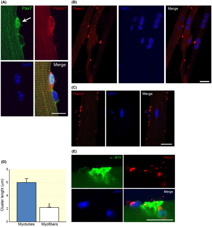
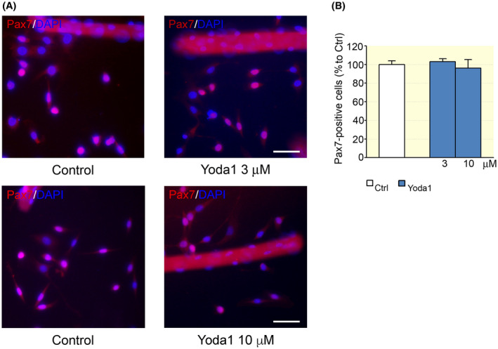
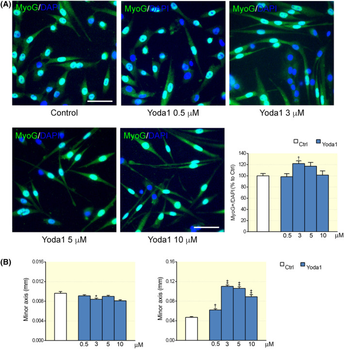
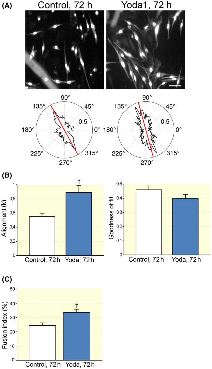
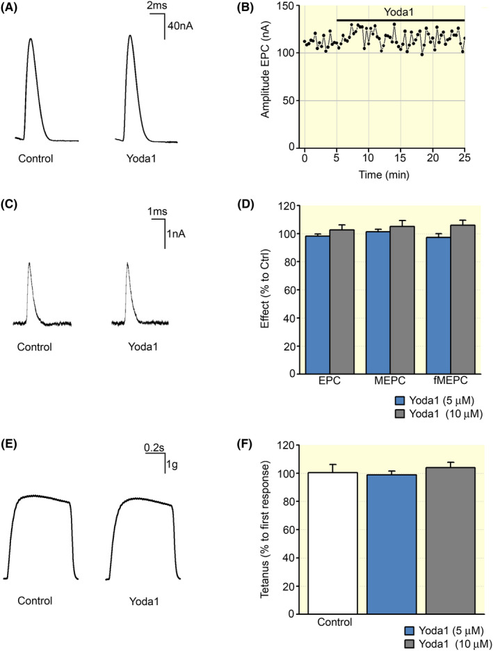
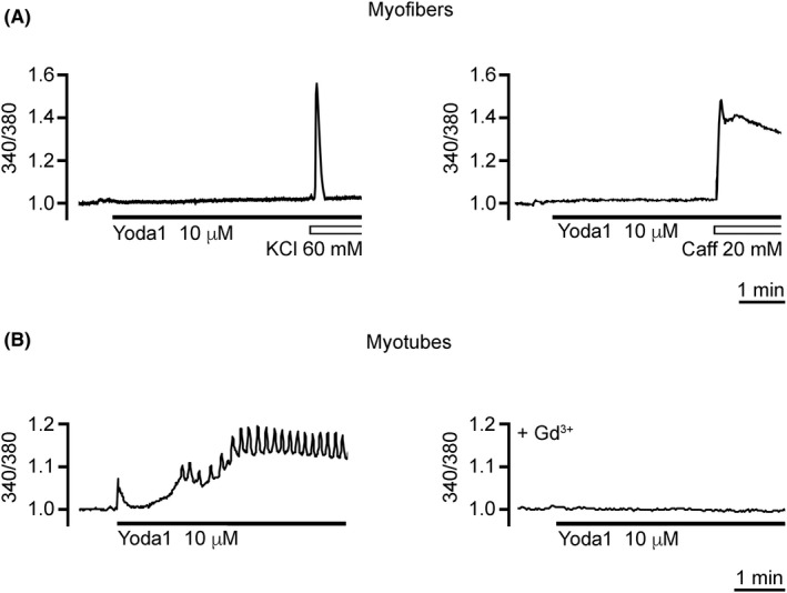
Results: Piezo1 channels were detected by immunostaining in satellite cells (SCs) and myotubes as well as in adult myofibres. In the skeletal muscle precursors, Yoda1 treatment stimulated the differentiation and cell fusion rather than the proliferation of SCs. Moreover, in myotubes, Yoda1 induced significant [Ca2+ ]i transients, without detectable [Ca2+ ]i response in adult myofibres. Furthermore, although expression of Piezo1 channels was detected around the muscle endplate region, Yoda1 application did not alter either the nerve-evoked or spontaneous synaptic activity or muscle contractions in adult myofibres.
Conclusion: Our data indicate that the chemical activation of Piezo1 channels specifically enhances the differentiation of skeletal muscle precursors, suggesting a possible new strategy to promote muscle regeneration.
Keywords: Piezo1 channels; Yoda1; myogenesis; myotubes; satellite cells; skeletal muscle myofibres.
© 2021 The Authors. Acta Physiologica published by John Wiley & Sons Ltd on behalf of Scandinavian Physiological Society.
Conflict of interest statement
The authors declare no conflict of interest.
Figures
Comment in
-
A crucial physiological role of Piezo1 channel in differentiation rather than proliferation during myogenesis.Acta Physiol (Oxf). 2021 Dec;233(4):e13728. doi: 10.1111/apha.13728. Epub 2021 Sep 12. Acta Physiol (Oxf). 2021. PMID: 34492170 No abstract available.
Similar articles
-
The Role of the Piezo1 Mechanosensitive Channel in the Musculoskeletal System.Int J Mol Sci. 2023 Mar 30;24(7):6513. doi: 10.3390/ijms24076513. Int J Mol Sci. 2023. PMID: 37047487 Free PMC article. Review.
-
Piezo1 channels enhance anabolic signaling activation induced by electrical stimulation of cultured myotubes.FEBS Open Bio. 2025 Jun;15(6):940-948. doi: 10.1002/2211-5463.70008. Epub 2025 Feb 17. FEBS Open Bio. 2025. PMID: 39961145 Free PMC article.
-
The State of the Art of Piezo1 Channels in Skeletal Muscle Regeneration.Int J Mol Sci. 2022 Jun 14;23(12):6616. doi: 10.3390/ijms23126616. Int J Mol Sci. 2022. PMID: 35743058 Free PMC article. Review.
-
A Pharmacological Investigation of the TMEM16A Currents in Murine Skeletal Myogenic Precursor Cells.Int J Mol Sci. 2024 Feb 13;25(4):2225. doi: 10.3390/ijms25042225. Int J Mol Sci. 2024. PMID: 38396901 Free PMC article.
-
Piezo1 channel activation mimics high glucose as a stimulator of insulin release.Sci Rep. 2019 Nov 14;9(1):16876. doi: 10.1038/s41598-019-51518-w. Sci Rep. 2019. PMID: 31727906 Free PMC article.
Cited by
-
The Role of the Piezo1 Mechanosensitive Channel in the Musculoskeletal System.Int J Mol Sci. 2023 Mar 30;24(7):6513. doi: 10.3390/ijms24076513. Int J Mol Sci. 2023. PMID: 37047487 Free PMC article. Review.
-
Mechanotransduction for Muscle Protein Synthesis via Mechanically Activated Ion Channels.Life (Basel). 2023 Jan 27;13(2):341. doi: 10.3390/life13020341. Life (Basel). 2023. PMID: 36836698 Free PMC article. Review.
-
Piezo1 channels enhance anabolic signaling activation induced by electrical stimulation of cultured myotubes.FEBS Open Bio. 2025 Jun;15(6):940-948. doi: 10.1002/2211-5463.70008. Epub 2025 Feb 17. FEBS Open Bio. 2025. PMID: 39961145 Free PMC article.
-
The emerging role of Piezo1 channels in skeletal muscle physiology.Biophys Rev. 2023 Sep 29;15(5):1171-1184. doi: 10.1007/s12551-023-01154-6. eCollection 2023 Oct. Biophys Rev. 2023. PMID: 37975010 Free PMC article. Review.
-
Mechanisms and Countermeasures for Muscle Atrophy in Microgravity.Cells. 2024 Dec 20;13(24):2120. doi: 10.3390/cells13242120. Cells. 2024. PMID: 39768210 Free PMC article. Review.
References
Publication types
MeSH terms
Substances
LinkOut - more resources
Full Text Sources
Miscellaneous
