

Maternal bone adaptation to mechanical loading during pregnancy, lactation, and post-weaning recovery
- PMID: 34098162
- PMCID: PMC8504362
- DOI: 10.1016/j.bone.2021.116031
Maternal bone adaptation to mechanical loading during pregnancy, lactation, and post-weaning recovery
Abstract
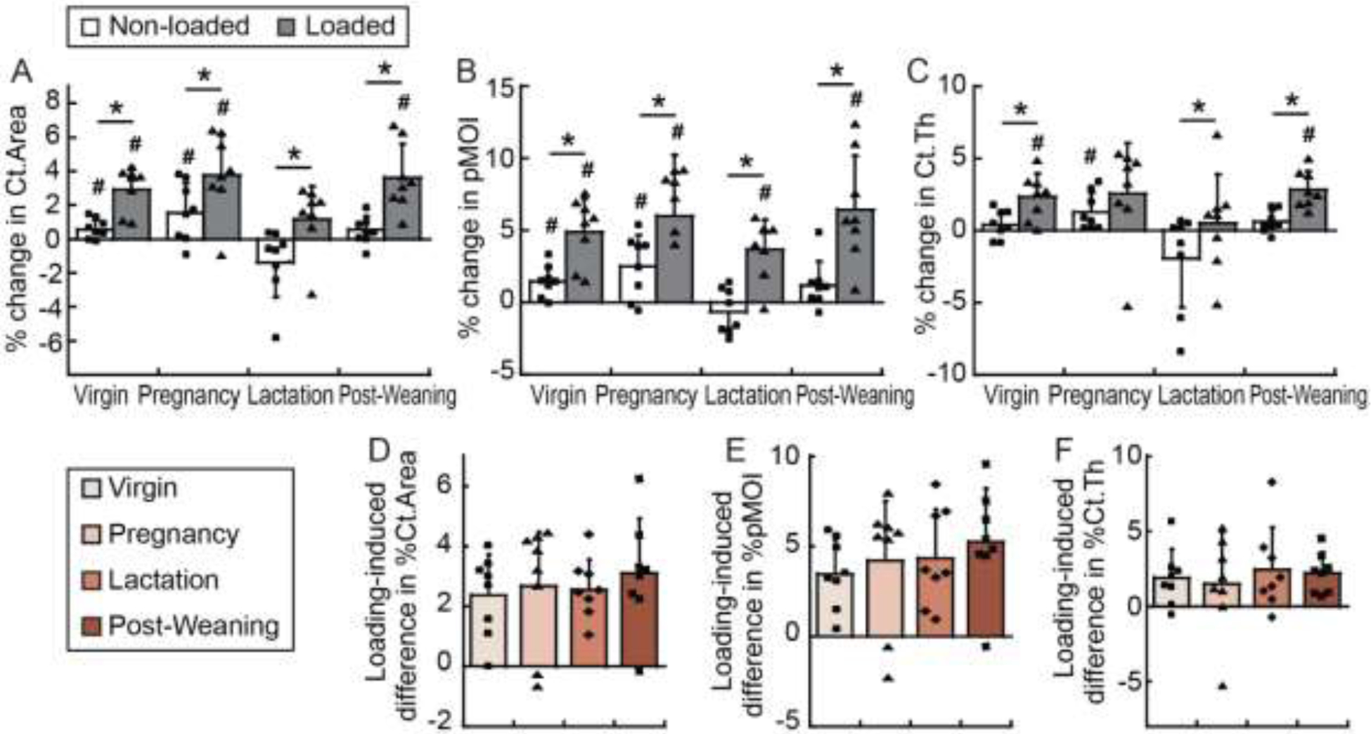
The maternal skeleton undergoes dramatic bone loss during pregnancy and lactation, and substantial bone recovery post-weaning. The structural adaptations of maternal bone during reproduction and lactation exert a better protection of the mechanical integrity at the critical load-bearing sites, suggesting the importance of physiological load-bearing in regulating reproduction-induced skeletal alterations. Although it is suggested that physical exercise during pregnancy and breastfeeding improves women's physical and psychological well-being, its effects on maternal bone health remain unclear. Therefore, the objective of this study was to investigate the maternal bone adaptations to external mechanical loading during pregnancy, lactation, and post-weaning recovery. By utilizing an in vivo dynamic tibial loading protocol in a rat model, we demonstrated improved maternal cortical bone structure in response to dynamic loading at tibial midshaft, regardless of reproductive status. Notably, despite the minimal loading responses detected in the trabecular bone in virgins, rat bone during lactation experienced enhanced mechano-responsiveness in both trabecular and cortical bone compartments when compared to rats at other reproductive stages or age-matched virgins. Furthermore, our study showed that the lactation-induced elevation in osteocyte peri-lacunar/canalicular remodeling (PLR) activities led to enlarged osteocyte lacunae. This may result in alterations in interstitial fluid flow-mediated mechanical stimulation on osteocytes and an elevation in solute transport through the lacunar-canalicular system (LCS) during high-frequency dynamic loading, thus enhancing mechano-responsiveness of maternal bone during lactation. Taken together, findings from this study provide important insights into the relationship between reproduction- and lactation-induced skeletal changes and external mechanical loading, emphasizing the importance of weight-bearing exercise on maternal bone health during reproduction and postpartum.
Keywords: Bone mechano-responsiveness; Lactation; Maternal bone adaptation; Osteocyte mechanosensing; Peri-lacunar/canalicular remodeling (PLR); Reproduction.
Copyright © 2021 Elsevier Inc. All rights reserved.
Conflict of interest statement
Conflict of Interest:
The authors declare that they have no conflict of interest.
Figures


Similar articles
-
Lactation alters fluid flow and solute transport in maternal skeleton: A multiscale modeling study on the effects of microstructural changes and loading frequency.Bone. 2021 Oct;151:116033. doi: 10.1016/j.bone.2021.116033. Epub 2021 Jun 5. Bone. 2021. PMID: 34102350 Free PMC article.
-
Mechanical Regulation of the Maternal Skeleton during Reproduction and Lactation.Curr Osteoporos Rep. 2019 Dec;17(6):375-386. doi: 10.1007/s11914-019-00555-5. Curr Osteoporos Rep. 2019. PMID: 31755029 Free PMC article. Review.
-
Adaptations in the Microarchitecture and Load Distribution of Maternal Cortical and Trabecular Bone in Response to Multiple Reproductive Cycles in Rats.J Bone Miner Res. 2017 May;32(5):1014-1026. doi: 10.1002/jbmr.3084. Epub 2017 Feb 9. J Bone Miner Res. 2017. PMID: 28109138 Free PMC article.
-
Lactation-Induced Changes in the Volume of Osteocyte Lacunar-Canalicular Space Alter Mechanical Properties in Cortical Bone Tissue.J Bone Miner Res. 2017 Apr;32(4):688-697. doi: 10.1002/jbmr.3044. Epub 2016 Dec 12. J Bone Miner Res. 2017. PMID: 27859586 Free PMC article.
-
Influence of Osteocyte Lacunar-Canalicular Morphology and Network Architecture on Osteocyte Mechanosensitivity.Curr Osteoporos Rep. 2023 Aug;21(4):401-413. doi: 10.1007/s11914-023-00792-9. Epub 2023 Jun 5. Curr Osteoporos Rep. 2023. PMID: 37273086 Review.
Cited by
-
Lactation alters fluid flow and solute transport in maternal skeleton: A multiscale modeling study on the effects of microstructural changes and loading frequency.Bone. 2021 Oct;151:116033. doi: 10.1016/j.bone.2021.116033. Epub 2021 Jun 5. Bone. 2021. PMID: 34102350 Free PMC article.
-
Methodological aspects of in vivo axial loading in rodents: a systematic review.J Musculoskelet Neuronal Interact. 2023 Jun 1;23(2):236-262. J Musculoskelet Neuronal Interact. 2023. PMID: 37259664 Free PMC article.
-
Function and Regulation of Bone Marrow Adipose Tissue in Health and Disease: State of the Field and Clinical Considerations.Compr Physiol. 2024 Jun 27;14(3):5521-5579. doi: 10.1002/cphy.c230016. Compr Physiol. 2024. PMID: 39109972 Free PMC article. Review.
-
Osteocyte Remodeling of the Lacunar-Canalicular System: What's in a Name?Curr Osteoporos Rep. 2023 Feb;21(1):11-20. doi: 10.1007/s11914-022-00766-3. Epub 2022 Dec 13. Curr Osteoporos Rep. 2023. PMID: 36512204 Free PMC article. Review.
-
Structural role of osteocyte lacunae on mechanical properties of bone matrix: A cohesive finite element study.J Mech Behav Biomed Mater. 2022 Jan;125:104943. doi: 10.1016/j.jmbbm.2021.104943. Epub 2021 Oct 28. J Mech Behav Biomed Mater. 2022. PMID: 34736032 Free PMC article.
References
-
- Kovacs CS, Maternal Mineral and Bone Metabolism During Pregnancy, Lactation, and Post-Weaning Recovery. Physiol Rev, 2016. 96(2): p. 449–547. - PubMed
-
- Black AJ, et al., A detailed assessment of alterations in bone turnover, calcium homeostasis, and bone density in normal pregnancy. J Bone Miner Res, 2000. 15(3): p. 557–63. - PubMed
-
- Moller UK, et al., Changes in bone mineral density and body composition during pregnancy and postpartum. A controlled cohort study. Osteoporos Int, 2012. 23(4): p. 1213–23. - PubMed
-
- Kaur M, et al., Longitudinal changes in bone mineral density during normal pregnancy. Bone, 2003. 32(4): p. 449–54. - PubMed
-
- Naylor KE, et al., The effect of pregnancy on bone density and bone turnover. J Bone Miner Res, 2000. 15(1): p. 129–37. - PubMed
Publication types
MeSH terms
Grants and funding
LinkOut - more resources
Full Text Sources
