. 2021 Jan-Dec:13:17590914211019886.
doi: 10.1177/17590914211019886.

Aspergillus versicolor Inhalation Triggers Neuroimmune, Glial, and Neuropeptide Transcriptional Changes

Affiliations

Aspergillus versicolor Inhalation Triggers Neuroimmune, Glial, and Neuropeptide Transcriptional Changes

Thatcher B Ladd et al. ASN Neuro. 2021 Jan-Dec.

Abstract

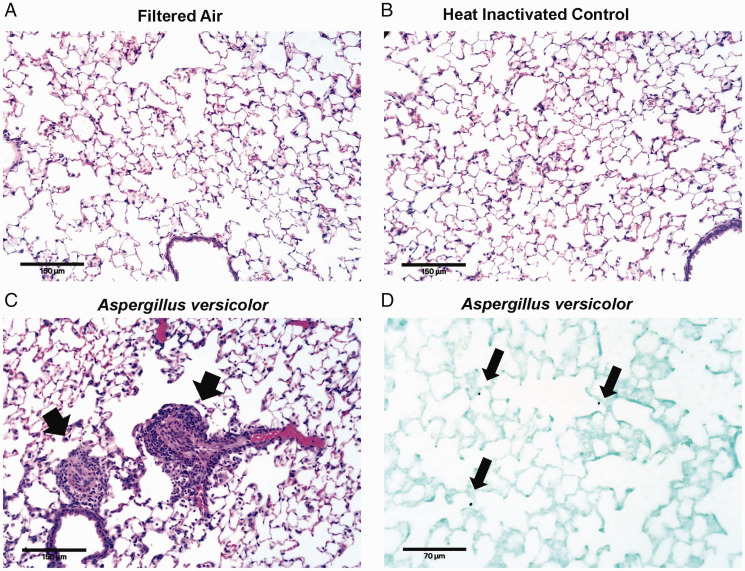
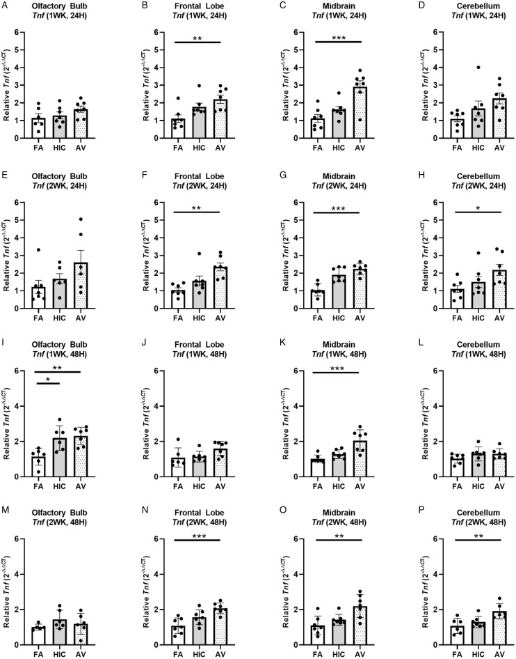
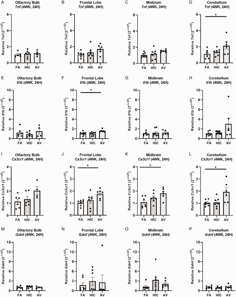
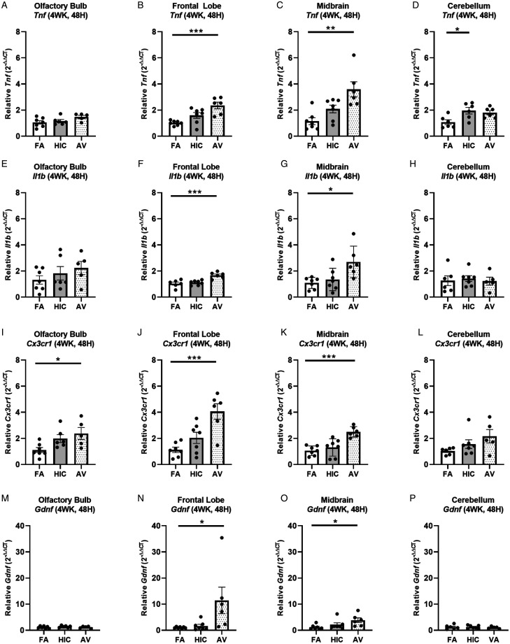
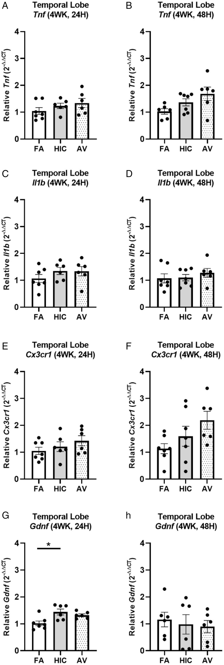
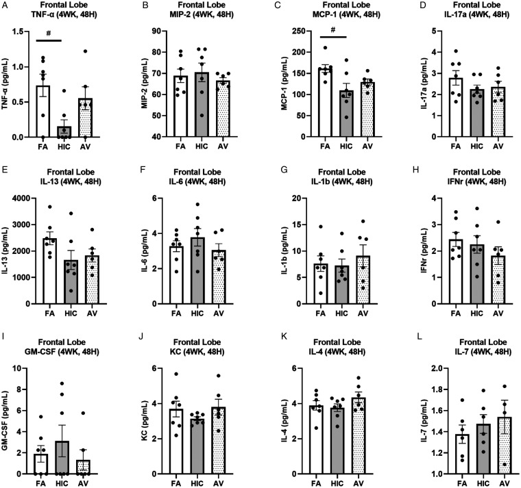
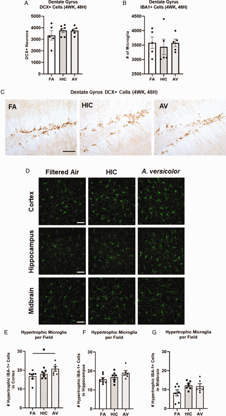
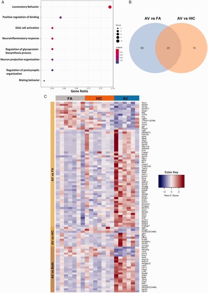
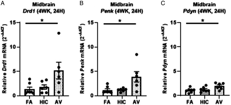
Increasing evidence associates indoor fungal exposure with deleterious central nervous system (CNS) health, such as cognitive and emotional deficits in children and adults, but the specific mechanisms by which it might impact the brain are poorly understood. Mice were exposed to filtered air, heat-inactivated Aspergillus versicolor (3 × 10⁵ spores), or viable A. versicolor (3 × 10⁵ spores) via nose-only inhalation exposure 2 times per week for 1, 2, or 4 weeks. Analysis of cortex, midbrain, olfactory bulb, and cerebellum tissue from mice exposed to viable A. versicolor spores for 1, 2, and 4 weeks revealed significantly elevated pro-inflammatory (Tnf and Il1b) and glial activity (Gdnf and Cxc3r1) gene expression in several brain regions when compared to filtered air control, with the most consistent and pronounced neuroimmune response 48H following the 4-week exposure in the midbrain and frontal lobe. Bulk RNA-seq analysis of the midbrain tissue confirmed that 4 weeks of A. versicolor exposure resulted in significant transcriptional enrichment of several biological pathways compared to the filtered air control, including neuroinflammation, glial cell activation, and regulation of postsynaptic organization. Upregulation of Drd1, Penk, and Pdyn mRNA expression was confirmed in the 4-week A. versicolor exposed midbrain tissue, highlighting that gene expression important for neurotransmission was affected by repeated A. versicolor inhalation exposure. Taken together, these findings indicate that the brain can detect and respond to A. versicolor inhalation exposure with changes in neuroimmune and neurotransmission gene expression, providing much needed insight into how inhaled fungal exposures can affect CNS responses and regulate neuroimmune homeostasis.

Keywords: Aspergillus versicolor; RNA-seq; filamentous fungi; microglia; neuroimmune homeostasis. neuropeptides; neuroinflammation.


Conflict of interest statement

Declaration of Conflicting Interests: The author(s) declared no potential conflicts of interest with respect to the research, authorship, and/or publication of this article.

Figures

Figure 1.
Figure 1.
Aspergillus versicolor exposure causes pulmonary inflammation. Eight week-old female B6C3F1/N mice were exposed in a nose only chamber to filtered air (FA), 3 × 105 spores of heat-inactivated Aspergillus versicolor conidia (HIC), or 3 × 105 spores of viable Aspergillus versicolor (AV) twice weekly, for 4 weeks. Representative photomicrographs of hematoxylin eosin staining of murine lung sections are shown (A–C). Airway inflammation (indicated by the black arrowheads) was observed following exposure to viable A. versicolor and not FA nor HIC. Images were captured using a 20× objective and the scale bar indicates 100 µM. A representative image of Grocott's methenamine silver staining in lung tissue from mice exposed to live AV (D). Black arrows indicate conidia deposited in the lung. Images were captured using a 40× objective and the scale bar indicates 70 µM.
Figure 2.
Figure 2.
Short term Aspergillus versicolor exposure elevates brain Tnf mRNA expression. Eight week-old female B6C3F1/N mice were exposed in a nose only chamber to filtered air (FA), 3 × 105 spores of heat-inactivated Aspergillus versicolor conidia (HIC), or 3 × 105 spores of viable Aspergillus versicolor (AV) twice weekly, for 1 (A–D) or 2 (E–H) weeks and Tnf mRNA expression was assessed 24H after the last exposure in several brain regions. At 48 H after the 1 (I–L) or 2 (M–P) week exposure, Tnf mRNA expression was assessed in several brain regions. Relative Tnf mRNA levels in the olfactory bulb (A, E, I, M), frontal lobe (B, F, J, N), midbrain (C, G, K, O), and cerebellum (G, H, L, P) were determined by qRT-PCR. Individual data points for an experimental animal are represented as black dots. Values were normalized to Gapdh using the 2-ΔΔCT method and are the mean ± SEM. *p < .05; **p < .01; ***p < .001 vs. filtered air control; n = 6–7.
Figure 3.
Figure 3.
Four-week Aspergillus versicolor exposure elevates neuroinflammation 24 hours after the final exposure. Eight week-old female B6C3F1/N mice were exposed in a nose only chamber to filtered air (FA), 3 × 105 spores of heat-inactivated Aspergillus versicolorconidia(HIC), or 3 × 105 spores of viable Aspergillus versicolor (AV) twice weekly, for 4 weeks and markers of neuroinflammation were assessed in the olfactory bulb, frontal lobe, midbrain, and cerebellum 24H after the last exposure. Relative Tnf (A–D), IL1b (E–H), Cx3cr1 (I–L), and Gdnf (M–P) mRNA levels in the olfactory bulb, frontal lobe, midbrain, and cerebellum were determined using qRT-PCR. Individual data points for an experimental animal are represented as black dots. Values were normalized to Gapdh using the 2-ΔΔCT method and are the mean ± SEM. *p<.05 vs. filtered air control; n = 6–7.
Figure 4.
Figure 4.
Four-week Aspergillus versicolor exposure elevates neuroinflammation gene expression 48 hours after the final exposure. Eight week-old female B6C3F1/N mice were exposed in a nose only chamber to filtered air (FA), 3 × 105 spores of heat-inactivated Aspergillus versicolor conidia (HIC), or 3 × 105 spores of viable Aspergillus versicolor (AV) twice weekly, for 4 weeks and markers of neuroinflammation were assessed in the olfactory bulb, frontal lobe, midbrain, and cerebellum 48H after the last exposure. Relative Tnf (A–D), IL1b (E–H), Cx3cr1 (I–L), and Gdnf (M–P) mRNA levels in the olfactory bulb, frontal lobe, midbrain, and cerebellum were determined using qRT-PCR. Individual data points for an experimental animal are represented as black dots. Values were normalized to Gapdh using the 2-ΔΔCT method and are the mean ± SEM. *p < .05; **p < .01; ***p < .001 vs. filtered air control; n = 6–7.
Figure 5.
Figure 5.
Four-week Aspergillus versicolor exposure fails to elevate neuroinflammation gene expression in the temporal lobe. Eight week-old female B6C3F1/N mice were exposed in a nose only chamber to filtered air (FA), 3 × 105 spores of heat-inactivated Aspergillus versicolor conidia (HIC), or 3 × 105 spores of viable Aspergillus versicolor (AV) twice weekly, for 4 weeks and markers of neuroinflammation were assessed in the temporal lobe at 24H or at 48H after the last exposure. Relative Tnf (A and B), IL1b (C and D), Cx3cr1 (E and F), and Gdnf (G and H) mRNA levels in the temporal lobe were determined using qRT-PCR. Individual data points for an experimental animal are represented as black dots. Values were normalized to Gapdh using the 2-ΔΔCT method and are the mean ± SEM. *p < .05 vs. filtered air control; n = 6–7.
Figure 6.
Figure 6.
Four-week Aspergillus versicolor exposure fails to elevate pro-inflammatory proteins in the frontal lobe 48 hours after the final exposure. Eight week-old female B6C3F1/N mice were exposed in a nose only chamber to filtered air (FA), 3 × 105 spores of heat-inactivated Aspergillus versicolor conidia (HIC), or 3 × 105 spores of viable Aspergillus versicolor (AV) twice weekly, for 4 weeks and pro-inflammatory cytokines in the frontal lobe were assessed with a cytokine multiplex assay for changes in 48H after the last exposure. Relative pro-inflammatory cytokines levels (A–L) in the frontal lobe were determined using multiplex assay. Individual data points for an experimental animal are represented as black dots. The mean ± SEM. *p < .05 vs. filtered air control; n = 6–7.
Figure 7.
Figure 7.
Four-week exposure to Aspergillus versicolor changes microglia morphology in the cortex, fails to affect morphology in other regions, and does not impact DCX+ neuron number. Eight week-old female B6C3F1/N mice were exposed in a nose only chamber to filtered air (Air), 3 × 105 spores of heat-inactivated Aspergillus versicolor conidia(HIC), or 3 × 105 spores of viable Aspergillus versicolor twice weekly for 4 weeks. The number of DCX+ neurons (A) and IBA1+ microglia (B) in the dentate gyrus were counted with unbiased stereology. Representative images at 40× are shown of DCX+ neurons in the dentate gyrus of the hippocampus (C). Representative images of 40× images of IBA1+ (green) changes in microglia morphology in the CA1 region of the hippocampus, substantial nigra of the midbrain, and cortex are depicted (D). The number of hypertrophic IBA1+ microglia cells were counted in the cortex (E), and hippocampus (F), and the midbrain (G). Hypertrophic cell = volume >500 µm3. The scale bar indicates 50 microns. Individual data points for an experimental animal are represented as black dots. *p < .05 vs. filtered air control; † p < .05 vs. heat-inactivated control. n = 6–7.
Figure 8.
Figure 8.
Four-week Aspergillus versicolor exposure results in a unique transcriptional signature in midbrain tissue. Eight week-old female B6C3F1/N mice were exposed in a nose only chamber to filtered air (FA), 3 × 105 spores of heat-inactivated Aspergillus versicolor conidia (HIC), or 3 × 105 spores of viable Aspergillus versicolor (AV) twice weekly, for 4 weeks and bulk RNA-seq analysis was performed on midbrain tissue 48H after the last exposure. Significantly enriched biological pathways were identified by GO analysis. Gene Ratio refers to the number of significant genes matched to the term/total number of significant genes (A), a Venn diagram illustrates the overlap of significantly modified genes by exposure (B), and hierarchical clustering of the differentially expressed genes, using the RNA‐seq data derived from the three exposures was assessed.
Figure 9.
Figure 9.
Four-week Aspergillus versicolor exposure elevates basal ganglia neurotransmission genes in the midbrain. Eight week-old female B6C3F1/N mice were exposed in a nose only chamber to filtered air (FA), 3 × 105 spores of heat-inactivated Aspergillus versicolor conidia (HIC), or 3 × 105 spores of viable Aspergillus versicolor (AV) twice weekly, for 4 weeks and markers of neuroinflammation were assessed in the midbrain 48H after the last exposure. Relative Drd1 (A), Penk (B), and PDyn (C) mRNA levels in the midbrain were determined using qRT-PCR. Individual data points for an experimental animal are represented as black dots. Values were normalized to Gapdh using the 2-ΔΔCT method and are the mean ± SEM. *p<.05 vs. filtered air control; n = 6–7.

References

    1. Baldo J. V., Ahmad L., Ruff R. (2002). Neuropsychological performance of patients following mold exposure. Appl Neuropsychol, 9(4), 193–202. - PubMed
    1. Barnes M. A., Croston T. L., Lemons A. R., Rush R. E., Beezhold D. H., Green B. J. (2020). Subchronic inhalation of Aspergillus versicolor conidia leads to sustained innate immune responses and is associated with irreversible remodeling of pulmonary arterial tissue following recovery. J Immunol, 204.
    1. Beguin H., Nolard N. (1994). Mould biodiversity in homes I. Air and surface analysis of 130 dwellings. Aerobiologia, 10(2–3), 157–166.
    1. Benjamini Y., Hochberg Y. (1995). Controlling the false discovery rate: A practical and powerful approach to multiple testing. J R Stat Soc Ser B, 57, 289–300.
    1. Breese M. R., Liu Y. (2013). NGSUtils: A software suite for analyzing and manipulating next-generation sequencing datasets. Bioinformatics, 29(4), 494–496. - PMC - PubMed

Publication types

Supplementary concepts

LinkOut - more resources