. 2021 Jun 7;28(1):43.
doi: 10.1186/s12929-021-00740-8.

Identification of COVID-19 B-cell epitopes with phage-displayed peptide library

Affiliations

Identification of COVID-19 B-cell epitopes with phage-displayed peptide library

Jing-You Guo et al. J Biomed Sci. .

Abstract

Background: Coronavirus disease 19 (COVID-19) first appeared in the city of Wuhan, in the Hubei province of China. Since its emergence, the COVID-19-causing virus, SARS-CoV-2, has been rapidly transmitted around the globe, overwhelming the medical care systems in many countries and leading to more than 3.3 million deaths. Identification of immunological epitopes on the virus would be highly useful for the development of diagnostic tools and vaccines that will be critical to limiting further spread of COVID-19.

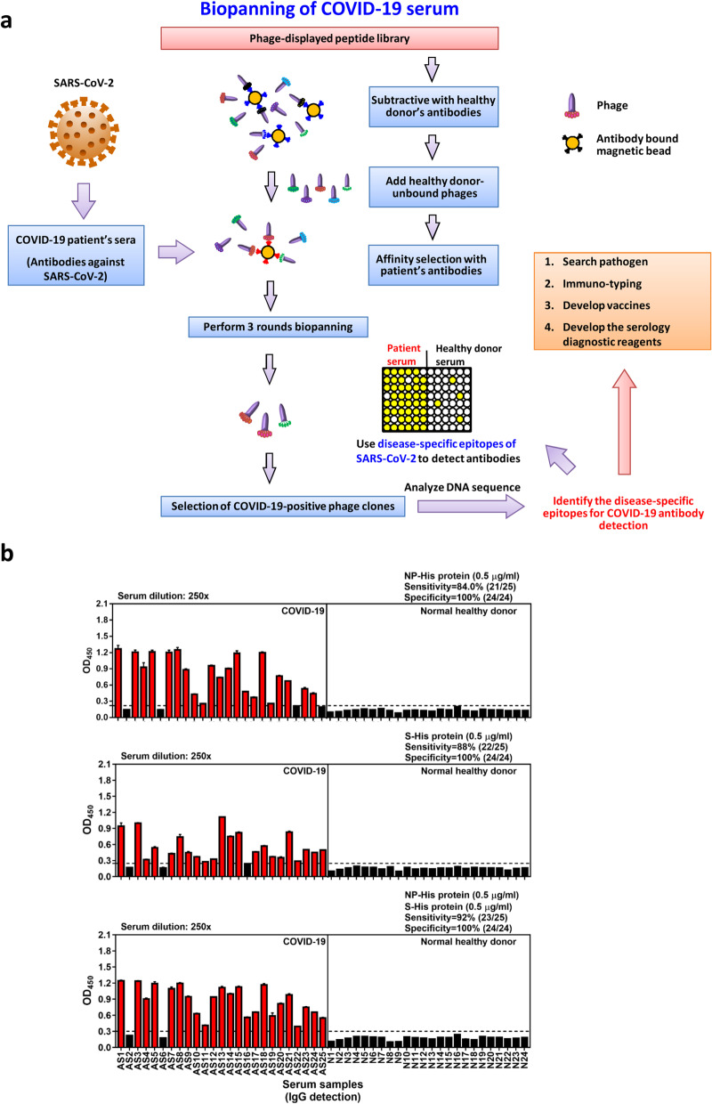
Methods: To find disease-specific B-cell epitopes that correspond to or mimic natural epitopes, we used phage display technology to determine the targets of specific antibodies present in the sera of immune-responsive COVID-19 patients. Enzyme-linked immunosorbent assays were further applied to assess competitive antibody binding and serological detection. VaxiJen, BepiPred-2.0 and DiscoTope 2.0 were utilized for B-cell epitope prediction. PyMOL was used for protein structural analysis.

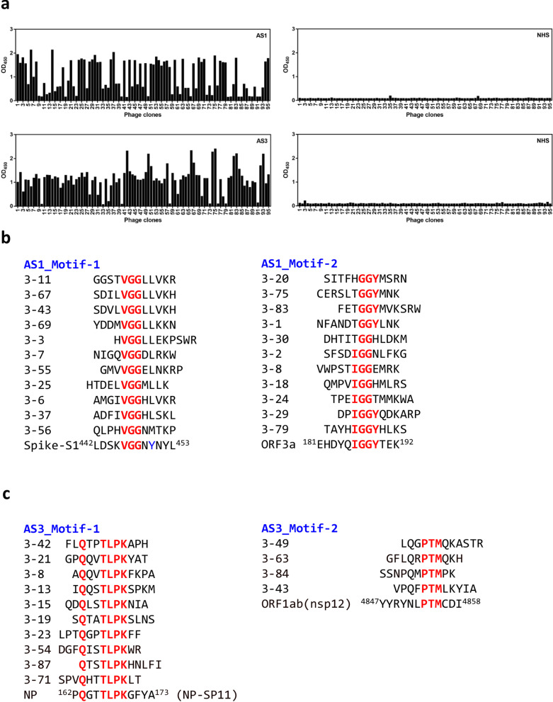
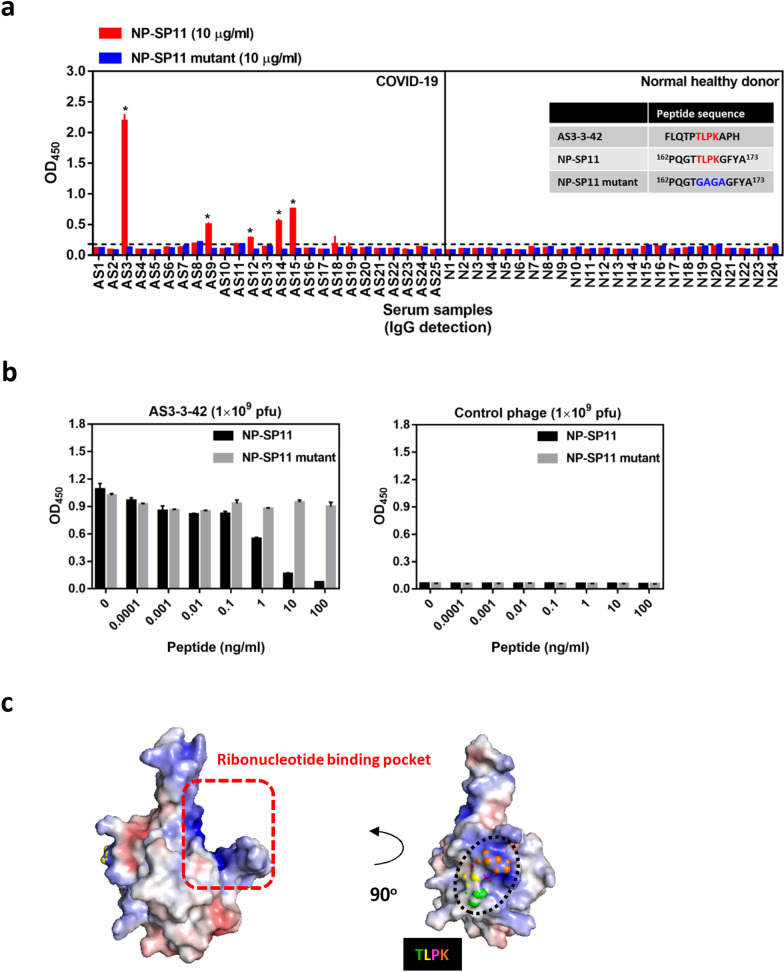
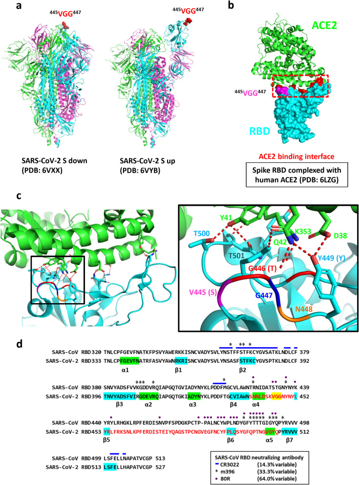
Results: 36 enriched peptides were identified by biopanning with antibodies from two COVID-19 patients; the peptides 4 motifs with consensus residues corresponding to two potential B-cell epitopes on SARS-CoV-2 viral proteins. The putative epitopes and hit peptides were then synthesized for validation by competitive antibody binding and serological detection.

Conclusions: The identified B-cell epitopes on SARS-CoV-2 may aid investigations into COVID-19 pathogenesis and facilitate the development of epitope-based serological diagnostics and vaccines.

Keywords: B-cell epitope; COVID-19; Diagnostic tool; Phage-displayed peptides; SARS-CoV-2; Serological detection.


Conflict of interest statement

The authors declare no competing interest.

Figures

Fig. 1
Fig. 1
Biopanning phage-displayed peptide library with COVID-19 patient serum samples. a Illustration shows the strategy for biopanning disease-specific B-cell epitopes. Serum samples of COVID-19 patients and normal healthy donors were used to prepare IgG-captured magnetic beads. The phage-displayed peptide library was pre-cleaned by normal healthy serum (NHS) IgG-coated magnetic beads, and three rounds of affinity selection of NHS-unbound phages were performed using COVID-19 IgG-magnetic beads. After biopanning, immunopositive phage clones were validated with ELISA and DNA sequenced. The disease-specific epitopes were further identified and characterized by bioinformatic prediction of B-cell epitopes, structural analysis and SARS-CoV-2 protein/peptide synthesis for binding and competitive-inhibition assays. Information about disease-specifc epitopes will be helpful for pathogen research, immune-typing, development of vaccines and serology diagnostic reagents. b Serological detection of COVID-19 convalescent patient antibodies using SARS-CoV-2 NP and extracellular domain of spike recombinant proteins. Horizontal dashed line denotes the cutoff value (calculated as mean OD450 + 4 × SD of normal healthy serum samples) for recombinant SARS-CoV-2 NP or extracellular domain of S protein. Immunopositive cases are colored red
Fig. 2
Fig. 2
Identified peptides from COVID19 positive reacted phage clones. a A phage-displayed random peptide library was screened with serum antibodies from patient AS1 and AS3, respectively. After three screening rounds, enriched phage clones were significantly reactive to antibodies in serum samples from COVID-19 patient but not to samples of normal human serum (NHS). b, c Alignment analysis of sequenced peptides from AS1 (b) and AS3 (c) biopanning was conducted, and identified the conserved residues corresponding to SARS-CoV-2 viral proteins
Fig. 3
Fig. 3
Validation of COVID-19 disease-specific B-cell epitopes derived from biopanning of phage-displayed random peptide libraries. a Serological detection of COVID-19 convalescent patients using NP-SP11 and NP-SP11 mutant peptides. Peptide (2.5 μg/well; 10 μg/ml) was coated overnight followed by serum incubation. Patient IgG binding to peptide was detected by anti-human IgG HRP secondary antibody, and the OD was measured at 450 nm. Sequence of AS3-3-42 phage-displayed peptide and synthetic SARS-CoV-2 NP protein peptides with TLPK motif (NP-SP11) or the GAGA-mutated motif (NP-SP11 mutant) are shown. b Peptide competition assay. IgG from AS3 patient serum was captured, followed by 1-h incubation with both 1 × 109 pfu AS3-3-42 phage (or control phage) and different amounts of synthetic NP peptide. Phage binding was detected by anti-M13 HRP secondary antibody and OD was measured at 450 nm. c Electrostatic surface of the SARS-CoV-2 NP N-terminal domain (NP-NTD, PDB ID: 6M3M). The potential charge distribution was calculated by PyMOL. Blue color indicates positive charge potential; red color indicates negative charge potential. Dashed line denotes the potential ribonucleotide binding pocket [14]. TLPK residues are shown as a sphere. Horizontal dashed line denotes the cutoff value (calculated as mean OD450 + 4 × SD of normal healthy serum samples) for AS3-3-42 phage or NP-SP11 peptide. * Indicates COVID-19-specific positive case
Fig. 4
Fig. 4
Location of VGG residues in SARS-CoV-2 spike glycoprotein. a Conserved VGG residues in the SARS-CoV-2 spike protein are shown as spheres in the prefusion conformation. b VGG residues in SARS-CoV-2 spike RBD are colored pink. ACE2 receptor is shown as a ribbon structure and colored green, while SARS-CoV-2-RBD is shown in the surface representation and colored cyan. RBD residues involved in receptor interaction [18] are colored red, and the ACE2 binding interface is marked by a red dashed outline. c Details of the polar contacts between SARS-CoV-2-RBD and ACE2 are indicated. Polar interactions are denoted as red dashed lines. Side chains of residues are shown as sticks (left panel). The position of VGG residues corresponds to SARS-CoV RBD serine (S), threonine (T) and glycine (G) (right panel). d Comparison of SARS-CoV and SARS-CoV-2 RBD sequence. The reported epitope residues of CR3022, m396 and 80R SARS-CoV neutralizing antibodies are denoted by blue lines, asterisks and purple points, respectively [18, 24]. VGG conserved residues derived from COVID-19 sera/phage-displayed peptide library biopanning are colored yellow

Similar articles

Cited by

References

    1. Center for Systems Science and Engineering (CSSE) at JHU. COVID-19 Dashboard. https://coronavirus.jhu.edu/map.
    1. Coronaviridae Study Group of the International Committee on Taxonomy of V The species Severe acute respiratory syndrome-related coronavirus: classifying 2019-nCoV and naming it SARS-CoV-2. Nat Microbiol. 2020;5(4):536–544. doi: 10.1038/s41564-020-0695-z. - DOI - PMC - PubMed
    1. Zhou P, Yang XL, Wang XG, Hu B, Zhang L, Zhang W, Si HR, Zhu Y, Li B, Huang CL, Chen HD, Chen J, Luo Y, Guo H, Jiang RD, Liu MQ, Chen Y, Shen XR, Wang X, Zheng XS, Zhao K, Chen QJ, Deng F, Liu LL, Yan B, Zhan FX, Wang YY, Xiao GF, Shi ZL. A pneumonia outbreak associated with a new coronavirus of probable bat origin. Nature. 2020;579(7798):270–273. doi: 10.1038/s41586-020-2012-7. - DOI - PMC - PubMed
    1. Lu R, Zhao X, Li J, Niu P, Yang B, Wu H, Wang W, Song H, Huang B, Zhu N, Bi Y, Ma X, Zhan F, Wang L, Hu T, Zhou H, Hu Z, Zhou W, Zhao L, Chen J, Meng Y, Wang J, Lin Y, Yuan J, Xie Z, Ma J, Liu WJ, Wang D, Xu W, Holmes EC, Gao GF, Wu G, Chen W, Shi W, Tan W. Genomic characterisation and epidemiology of 2019 novel coronavirus: implications for virus origins and receptor binding. Lancet. 2020;395(10224):565–574. doi: 10.1016/S0140-6736(20)30251-8. - DOI - PMC - PubMed
    1. Helmy YA, Fawzy M, Elaswad A, Sobieh A, Kenney SP, Shehata AA. The COVID-19 pandemic: a comprehensive review of taxonomy, genetics, epidemiology, diagnosis, treatment, and control. J Clin Med. 2020;9(4):1225. doi: 10.3390/jcm9041225. - DOI - PMC - PubMed