Review
. 2021 Jun 7;40(1):186.
doi: 10.1186/s13046-021-01961-3.

Determinants of resistance to VEGF-TKI and immune checkpoint inhibitors in metastatic renal cell carcinoma

Affiliations
Review

Determinants of resistance to VEGF-TKI and immune checkpoint inhibitors in metastatic renal cell carcinoma

Revati Sharma et al. J Exp Clin Cancer Res. .

Abstract

Vascular endothelial growth factor tyrosine kinase inhibitors (VEGF-TKIs) have been the mainstay of treatment for patients with advanced renal cell carcinoma (RCC). Despite its early promising results in decreasing or delaying the progression of RCC in patients, VEGF-TKIs have provided modest benefits in terms of disease-free progression, as 70% of the patients who initially respond to the treatment later develop drug resistance, with 30% of the patients innately resistant to VEGF-TKIs. In the past decade, several molecular and genetic mechanisms of VEGF-TKI resistance have been reported. One of the mechanisms of VEGF-TKIs is inhibition of the classical angiogenesis pathway. However, recent studies have shown the restoration of an alternative angiogenesis pathway in modulating resistance. Further, in the last 5 years, immune checkpoint inhibitors (ICIs) have revolutionized RCC treatment. Although some patients exhibit potent responses, a non-negligible number of patients are innately resistant or develop resistance within a few months to ICI therapy. Hence, an understanding of the mechanisms of VEGF-TKI and ICI resistance will help in formulating useful knowledge about developing effective treatment strategies for patients with advanced RCC. In this article, we review recent findings on the emerging understanding of RCC pathology, VEGF-TKI and ICI resistance mechanisms, and potential avenues to overcome these resistance mechanisms through rationally designed combination therapies.

Keywords: Clear cell renal carcinoma; Epithelial mesenchymal transition; Hypoxia; Metastatic renal cell carcinoma; Sunitinib; Vascular endothelial growth factor, tyrosine kinase inhibitor.


Conflict of interest statement

The authors declare no competing interests.

Figures

Fig. 1
Fig. 1
Haematoxylin and eosin (H and E) stained paraffin embedded ccRCC section and its corresponding normal kidney tissue. Histopathological slides showing H and E images of normal kidney with well-defined glomerulus and tubules, and conventional ccRCC with typical histological appearance of epithelial nests of large uniform cells with clear cytoplasm and distinct cell membrane (blue arrow). Delicate branches of blood vessels (red arrow) surround the nests of cells. Magnification: 40X, Scale bar: 75 μm
Fig. 2
Fig. 2
Representative immunohistochemistry images of RCC patient tumour sections and adjacent normal kidney tissues stained with HIF-1α target genes GLUT1, HK, LDH, PDK1 and PKM2. Tumours/tissues were stained as described previously [14]. GLUT1 (abcam: ab652): negative staining in glomerulus and moderate cytoplasmic staining in the tubules of adjacent normal kidney tissue while RCC tissues shows a distinct membranous staining. HK (OriGene Technologies, Inc.; AM05641PU-S): adjacent normal kidney tissue shows strong cytoplasmic staining in the tubules and low cytoplasmic staining in the glomerulus while RCC demonstrates a strong cytoplasmic and membranous staining. LDH (abcam; ab52488): adjacent normal kidney is strongly positive for cytoplasmic staining in the tubules and low cytoplasmic staining in the glomerulus; RCC tissues presents an intense positive membranous and nuclear staining. PDK1 (GeneTex; GTX60386): adjacent normal kidney is negative in glomerulus staining and shows very low cytoplasmic staining in the tubules while RCC tissues illustrates distinct membranous and moderate cytoplasmic staining. PKM2 (abcam; ab150377) staining is negative in the adjacent normal kidney tissue while uniform moderate cytoplasmic staining is present in the RCC tissue. Magnification: 40X, Scale bar: 75 μm. Representative images of n = 5 tissues for each antibody
Fig. 3
Fig. 3
Representative immunohistochemistry images of RCC patient tumour sections and adjacent normal kidney tissues stained with E-cadherin and N-cadherin. Immunohistochemistry was performed as described in Fig. 2. E-cadherin (Cell Signaling; 14,472), images show adjacent normal kidney tissue to be slightly positively stained in the tubules while RCC tissue is negative for any staining. N cadherin (Cell Signaling; 13,116): increase in the membranous expression of N-cadherin in the RCC tumour compared to normal adjacent kidney tissue. Magnification: 40X, Scale bar: 75 μm. Representative images of n = 5 tissues for each antibody
Fig. 4
Fig. 4
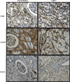
Representative immunohistochemistry images of RCC patient tumour sections and adjacent normal kidney tissues stained with CSC markers CD44, CD105 and CD133. Immunohistochemistry was performed as described in Fig. 2. CD44 (abcam; ab51037): normal kidney tissue shows negative staining in glomerulus and moderate cytoplasmic staining in the tubules; while RCC tissues shows distinct membranous and moderate cytoplasmic staining. CD105 (abcam; ab169545): adjacent normal kidney tissue shows strong cytoplasmic staining in the tubules and low cytoplasmic staining in the glomerulus; RCC tissues shows an intense positive membranous staining. CD133 (abcam; ab19898): adjacent normal kidney showing negative staining; RCC tumour shows a specific membranous staining. Magnification: 40X, Scale bar: 75 μm. Representative images of n = 5 tissues for each antibody
Fig. 5
Fig. 5
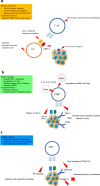
Tumour resistance mechanisms against anti-angiogenic treatments. The upper image shows the various innate resistance mechanisms, which include a) presence of different targets, b) reduced apoptosis due to overproduction of Bcl2/Bcl-xl, and c) over expression of EZH2. In the lower image, the tumour responds initially in response to therapy by shrinking in size but undergoes acquired resistance which facilitates cancer re-growth. Some of the resistance mechanisms are a) initiation of alternative or complementary pathways, b) lysosomal sequestration of the drugs, c) SNPs and micro RNAs, d) recruitment of BMDCs, e) increased pericyte coverage of tumour vessels, f) EMT and g) establishment of new-pro-angiogenic pathways
Fig. 6
Fig. 6
Resistance to ICI therapy. Various putative or acquired immune escape mechanisms can result in tumour evading the immune checkpoint inhibitor therapy. These include; a) Lack of T cell priming through impaired antigen presentation via inadequate DC maturation or through appropriate MHC1 expression; b) decreased T cell activity in the tumour microenvironment through increased infiltration of Tregs, expression of T cell suppressive molecules on tumour cells such as IDO, PD-L1, or mutational changes in JAK2 pathway, loss of tumour suppressor genes such as PTEN or increased expression of T cell exhaustion markers such as TIM-3, PD-1, etc.; c) lack of T cell infiltration in the tumour microenvironment due to enhanced LDH accumulation, low expression of T cell migratory chemokines, increased expression of tumour suppressive cytokines such as TGF-β, IL-6, IL-8, etc.

Similar articles

Cited by

References

    1. Bray F, Ferlay J, Soerjomataram I, Siegel RL, Torre LA, Jemal A. Global cancer statistics 2018: GLOBOCAN estimates of incidence and mortality worldwide for 36 cancers in 185 countries. CA Cancer J Clin. 2018;68(6):394–424. doi: 10.3322/caac.21492. - DOI - PubMed
    1. Koul H, Huh J-S, Rove KO, Crompton L, Koul S, Meacham RB, et al. Molecular aspects of renal cell carcinoma: a review. Am J Cancer Res. 2011;1(2):240–254. - PMC - PubMed
    1. Delahunt B, Srigley JR, Egevad L, Montironi R. International Society of Urological Pathology Grading and Other Prognostic Factors for renal Neoplasia. Eur Urol. 2014;66(5):795–798. doi: 10.1016/j.eururo.2014.05.027. - DOI - PubMed
    1. Mlcochova H, Machackova T, Rabien A, Radova L, Fabian P, Iliev R, et al. Epithelial-mesenchymal transition-associated microRNA/mRNA signature is linked to metastasis and prognosis in clear-cell renal cell carcinoma. Sci Rep. 2016;6(1):31852. doi: 10.1038/srep31852. - DOI - PMC - PubMed
    1. Bhat S. Role of surgery in advanced/metastatic renal cell carcinoma. Indian J Urol. 2010;26(2):167–176. doi: 10.4103/0970-1591.65381. - DOI - PMC - PubMed

MeSH terms

Substances

LinkOut - more resources