. 2021 Jun 7;11(1):106.
doi: 10.1186/s13578-021-00598-1.

Crucial mutation in the exoribonuclease domain of nsp14 of PEDV leads to high genetic instability during viral replication

Affiliations

Crucial mutation in the exoribonuclease domain of nsp14 of PEDV leads to high genetic instability during viral replication

Xiaoyu Niu et al. Cell Biosci. .

Abstract

Background: Coronavirus (CoV) nonstructural protein 14 (nsp14) has exoribonuclease (ExoN) activity, responsible for proofreading and contributing to replication fidelity. It has been reported that CoVs exhibit variable sensitivity to nsp14-ExoN deficiency. Betacoronavirus murine hepatitis virus (MHV) and severe acute respiratory syndrome (SARS)-CoV were viable upon nsp14-ExoN deficiency. While betacoronavirus Middle East respiratory syndrome (MERS)-CoV and SARS-CoV-2 were non-viable with disabled nsp14-ExoN. In this study, we investigated the nsp14-ExoN deficiency of alphacoronavirus porcine epidemic diarrhea virus (PEDV) in viral pathogenesis using reverse genetics.

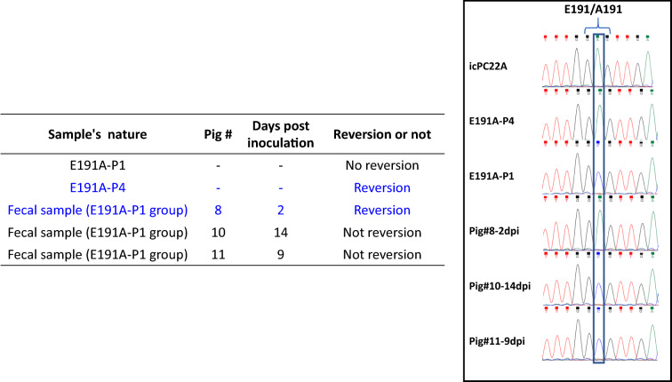
Results: Eight nsp14-ExoN deficient mutants, targeting the predicted active sites and the Zinc finger or mental-coordinating sites, of PEDV were designed. Only one mutant E191A with a mutation in the Mg2+-binding site was rescued using the infectious clone of PEDV PC22A strain (icPC22A). The passage no.1-3 (P1-3) of E191A grew to very low titers in Vero cells. To evaluate the pathogenesis of the E191A, 4 or 5-day-old gnotobiotic pigs were inoculated orally with 100 TCID50/pig of the E191A-P1, icPC22A, or mock. All mock pigs did not shed virus in feces or show clinical signs. All pigs inoculated with icPC22A shed high viral RNA levels, had severe diarrhea, and died by 6 days post-inoculation (dpi). In contrast, only 3 pigs (3/4, 75%) in the E191A-P1 group shed low levels of viral RNA and 2 pigs had moderate diarrhea at acute infection phase. At 22 dpi, each pig was challenged orally with 106 plaque forming unit of virulent icPC22A. All pigs in the mock group developed severe diarrhea and 2 of the 5 pigs died. Pigs in the E191A-P1 group had less severe diarrhea and no pigs died. Sanger sequencing analysis revealed that the viral genome in the fecal sample of one E191A-P1-inoculated pig and the P4 virus passaged in vitro lost the E191A mutation, suggesting the genetic instability of the E191A mutant.

Conclusion: The recombinant PEDV variants carrying mutations at the essential functional sites within nsp14-ExoN were either lethal or genetically unstable. Our finding further confirmed the critical role of nsp14-ExoN in CoV life cycle, suggesting that it may be a target for the design of universal anti-CoV drugs.

Keywords: Coronavirus; Exoribonuclease; Infectious clone; Nsp14; Porcine epidemic diarrhea virus; Reverse genetics.


Conflict of interest statement

The authors declare that they have no competing interests.

Figures

Fig. 1
Fig. 1
Design of recombinant PEDVs with mutations at different active sites of nsp14-ExoN. a Alignment of nsp14-ExoN amino acid sequences from selected coronaviruses. Sequences of the ExoN domains in PEDV (KY499262), IBV (NP_040829), MHV (NP_045298), MERS-CoV (NC-019843), SARS-CoV (NC_004718), and SARS-CoV-2 (NC_045512.2) were used for the analysis. b Alignment of partial nsp14 amino acid sequences of the infectious clone derived PC22A (icPC22A) and the eight PEDV nsp14 mutants designed in this study. Motifs I, II, and III conferring the ExoN active site are shown in green boxes. The Zinc fingers region is indicated in black box. The metal ion cooperating residues (D90, E92, E191, and D272), Zinc finger-related sites (C209 and H228), as well as other active sites involved in the study (N237, D242, and H267) are labeled in red. The introduced mutations are shown in blue. The numbers in gray indicate how many residues in PEDV-nsp14 are not shown. c 3D structural models for icPC22A nsp14. ExoN domain is circled. The metal ion cooperating residues (D90, E92, E191, and D272), Zinc finger-related sites (C209 and H228), as well as other active sites involved in the study (N237, D242, and H267) are labeled in red circles. d The predicted structural models for each mutation site and surrounding amino acids of wild type nsp14 and the mutants designed in this study
Fig. 2
Fig. 2
In vitro Characterization of the recombinant nsp14-ExoN mutant E191A. a, b Multi-step growth curves of recombinant PEDVs (E191A-P1, E191A-P4, and the parental icPC22A) in Vero cells using an MOI of 0.01. Due to the very low infectious titers of E191A-P1, the RNA titers presented in b was employed to interpret the growth kinetics of recombinant PEDVs; c plaques of recombinant PEDVs in Vero cells overlayed with 1.5% agarose. The cells were fixed and stained with crystal violet at 72 hpi. d, e Vero cells infected with recombinant PEDVs at a MOI of 0.01 was harvested at 24 hpi. The expression level of sgmRNA-3 (left Y axis), sgmRNA-N (left Y axis), and genomic RNA (right Y axis) were presented in d and the sgmRNAs/TCID50 (left Y axis) or genomic RNA/TCID50 (right Y axis) ratios were shown in e. **, p < 0.01; ***, p < 0.005; ****, p < 0.001; ns, no significance
Fig. 3
Fig. 3
Pathogenicity of the recombinant PEDVs in Gn piglets. a Fecal consistency scores of pigs within 18 dpi. Fecal consistency was scored as follows: 0, solid; 1, pasty; 2, semiliquid; and 3, liquid. Scores of ≥ 2 and 3 were considered diarrhea and severe diarrhea, respectively. Each dot represents the score of an individual pig; each line indicates the mean scores of a group. Pigs with fecal scores greater than 2 were defined as having diarrhea and a score of 3 was severe diarrhea. All pigs in the icPC22A group died by 6 dpi. b PEDV RNA (N gene) shedding titers in rectal swabs within 18 dpi. Each symbol represents the titer of an individual piglet; each line indicates the mean values of a group. The dash line at 4.8 log10 copies/mL indicates the detection limit. c Survival curves of pigs after inoculation by 21dpi. d Villous height to crypt depth (VH: CD) ratios of ileum of piglets euthanized at 3 dpi. Groups with significant differences are indicated with different letters. e Immunohistochemistry staining of PEDV N proteins in the enterocytes of ileum of piglets (magnification, 100 ×). The brown signals, indicated by arrows, represented the PEDV antigens in enterocytes and were observed in the icPC22A- and E191A-P1-inoculated, but not in mock piglets
Fig. 4
Fig. 4
Induction of partial protection by E191A-P1 mutant in Gn pigs against icPC22A challenge. a Fecal consistency scores of pigs post challenge. Fecal consistency was scored as follows: 0, solid; 1, pasty; 2, semiliquid; and 3, liquid. Scores of ≥ 2 and 3 were considered diarrhea and severe diarrhea, respectively. Each dot represents the score of an individual pig; each line indicates the mean scores of a group. A pig with a score of ≥ 2 was defined as having diarrhea and a score of 3 was severe diarrhea. b Fecal PEDV RNA shedding profile of pigs post challenge. Each line indicates the mean values of a group. The dash line at 4.8 log10 copies/mL indicates the detection limit. c Survival curves of pigs by 9 dpc. d VN antibody titers in serum samples collected at different time points
Fig. 5
Fig. 5
Genetic instability of E191A mutant. Sanger sequencing showed that E191A-P4 and the fecal sample collected at 2 dpi from the E191A-P1-inoculated pig#8 reverted to wildtype ExoN (GAG)

Similar articles

Cited by

References

    1. Li W, Li H, Liu Y, Pan Y, Deng F, Song Y, et al. New variants of porcine epidemic diarrhea virus, China, 2011. Emerg Infect Dis. 2012;18(8):1350. doi: 10.3201/eid1803.120002. - DOI - PMC - PubMed
    1. Stevenson GW, Hoang H, Schwartz KJ, Burrough ER, Sun D, Madson D, et al. Emergence of Porcine epidemic diarrhea virus in the United States: clinical signs, lesions, and viral genomic sequences. J Vet Diagn Invest. 2013;25(5):649–654. doi: 10.1177/1040638713501675. - DOI - PubMed
    1. Sun RQ, Cai RJ, Chen YQ, Liang PS, Chen DK, Song CX. Outbreak of porcine epidemic diarrhea in suckling piglets, China. Emerg Infect Dis. 2012;18(1):161. doi: 10.3201/eid1801.111259. - DOI - PMC - PubMed
    1. Schulz LL, Tonsor GT. Assessment of the economic impacts of porcine epidemic diarrhea virus in the United States. J Anim Sci. 2015;93(11):5111–5118. doi: 10.2527/jas.2015-9136. - DOI - PMC - PubMed
    1. Paarlberg PL. Updated Estimated Economic Welfare Impacts of Porcine Epidemic Diarrhea Virus (PEDV). 2014. Available online:http://ageconsearch.umn.edu/bitstream/174517/2/14-4.Updated%20Estimated%.... Accessed 25 Sept 2015.

LinkOut - more resources