

Pulmonary Vein Thrombosis in COVID-19
- PMID: 34099150
- PMCID: PMC8175944
- DOI: 10.1016/j.chest.2020.11.064
Pulmonary Vein Thrombosis in COVID-19
Abstract
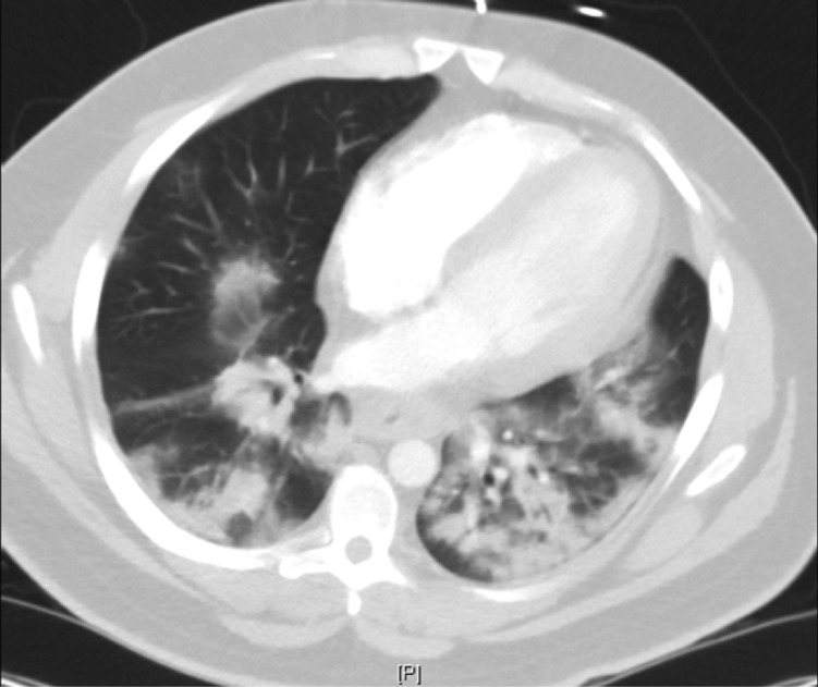
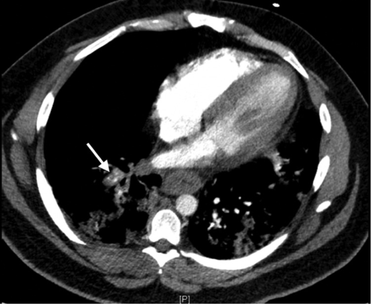
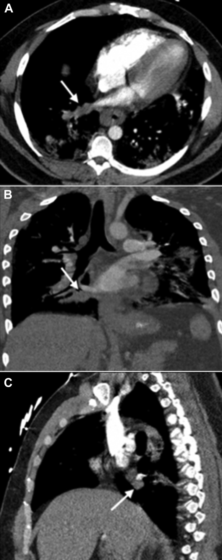
Research on COVID-19, the cause of a rapidly worsening pandemic, has led to the observation of laboratory derangements such as a propensity towards a hypercoagulable state. However, there are currently no reports on the incidence of pulmonary venous thrombosis in the setting of COVID-19. We report a case in which follow-up chest CT scans revealed an expansile filling defect in a branch of the right inferior pulmonary vein, which is consistent with pulmonary venous thrombosis. Our objective was to provide insight into an uncommon sequela of COVID-19 and consequently garner increased clinical suspicion for pulmonary VTE during hospitalization.
Keywords: COVID-19; imaging; pulmonary; thrombosis.
Copyright © 2021 American College of Chest Physicians. Published by Elsevier Inc. All rights reserved.
Figures
References
-
- Zhang Y., Cao W., Xiao M., et al. Clinical and coagulation characteristics of 7 patients with critical COVID 2019 pneumonia and acro-ischemia. Zhonghua Xue Ye Xue Za Zhi. 2020;41(0):E006. - PubMed
-
- Kim N.H., Roldan C.A., Shively B.K., et al. Pulmonary vein thrombosis. Chest. 1993;104(2):624–626. - PubMed
Publication types
MeSH terms
LinkOut - more resources
Full Text Sources
Medical
