

Production and purification of ATP-sensitive potassium channel particles for cryo-electron microscopy
- PMID: 34099169
- PMCID: PMC9719412
- DOI: 10.1016/bs.mie.2021.02.008
Production and purification of ATP-sensitive potassium channel particles for cryo-electron microscopy
Abstract
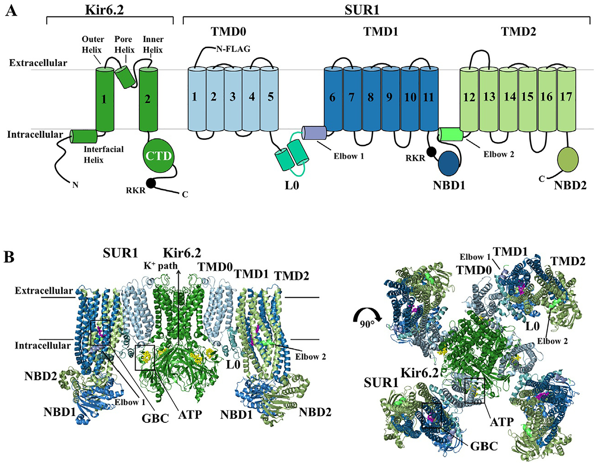
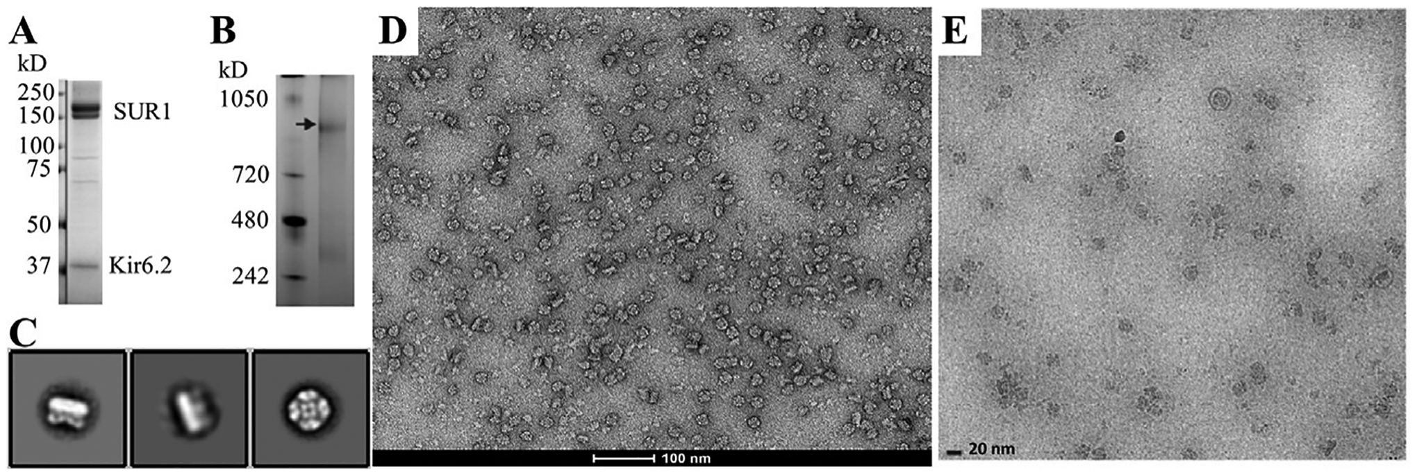
ATP-sensitive potassium (KATP) channels are multimeric protein complexes made of four inward rectifying potassium channel (Kir6.x) subunits and four ABC protein sulfonylurea receptor (SURx) subunits. Kir6.x subunits form the potassium ion conducting pore of the channel, and SURx functions to regulate Kir6.x. Kir6.x and SURx are uniquely dependent on each other for expression and function. In pancreatic β-cells, channels comprising SUR1 and Kir6.2 mediate glucose-stimulated insulin secretion and are the targets of antidiabetic sulfonylureas. Mutations in genes encoding SUR1 or Kir6.2 are linked to insulin secretion disorders, with loss- or gain-of-function mutations causing congenital hyperinsulinism or neonatal diabetes mellitus, respectively. Defects in the KATP channel in other tissues underlie human diseases of the cardiovascular and nervous systems. Key to understanding how channels are regulated by physiological and pharmacological ligands and how mutations disrupt channel assembly or gating to cause disease is the ability to observe structural changes associated with subunit interactions and ligand binding. While recent advances in the structural method of single-particle cryo-electron microscopy (cryoEM) offers direct visualization of channel structures, success of obtaining high-resolution structures is dependent on highly concentrated, homogeneous KATP channel particles. In this chapter, we describe a method for expressing KATP channels in mammalian cell culture, solubilizing the channel in detergent micelles and purifying KATP channels using an affinity tag to the SURx subunit for cryoEM structural studies.
Keywords: K(ATP) channel; Kir6.2; Structure; Sulfonylurea receptor; cryoEM.
Copyright © 2021 Elsevier Inc. All rights reserved.
Figures
Similar articles
-
Structure of an open KATP channel reveals tandem PIP2 binding sites mediating the Kir6.2 and SUR1 regulatory interface.Nat Commun. 2024 Mar 20;15(1):2502. doi: 10.1038/s41467-024-46751-5. Nat Commun. 2024. PMID: 38509107 Free PMC article.
-
Dynamic duo: Kir6 and SUR in KATP channel structure and function.Channels (Austin). 2024 Dec;18(1):2327708. doi: 10.1080/19336950.2024.2327708. Epub 2024 Mar 15. Channels (Austin). 2024. PMID: 38489043 Free PMC article. Review.
-
Molecular biology of adenosine triphosphate-sensitive potassium channels.Endocr Rev. 1999 Apr;20(2):101-35. doi: 10.1210/edrv.20.2.0361. Endocr Rev. 1999. PMID: 10204114 Review.
-
New insights into KATP channel gene mutations and neonatal diabetes mellitus.Nat Rev Endocrinol. 2020 Jul;16(7):378-393. doi: 10.1038/s41574-020-0351-y. Epub 2020 May 6. Nat Rev Endocrinol. 2020. PMID: 32376986 Review.
-
ATP binding without hydrolysis switches sulfonylurea receptor 1 (SUR1) to outward-facing conformations that activate KATP channels.J Biol Chem. 2019 Mar 8;294(10):3707-3719. doi: 10.1074/jbc.RA118.005236. Epub 2018 Dec 26. J Biol Chem. 2019. PMID: 30587573 Free PMC article.
Cited by
-
HCV affects KATP channels through GnT-IVa-mediated N-glycosylation of GLUT2 on the surface of pancreatic β-cells leading to impaired insulin secretion.Endocrine. 2024 May;84(2):427-440. doi: 10.1007/s12020-023-03589-z. Epub 2023 Nov 14. Endocrine. 2024. PMID: 37962815 Free PMC article.
-
AI-based discovery and cryoEM structural elucidation of a KATP channel pharmacochaperone.Elife. 2025 Mar 26;13:RP103159. doi: 10.7554/eLife.103159. Elife. 2025. PMID: 40135739 Free PMC article.
-
Structure of an open KATP channel reveals tandem PIP2 binding sites mediating the Kir6.2 and SUR1 regulatory interface.Nat Commun. 2024 Mar 20;15(1):2502. doi: 10.1038/s41467-024-46751-5. Nat Commun. 2024. PMID: 38509107 Free PMC article.
-
Vascular KATP channel structural dynamics reveal regulatory mechanism by Mg-nucleotides.Proc Natl Acad Sci U S A. 2021 Nov 2;118(44):e2109441118. doi: 10.1073/pnas.2109441118. Proc Natl Acad Sci U S A. 2021. PMID: 34711681 Free PMC article.
-
Identification of a novel KCNT2 variant in a family with developmental and epileptic encephalopathies: a case report and literature review.Front Genet. 2024 Mar 6;15:1371282. doi: 10.3389/fgene.2024.1371282. eCollection 2024. Front Genet. 2024. PMID: 38510274 Free PMC article.
References
-
- Aguilar-Bryan L, & Bryan J (1999). Molecular biology of adenosine triphosphate-sensitive potassium channels. Endocrine Reviews, 20(2), 101–135. - PubMed
-
- Aguilar-Bryan L, Clement J. P.t., Gonzalez G, Kunjilwar K, Babenko A, & Bryan J (1998). Toward understanding the assembly and structure of KATP channels. Physiological Reviews, 78(1), 227–245. - PubMed
-
- Aguilar-Bryan L, Nichols CG, Rajan AS, Parker C, & Bryan J (1992). Co-expression of sulfonylurea receptors and KATP channels in hamster insulinoma tumor (HIT) cells. Evidence for direct association of the receptor with the channel. The Journal of Biological Chemistry, 267(21), 14934–14940. - PubMed
-
- Alekseev AE, Kennedy ME, Navarro B, & Terzic A (1997). Burst kinetics of co-expressed Kir6.2/SUR1 clones: Comparison of recombinant with native ATP-sensitive K+ channel behavior. The Journal of Membrane Biology, 159(2), 161–168. - PubMed
Publication types
MeSH terms
Substances
Grants and funding
LinkOut - more resources
Full Text Sources
