Review
. 2021 Aug;44(8):669-686.
doi: 10.1016/j.tins.2021.05.002. Epub 2021 Jun 4.

To look or not to look: dissociating presaccadic and covert spatial attention

Affiliations
Review

To look or not to look: dissociating presaccadic and covert spatial attention

Hsin-Hung Li et al. Trends Neurosci. 2021 Aug.

Abstract

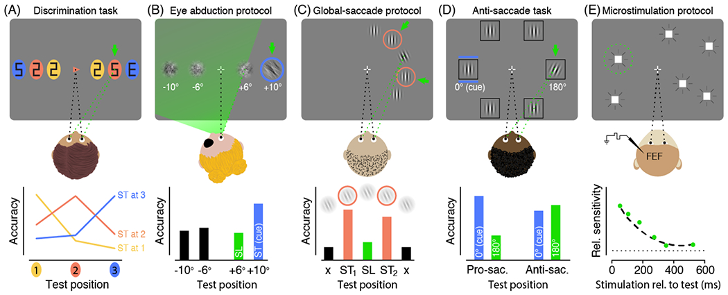
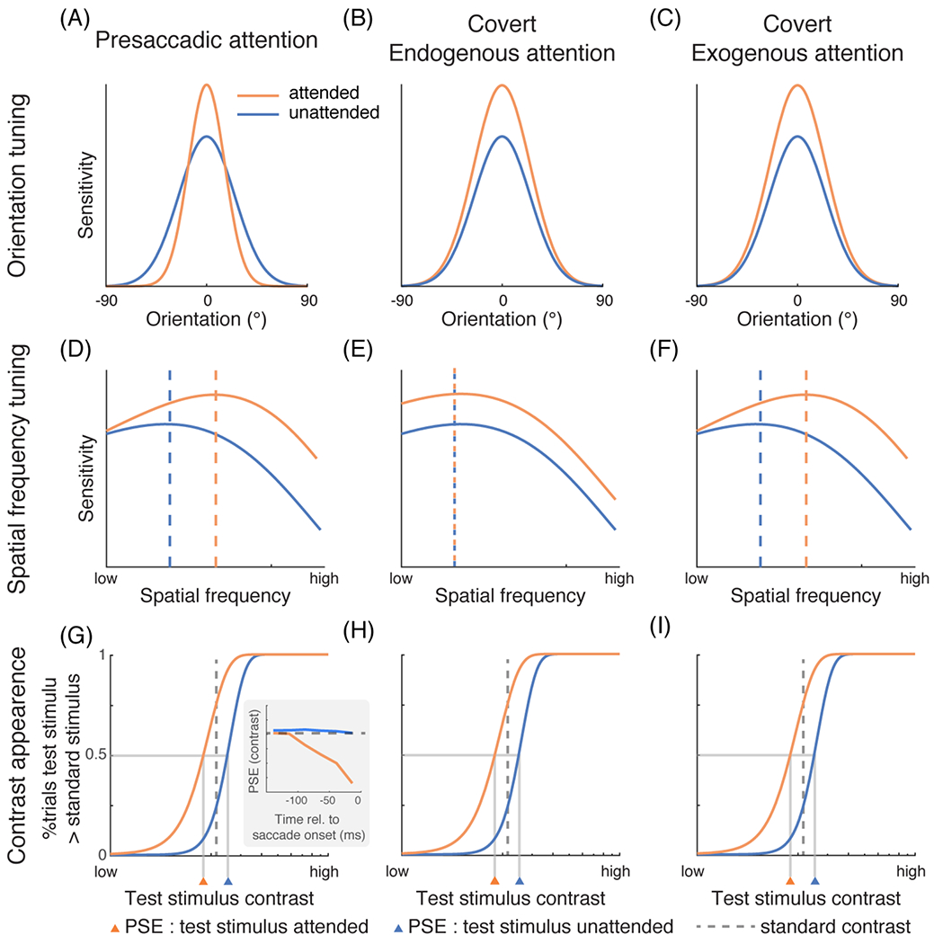
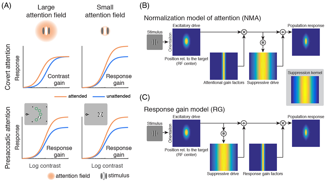
Attention is a central neural process that enables selective and efficient processing of visual information. Individuals can attend to specific visual information either overtly, by making an eye movement to an object of interest, or covertly, without moving their eyes. We review behavioral, neuropsychological, neurophysiological, and computational evidence of presaccadic attentional modulations that occur while preparing saccadic eye movements, and highlight their differences from those of covert spatial endogenous (voluntary) and exogenous (involuntary) attention. We discuss recent studies and experimental procedures on how these different types of attention impact visual performance, alter appearance, differentially modulate the featural representation of basic visual dimensions (orientation and spatial frequency), engage different neural computations, and recruit partially distinct neural substrates. We conclude that presaccadic attention and covert attention are dissociable.

Keywords: contrast; endogenous attention; exogenous attention; eye movements; featural representation; orientation; spatial frequency.


Conflict of interest statement

Declaration of interests The authors declare no competing interests.

Figures

Figure 1.
Figure 1.. Prominent experimental procedures used to investigate the effects of presaccadic attention on visual performance.
Top row: Depiction of the respective task (saccade targets indicated by green arrows, saccade endpoints indicated by dashed green lines). Bottom row: exemplary results. ST: Saccade target; SL: Saccade landing position; (A) Discrimination task. Participants prepare a saccade to an instructed saccade target (here the central arrow cue indicates the orange item on the right). A test stimulus (“E”) is presented briefly before eye movement onset along with several distractors (“2”s and “5”s), at a location coinciding with the saccade target or not. Participants are asked to discriminate the test stimulus (“E” versus “mirror-E”). While the eyes are still fixating the center, discrimination accuracy is selectively enhanced at the respective saccade target–indicating a presaccadic shift of attention. Adapted from [7]. (B) Eye abduction protocol. Participants view the display with one eye patched and their head rotated so that a portion of the display is still visible but outside the range reachable by the eyes (indicated by the green area). Saccades aimed at a cued target outside this range will fall short. Presaccadic discrimination accuracy (assessed with an oriented 1/f noise patch) is nonetheless highest at the unreachable saccade target (and not enhanced at the actual saccade landing)–demonstrating that attention is not limited to the eye movement range. Adapted from [34]. (C) Global-saccade protocol. Participants are instructed to saccade to one of two nearby items at free choice (either of the orange frames), in which case the eyes will frequently land in between the two salient targets. Presaccadic discrimination accuracy (assessed with an oriented Gabor patch) is not enhanced at the endpoint of such global-saccades, but instead equally enhanced at the two targets–showing that presaccadic attention is not necessarily coupled to the saccade endpoint. Adapted from [42]. (D) Anti-saccade task. Participants are instructed to saccade to the location in the opposite direction of a peripheral cue. Presaccadic discrimination accuracy (assessed with a tilted Gabor patch) before anti-saccades is equally enhanced at the cued location and the anti-saccade goal indicating attentional selection at both locations. Adapted from [44]. (E) Microstimulation protocol. Monkeys have to detect a luminance change of a peripheral target. Subthreshold frontal eye field stimulation increases visual sensitivity at the movement field (to which saccades would be evoked by stimulation currents above threshold; marked by the green circle), suggesting that saccade related mechanisms provide a source of spatial attention. Adapted from [46].
Figure 2.
Figure 2.. The effects of presaccadic and covert attention on featural representations.
(A) In psychophysical experiments, participants were required to detect a grating at a fixed orientation (represented by 0° in the figure), and a reverse correlation technique was used to characterize the orientation tuning employed by the visual system. Presaccadic attention increases the gain and reduces the width of orientation tuning [18,50]. Adapted from [18]. (B-C) Covert endogenous and exogenous attention only increase the gain [–55]. Adapted from [55]. (D-F) Presaccadic attention [18,47] and covert exogenous attention preferentially increase the sensitivity of high-SF information by shifting the SF tuning curve rightward [55,58,59,64]. whereas covert endogenous attention enhances a broad range of SFs uniformly [,–64]. The dashed vertical lines indicate the peak of the tuning functions. See details of the experimental protocols in BOX 1. (D) adapted from [18]. (E) and (F) adapted from [55]. (G-I) Subjective contrast appearance of visual stimuli can be estimated by measuring the point of subjective equality (PSE) in tasks requiring participants to compare the contrast of two stimuli. In the experiments, the contrast of a test stimulus typically varies across trials while the contrast of a standard stimulus is fixed. Here, the percentage of trials in which participants judge the test stimulus to have a higher contrast than the standard (y-axis) is plotted against the contrast of the test stimulus (x-axis). The orange curves represent the condition in which the test stimulus is cued (attended). Both presaccadic attention [15] as well as endogenous [74] and exogenous [–71] covert attention enhance perceived contrast. The enhancement by presaccadic attention exhibits a gradual trend right before saccade onset (inset in G, adapted from [15]). (H) adapted from [65]. (I) adapted from [66].
Figure 3.
Figure 3.. Attentional modulations on contrast response functions.
(A) Illustrations of how attentional field size (relative to the stimulus size) affects the form of attentional modulations. The left column represents the condition in which the size of the attentional field is large whereas the right column represents the condition in which the size of the attentional field is small. Covert attention (endogenous and exogenous; top row) generates contrast gain when the size of the attentional field is large and response gain when the size of the attentional field is small [77,84]. In contrast, presaccadic attention (bottom row) generates response gain changes regardless of the attentional field size [49]. The insets in the bottom row illustrate how the size of attention field was manipulated by the location uncertainty of the target in a study of presaccadic attention [49]. Only the fixation point with the pre-cue and the right half of the screen are shown. The left half of the screen contains similar stimuli (an aperture outlined by four black dots and a test stimulus). In the experiments, participants made a saccadic eye movement toward the center of the cued aperture (outlined by the four black dots). In the large attention field size condition, the aperture was large and the test stimulus (a grating) was presented at one of five possible locations (indicated by green circles for illustration purpose only) with equal probability. In the small attention field size condition, the test stimulus was presented at a fixed location [49,77,84] (attentional field size was also manipulated by location uncertainty in covert attention experiments [77,84]). (B) The Reynolds-Heeger normalization model of attention (NMA) explains these size-dependent gain changes. Blue-yellow 2-dimensional images represent a population of visual neurons selective for different orientations and receptive field centers. The response of visual neurons is computed by normalization: dividing neurons’ excitatory drive by their suppressive drive. The excitatory drive is determined by the preferred orientation and position of each neuron. The suppressive drive is computed by convolving the excitatory drive with the suppression kernel. The suppression kernel is uniform in the orientation domain. For simulating surround suppression of visual neurons, the suppression kernel is a Gaussian with a wide width in its spatial domain. In NMA, attention is modeled as attentional gain factors that multiplicatively modulate the excitatory drive before computing the suppressive drive and normalization. (C) The modulation of presaccadic attention is better explained by response gain factors that scale the neurons’ responses after normalization. In both (B) and (C), it is assumed that attention is deployed to the center of the target stimulus (the vertical grating). (A-C) adapted from [49].
Figure I.
Figure I.
Texture segmentation tasks. Top: Example stimuli. Bottom: Exogenous attention impairs or improves performance as a function of stimulus eccentricity; endogenous attention improves performance throughout eccentricity. Adapted from [56].
Figure II.
Figure II.
Masking experiments. Left: Example stimuli used in [47]. Right: Presaccadic attention impairs performance when the SF of the mask is higher that the SF of the target. Adapted from [47]

Similar articles

Cited by

References

    1. Carrasco M Visual attention: the past 25 years. Vision Res. 2011. ;51: 1484–1525. - PMC - PubMed
    1. Anton-Erxleben K, Carrasco M. Attentional enhancement of spatial resolution: linking behavioural and neurophysiological evidence. Nat Rev Neurosci. 2013;14: 188–200. - PMC - PubMed
    1. Binda P, Morrone MC. Vision During Saccadic Eye Movements. Annu Rev Vis Sci. 2018;4: 193–213. - PubMed
    1. Marino AC, Mazer JA. Perisaccadic Updating of Visual Representations and Attentional States: Linking Behavior and Neurophysiology. Front Syst Neurosci. 2016;10: 3. - PMC - PubMed
    1. Kowler E, Anderson E, Dosher B, Blaser E. The role of attention in the programming of saccades. Vision Res. 1995;35: 1897–1916. - PubMed

Publication types