

Assessment of stunned and viable myocardium using manganese-enhanced MRI
- PMID: 34099530
- PMCID: PMC8186753
- DOI: 10.1136/openhrt-2021-001646
Assessment of stunned and viable myocardium using manganese-enhanced MRI
Abstract
Objective: In a proof-of-concept study, to quantify myocardial viability in patients with acute myocardial infarction using manganese-enhanced MRI (MEMRI), a measure of intracellular calcium handling.
Methods: Healthy volunteers (n=20) and patients with ST-elevation myocardial infarction (n=20) underwent late gadolinium enhancement (LGE) using gadobutrol and MEMRI using manganese dipyridoxyl diphosphate. Patients were scanned ≤7 days after reperfusion and rescanned after 3 months. Differential manganese uptake was described using a two-compartment model.
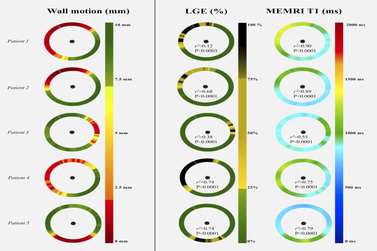
Results: After manganese administration, healthy control and remote non-infarcted myocardium showed a sustained 25% reduction in T1 values (mean reductions, 288±34 and 281±12 ms). Infarcted myocardium demonstrated less T1 shortening than healthy control or remote myocardium (1157±74 vs 859±36 and 835±28 ms; both p<0.0001) with intermediate T1 values (1007±31 ms) in peri-infarct regions. Compared with LGE, MEMRI was more sensitive in detecting dysfunctional myocardium (dysfunctional fraction 40.5±11.9 vs 34.9%±13.9%; p=0.02) and tracked more closely with abnormal wall motion (r2=0.72 vs 0.55; p<0.0001). Kinetic modelling showed reduced myocardial manganese influx between remote, peri-infarct and infarct regions, enabling absolute discrimination of infarcted myocardium. After 3 months, manganese uptake increased in peri-infarct regions (16.5±3.5 vs 22.8±3.5 mL/100 g/min, p<0.0001), but not the remote (23.3±2.8 vs 23.0±3.2 mL/100 g/min, p=0.8) or infarcted (11.5±3.7 vs 14.0±1.2 mL/100 g/min, p>0.1) myocardium.
Conclusions: Through visualisation of intracellular calcium handling, MEMRI accurately differentiates infarcted, stunned and viable myocardium, and correlates with myocardial dysfunction better than LGE. MEMRI holds major promise in directly assessing myocardial viability, function and calcium handling across a range of cardiac diseases.
Trial registration numbers: NCT03607669; EudraCT number 2016-003782-25.
Keywords: heart failure; magnetic resonance imaging; myocardial infarction.
© Author(s) (or their employer(s)) 2021. Re-use permitted under CC BY-NC. No commercial re-use. See rights and permissions. Published by BMJ.
Conflict of interest statement
Competing interests: SIS has a consultancy agreement with GSK. DEN and SIS hold unrestricted educational grants from Siemens Healthineers.
Figures


Similar articles
-
Manganese-Enhanced T1 Mapping in the Myocardium of Normal and Infarcted Hearts.Contrast Media Mol Imaging. 2018 Oct 25;2018:9641527. doi: 10.1155/2018/9641527. eCollection 2018. Contrast Media Mol Imaging. 2018. PMID: 30498403 Free PMC article.
-
Manganese-enhanced MRI of the myocardium.Heart. 2019 Nov;105(22):1695-1700. doi: 10.1136/heartjnl-2019-315227. Epub 2019 Jul 23. Heart. 2019. PMID: 31337670 Free PMC article. Review.
-
Myocardial viability of the peri-infarct region measured by T1 mapping post manganese-enhanced MRI correlates with LV dysfunction.Int J Cardiol. 2019 Apr 15;281:8-14. doi: 10.1016/j.ijcard.2019.01.101. Epub 2019 Jan 31. Int J Cardiol. 2019. PMID: 30739802 Free PMC article.
-
Manganese dipyridoxyl-diphosphate (MnDPDP) as a viability marker in patients with myocardial infarction.J Magn Reson Imaging. 2007 Sep;26(3):720-7. doi: 10.1002/jmri.21065. J Magn Reson Imaging. 2007. PMID: 17729351
-
Contrast-enhanced MRI for quantification of myocardial viability.J Magn Reson Imaging. 1999 Nov;10(5):694-702. doi: 10.1002/(sici)1522-2586(199911)10:5<694::aid-jmri12>3.0.co;2-j. J Magn Reson Imaging. 1999. PMID: 10548777 Review.
Cited by
-
In silico evaluation of cell therapy in acute versus chronic infarction: role of automaticity, heterogeneity and Purkinje in human.Sci Rep. 2024 Sep 16;14(1):21584. doi: 10.1038/s41598-024-67951-5. Sci Rep. 2024. PMID: 39284812 Free PMC article.
-
Manganese-Enhanced Magnetic Resonance Imaging of the Heart.J Magn Reson Imaging. 2023 Apr;57(4):1011-1028. doi: 10.1002/jmri.28499. Epub 2022 Oct 31. J Magn Reson Imaging. 2023. PMID: 36314991 Free PMC article. Review.
-
Repeatability and reproducibility of cardiac manganese-enhanced magnetic resonance imaging.Sci Rep. 2023 Feb 27;13(1):3366. doi: 10.1038/s41598-023-29591-z. Sci Rep. 2023. PMID: 36849509 Free PMC article.
-
Assessment of Cardiac Toxicity of Manganese Chloride for Cardiovascular Magnetic Resonance.Front Physiol. 2022 Jul 7;13:952043. doi: 10.3389/fphys.2022.952043. eCollection 2022. Front Physiol. 2022. PMID: 35874541 Free PMC article.
-
Myocardial Calcium Handling in Type 2 Diabetes: A Novel Therapeutic Target.J Cardiovasc Dev Dis. 2023 Dec 30;11(1):12. doi: 10.3390/jcdd11010012. J Cardiovasc Dev Dis. 2023. PMID: 38248882 Free PMC article. Review.
References
Publication types
MeSH terms
Substances
Associated data
Grants and funding
LinkOut - more resources
Full Text Sources
Medical