. 2021 Jun 15;118(24):e2105465118.
doi: 10.1073/pnas.2105465118.

STING inhibitors target the cyclic dinucleotide binding pocket

Affiliations

STING inhibitors target the cyclic dinucleotide binding pocket

Ze Hong et al. Proc Natl Acad Sci U S A. .

Abstract

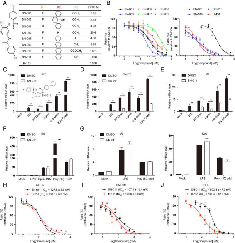
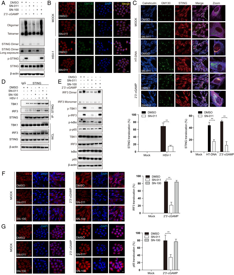
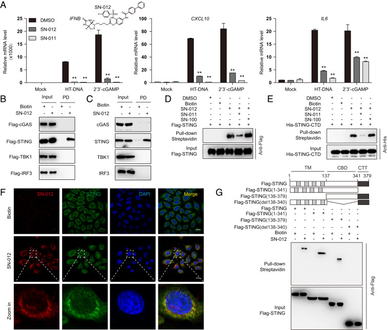
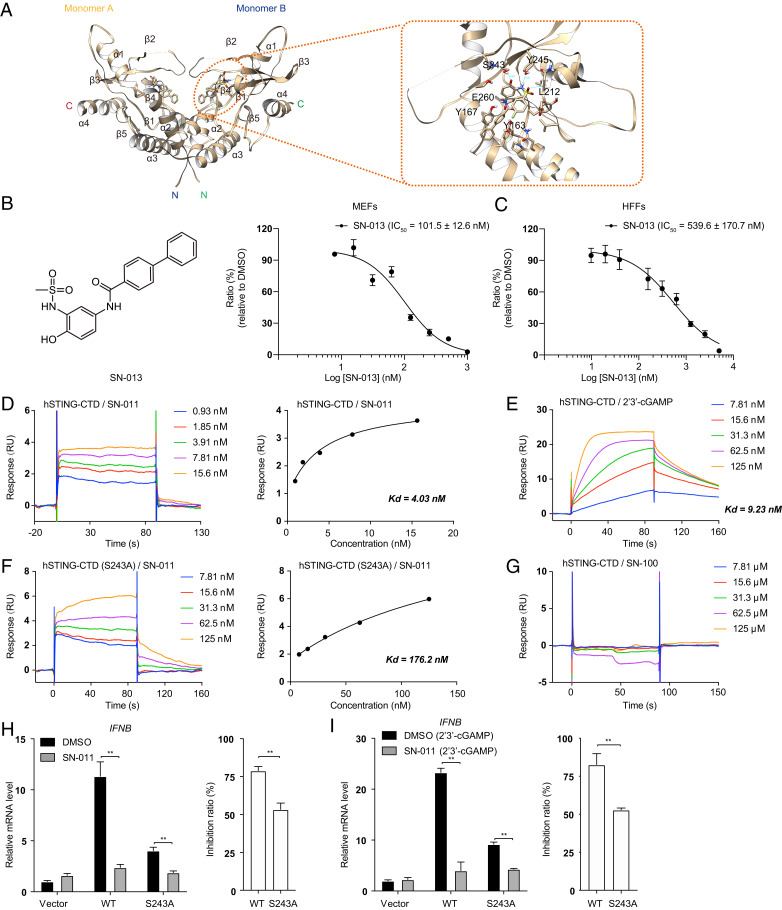
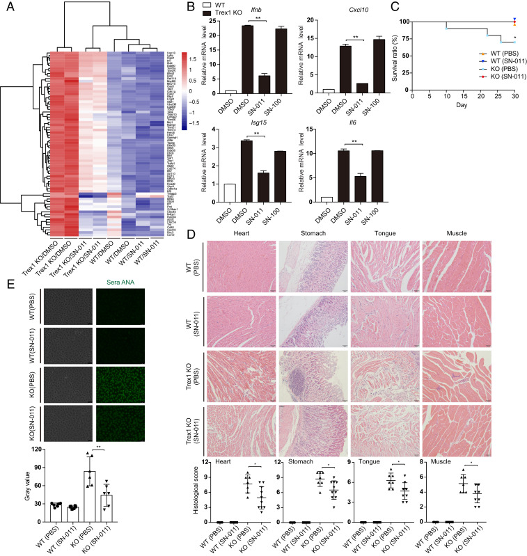
Cytosolic DNA activates cGAS (cytosolic DNA sensor cyclic AMP-GMP synthase)-STING (stimulator of interferon genes) signaling, which triggers interferon and inflammatory responses that help defend against microbial infection and cancer. However, aberrant cytosolic self-DNA in Aicardi-Goutière's syndrome and constituently active gain-of-function mutations in STING in STING-associated vasculopathy with onset in infancy (SAVI) patients lead to excessive type I interferons and proinflammatory cytokines, which cause difficult-to-treat and sometimes fatal autoimmune disease. Here, in silico docking identified a potent STING antagonist SN-011 that binds with higher affinity to the cyclic dinucleotide (CDN)-binding pocket of STING than endogenous 2'3'-cGAMP. SN-011 locks STING in an open inactive conformation, which inhibits interferon and inflammatory cytokine induction activated by 2'3'-cGAMP, herpes simplex virus type 1 infection, Trex1 deficiency, overexpression of cGAS-STING, or SAVI STING mutants. In Trex1-/- mice, SN-011 was well tolerated, strongly inhibited hallmarks of inflammation and autoimmunity disease, and prevented death. Thus, a specific STING inhibitor that binds to the STING CDN-binding pocket is a promising lead compound for STING-driven disease.

Keywords: Aicardi–Goutières syndrome; SAVI; STING; antagonist; type I interferons.


Conflict of interest statement

The authors declare no competing interest.

Figures

Fig. 1.
Fig. 1.
SN-011 is a potent STING inhibitor. (A and B) Chemical structures of SN-001, SN-005 to SN-011 (A), and dose-dependent inhibitory curves (B). L929 cells, pretreated with different concentrations of the indicated compounds for 6 h, were stimulated by transfection of HT-DNA and Ifnb mRNA was measured by quantitative PCR 6 h later. The dose-dependent inhibitory curve was fit to calculate the IC50s of SN-001, SN-005 to SN-011, and H-151. (CE) MEFs, pretreated for 6 h with 1 μM SN-011, were stimulated by transfection of ISD or HT-DNA or HSV-1 infection for 6 h, or addition of c-di-GMP or 2′3′-cGAMP for 3 h. Induction of Ifnb (C), Cxcl10 (D), Il6 (E) mRNA was measured by quantitative PCR. (F and G) Tmem173−/− MEFs, pretreated with 1 μM SN-011 for 6 h, were stimulated by adding LPS or poly (I and C) to the medium for 1.5 h or transfection of CpG-DNA or poly (I and C) or infection with SeV for 6 h. Induction of Ifnb (F), Il6, and Tnfa (G) mRNA was measured by quantitative PCR. (HJ) MEFs (H), BMDMs (I), and HFFs (J) were pretreated with the indicated concentrations of SN-011 or H-151 before 2′3′-cGAMP stimulation for 3 h. Induction of Ifnb mRNA was measured by quantitative PCR. The dose-dependent inhibitory curve was fit to calculate the IC50s. Data shown are mean ± SD from three independent experiments. **P < 0.01. Gene expression was normalized to Gapdh.
Fig. 2.
Fig. 2.
SN-011 inhibits STING activation and signal transduction. (A) HFFs were pretreated with SN-011 or SN-100 (1 μM) and then stimulated with 2′3′-cGAMP for 1 h. STING phosphorylation and oligomerization were analyzed by immunoblot probed for STING. (B) HeLa, pretreated with SN-011 or SN-100 (1 μM) or DMSO control, were infected with HSV-1 for 4 h before immunostaining for STING and GM130 (Golgi autoantigen) and confocal microscopy imaging. (C) HFFs, pretreated with SN-011, were stimulated with HT-DNA for 1 h or 2′3′-cGAMP for 30 min before immunostaining for STING, calreticulin, and GM130 and subsequent confocal microscopy imaging analysis (Upper). Quantification of cells with STING puncta that colocalize with the Golgi (Lower). (D) HFFs were pretreated with SN-011 or SN-100 (1 μM) before infecting with HSV-1 for 3 h. Cell lysates were immunoprecipitated with STING antibody and immunoblotted with indicated antibodies. (E) HFFs, pretreated or not with SN-011 or SN-100 (1 μM) were stimulated with 2′3′-cGAMP for 2 h and then analyzed for phosphorylated (p)- IRF3, TBK1, IκBα, and p65, and IRF3 dimerization by immunoblot. (F) HFFs, pretreated or not with SN-011 or SN-100 (1 μM), were stimulated with 2′3′-cGAMP for 2 h. Cells were then immunostained with anti-IRF3 and imaged by confocal microscopy. (Left and Center) Representative images; (Right) quantification of cells with nuclear IRF3. (G) HeLa, pretreated or not with SN-011 or SN-100 (1 μM), were stimulated with 2′3′-cGAMP for 2 h and immunostained with anti-p65 and imaged by confocal microscopy. (Left and Center) Representative images; (Right) quantification of cells with nuclear p65. Data in B, C, F, and G are mean ± SD from three independent experiments. The images are 63× magnification with a digital scan zoom of 3.0 that was used to enhance magnification. **P < 0.01. (Scale bars, 25 μm.)
Fig. 3.
Fig. 3.
Biotinylated SN-011 specifically binds to the STING CDN binding domain. (A) HFFs, pretreated with SN-011 or biotinylated SN-011 (SN-012; 1 μM), were transfected with HT-DNA for 6 h or treated with 2′3′-cGAMP for 3 h. Induction of IFNB, CXCL10, and IL6 mRNA was measured by quantitative PCR assay. (B) Cell lysates of HEK293T cells, transfected for 24 h with expression plasmids for the indicated Flag-tagged proteins, were incubated with biotin (5 μM) or SN-012 (5 μM) for 1 h, followed by pull-down with streptavidin-conjugated beads and immunoblot with anti-Flag. (C) HFF lysates were incubated with biotin (5 μM) or SN-012 (5 μM) for 1 h, followed by pull-down with streptavidin-conjugated beads, and immunoblot with the indicated antibodies. (D) Cell lysates of HEK293T cells overexpressing Flag-hSTING were incubated with SN-012 (5 μM) in the absence or presence of a 10-fold excess (50 μM) of SN-011 or SN-100, before pull-down with streptavidin-conjugated beads and immunoblot with anti-Flag. (E) Recombinant His-tagged hSTING-CTD (149-379) protein was incubated with SN-012 (5 μM) in the absence or presence of a 10-fold excess (50 μM) of SN-011 or SN-100, before pull-down with streptavidin-conjugated beads and immunoblot with anti-His. (F) HeLa cells were incubated with Biotin or SN-012 (1 μM) and then immunostained with anti-STING and Alexa Fluor 594-conjugated streptavidin. Nuclei were stained with DAPI. (Scale bars, 25 μm.) (G) Schematic of STING and its truncation mutants (Upper). HEK293T cells were transfected to express Flag-tagged hSTING or hSTING-CTD (138–379), hSTING-∆CTT (1–341) or hSTING-∆CBD (∆138–340) truncations and 24 h later, cell lysates were collected and incubated with biotin or SN-012 (5 μM), followed by pull-down with streptavidin-conjugated beads, and immunoblot with anti-Flag (Lower). Data in A are mean ± SD from three independent experiments. The images in F are 63× magnification with a digital scan zoom of 3.0 that was used to enhance magnification. **P < 0.01. Gene expression was normalized to Gapdh.
Fig. 4.
Fig. 4.
Mutations of the STING-SN-011 interface reduce inhibition by SN-011. (A) Intermolecular contacts of SN-011 bound to hSTING-CTD (amino acids 149 to 379) in virtual docking structure (Left). SN-011 and contacted STING amino acids are shown as a stick model. Hydrogen-bounds with distance are labeled in green (Right). (B and C) MEFs (B) and HFFs (C) were pretreated with the indicated concentrations of SN-013 before 2′3′-cGAMP stimulation for 3 h. Induction of Ifnb mRNA was measured by quantitative PCR. The dose-dependent inhibitory curve was fit to calculate the IC50s. (D) Biacore analysis of hSTING-CTD and SN-011 binding (Left). The binding affinity (Kd) was determined by fitting the binding data to a steady-state 1:1 binding model (Right). RU, response units. (E) Biacore analysis of hSTING-CTD and 2′3′-cGAMP binding. The binding affinity (Kd) was determined by fitting the binding data to a kinetics 1:1 binding model. (F) Biacore analysis of hSTING-CTD (S243A) and SN-011 binding (Left). Kd was determined by fitting the binding data to a steady-state 1:1 binding model (Right). (G) Biacore analysis of hSTING-CTD and SN-100 binding. (H) HEK293T cells, transfected with Flag-hSTING or its mutants, were treated 12 h later with DMSO or SN-011 (10 μM) for 12 h. mRNA was measured by quantitative PCR assay (Left). The inhibition ratio of SN-011 relative to DMSO control was calculated based on Ifnb mRNA levels (Right). (I) HEK293T cells, transfected with Flag-hSTING or its mutant, were treated 12 h later with DMSO or SN-011 (10 μM) for 9 h and then stimulated with 2′3′-cGAMP for 3 h. mRNA was measured by quantitative PCR assay (Left). The inhibition ratio of SN-011 relative to DMSO was calculated based on Ifnb mRNA levels (Right). Data from B, C, H, and I are mean ± SD from three independent experiments. **P < 0.01. Gene expression was normalized to Gapdh.
Fig. 5.
Fig. 5.
SN-011 suppresses systemic inflammation and death in Trex1−/− mice. (A) Heat map of RNA-seq of BMDMs from WT or Trex1−/− mice that were treated or not with 0.5 μM SN-011. The top 72 differentially expressed ISGs are shown. (B) Trex1−/− BMDMs were treated with SN-011 (0.5 μM), SN-100 (0.5 μM) or DMSO. Induction of Ifnb, Cxcl10, Isg15, and Il6 mRNA was measured by quantitative PCR. Fold-changes are relative to WT BMDM controls. (C) Survival curves of WT and Trex1−/− mice treated or not with SN-011 (5 mg/kg) three times per week for a month. *P < 0.05, statistical analysis was performed using the log-rank test (10 mice per group). (D) Representative H&E-stained tissue sections from WT or Trex1−/− mice treated or not with SN-011 (5 mg/kg) three times per week for a month (Upper). Results of blinded histological scoring of individual animal samples is shown (Lower). (E) Antinuclear antibodies (ANA) in the serum of WT or Trex1−/− mice treated or not with SN-011 (5 mg/kg) detected by antinuclear antibody antigen substrate slide kit (Upper). Fluorescence intensity was analysis by calculating mean gray value of individual animal sera (Lower). Data in B are mean ± SD from three independent experiments. Data from D and E are presented as mean ± SD from at least six mice in each group. Images in D and E are 20× magnification. *P < 0.05; **P < 0.01. Gene expression was normalized to Gapdh.

Similar articles

Cited by

References

    1. Soni C., Reizis B., DNA as a self-antigen: Nature and regulation. Curr. Opin. Immunol. 55, 31–37 (2018). - PMC - PubMed
    1. Sun L., Wu J., Du F., Chen X., Chen Z. J., Cyclic GMP-AMP synthase is a cytosolic DNA sensor that activates the type I interferon pathway. Science 339, 786–791 (2013). - PMC - PubMed
    1. Ablasser A., et al. ., cGAS produces a 2′-5′-linked cyclic dinucleotide second messenger that activates STING. Nature 498, 380–384 (2013). - PMC - PubMed
    1. Gao P., et al. ., Cyclic [G(2′,5′)pA(3′,5′)p] is the metazoan second messenger produced by DNA-activated cyclic GMP-AMP synthase. Cell 153, 1094–1107 (2013). - PMC - PubMed
    1. Wu J., et al. ., Cyclic GMP-AMP is an endogenous second messenger in innate immune signaling by cytosolic DNA. Science 339, 826–830 (2013). - PMC - PubMed

Publication types