Multicenter Study
. 2021 Jun 7;12(1):3400.
doi: 10.1038/s41467-021-23620-z.

A multicentre validation study of the diagnostic value of plasma neurofilament light

Affiliations
Multicenter Study

A multicentre validation study of the diagnostic value of plasma neurofilament light

Nicholas J Ashton et al. Nat Commun. .

Abstract

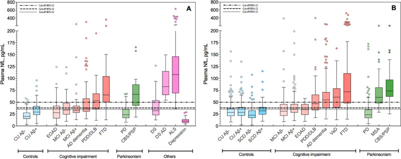
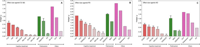
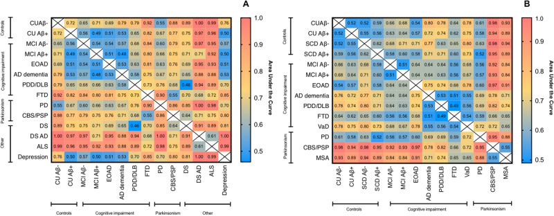
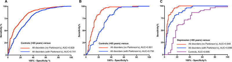
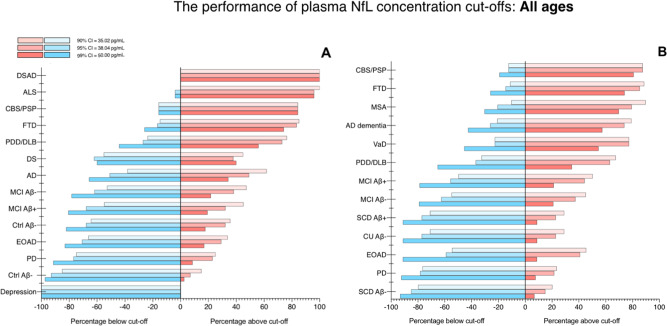
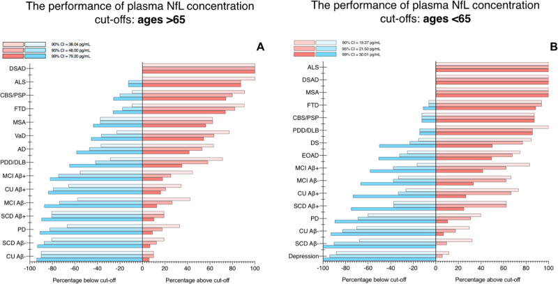
Increased cerebrospinal fluid neurofilament light (NfL) is a recognized biomarker for neurodegeneration that can also be assessed in blood. Here, we investigate plasma NfL as a marker of neurodegeneration in 13 neurodegenerative disorders, Down syndrome, depression and cognitively unimpaired controls from two multicenter cohorts: King's College London (n = 805) and the Swedish BioFINDER study (n = 1,464). Plasma NfL was significantly increased in all cortical neurodegenerative disorders, amyotrophic lateral sclerosis and atypical parkinsonian disorders. We demonstrate that plasma NfL is clinically useful in identifying atypical parkinsonian disorders in patients with parkinsonism, dementia in individuals with Down syndrome, dementia among psychiatric disorders, and frontotemporal dementia in patients with cognitive impairment. Data-driven cut-offs highlighted the fundamental importance of age-related clinical cut-offs for disorders with a younger age of onset. Finally, plasma NfL performs best when applied to indicate no underlying neurodegeneration, with low false positives, in all age-related cut-offs.


Conflict of interest statement

J.L. has received travel support and/or lecture honoraria from Biogen, Novartis, Merck, Roche, and Sanofi Genzyme; has served on scientific advisory boards for Biogen, Novartis, Merck, Alexion, Roche, and Sanofi Genzyme; serves on the editorial board of the Acta Neurologica Scandinavica; has received unconditional research grants from Biogen and Novartis. A.A.C. has served as a consultant or on advisory boards for Amylyx, Apellis, Biogen Idec, Brainstorm, Cytokinetics, GSK, Lilly, Mitsubishi Tanabe Pharma, Novartis, OrionPharma, Quralis, and Wave Pharmaceuticals. A.S. has been a consultant for AC-Immune and is a member of the scientific advisory board of ProMIS Neurosciences. P.S. has received speaker fees for Shire/Takeda and Sanofi Genzyme. H.Z. has served at scientific advisory boards for Denali, Roche Diagnostics, Wave, Samumed, and CogRx; has given lectures in symposia sponsored by Alzecure and Biogen; and is a co-founder of Brain Biomarker Solutions in Gothenburg AB, a GU Ventures-based platform company at the University of Gothenburg. K.B. has served as a consultant or at advisory boards for Alector, Alzheon, CogRx, Biogen, Lilly, Novartis, and Roche Diagnostics, and is a co-founder of Brain Biomarker Solutions in Gothenburg AB, a GU Venture-based platform company at the University of Gothenburg, all unrelated to the work presented in this paper. O.H. has acquired research support (for the institution) from Roche, GE Healthcare, Biogen, AVID Radiopharmaceuticals, and Euroimmun. In the past 2 years, he has received consultancy/speaker fees (paid to the institution) from Biogen and Roche. The remaining authors declare no competing interests.

Figures

Fig. 1
Fig. 1. The concentrations of plasma NfL for different diagnostic and controls groups in the KCL and Lund cohorts.
Plasma neurofilament light (NfL) in different diagnostic groups; KCL (A n  =  805) and Lund (B n  =  1464) cohorts. For each plot, the horizontal bar shows the median, and the upper and lower boundaries show the 25th and 75th percentiles, respectively. Source data are provided as a Source Data file. KCL Cohort—AD    Alzheimer’s disease (n = 102), ALS   amyotrophic lateral sclerosis (n = 50), CU Aβ− cognitively unimpaired without Aβ pathology (n = 130), CU Aβ+ cognitively unimpaired with Aβ pathology (n = 28), CBS/PSP   corticobasal syndrome and progressive supranuclear palsy (n = 19), depression (n = 37), DS    Down syndrome (n = 29), DSAD   Down syndrome Alzheimer’s disease (n = 12), EOAD   early-onset Alzheimer’s disease (n = 59), FTD   frontotemporal dementia (n = 54), MCI Aβ− mild cognitive impairment without Aβ pathology (n = 55), MCI Aβ+ mild cognitive impairment with Aβ pathology (n = 31), PD   Parkinson’s disease (n = 140), PDD/DLB   Parkinson’s disease dementia and dementia with Lewy bodies (n = 59). Lund Cohort—AD   Alzheimer’s disease (n = 134), CU Aβ− cognitively unimpaired without Aβ pathology (n = 273), CU Aβ+ cognitively unimpaired with Aβ pathology (n = 103), CBS/PSP   corticobasal syndrome and progressive supranuclear palsy (n = 24), EOAD   early-onset Alzheimer’s disease (n = 23), FTD   frontotemporal dementia (n = 150), MCI Aβ− mild cognitive impairment without Aβ pathology (n = 115), MCI Aβ+ mild cognitive impairment with Aβ pathology (n = 165), MSA   multiple system atrophy (n = 29), PD    Parkinson’s disease (n = 171), PDD/DLB   Parkinson’s disease dementia and dementia with Lewy bodies (n = 46), SCD Aβ− subjective cognitive decline without Aβ pathology (n = 134), SCD Aβ+ subjective cognitive decline with Aβ pathology (n = 75), VaD vascular dementia (n = 22).
Fig. 2
Fig. 2. Effect sizes of neurodegenerative disorders as compared to amyloid-negative cognitively unimpaired controls, Parkinson’s disease and Alzheimer’s disease.
Effect sizes (Hedges’s g) of different neurodegenerative disorders as compared to amyloid-negative cognitively unimpaired controls (A n  =  403), Parkinson’s disease (B n  =  311), and Alzheimer’s disease (C n  =  236). The bars represent the mean effect size for the cohort, whereas the error bars represent the standard deviation of effect size when considering the KCL and Lund cohorts separately. Those without error bars (e.g., VaD) are only included in one cohort. AD    Alzheimer’s disease (n = 236), ALS   amyotrophic lateral sclerosis (n = 50), CU Aβ− cognitively unimpaired without Aβ pathology (n = 403), CBS/PSP   corticobasal syndrome and progressive supranuclear palsy (n = 43), depression (n = 37), DS   Down syndrome (n = 29), DSAD   Down syndrome Alzheimer’s disease (n = 12), EOAD   early-onset Alzheimer’s disease (n = 82), FTD   frontotemporal dementia (n = 204), MCI Aβ− mild cognitive impairment without Aβ pathology (n = 170), MCI Aβ + mild cognitive impairment with Aβ pathology (n = 196), MSA   multiple system atrophy (n = 29), PD   Parkinson’s disease (n = 311), PDD/DLB   Parkinson’s disease dementia and dementia with Lewy bodies (n = 105), VaD vascular dementia (n = 22).
Fig. 3
Fig. 3. The diagnsotic accuracy of plasma NfL in neurodegenerative disorders.
Heatmaps to demonstrate the accuracy (AUC) of plasma NfL to distinguish CU and neurodegenerative disorders in the KCL (A) and Lund (B) cohorts. Heatmaps tables that demonstrate sensitivity, specificity, and 95% CI of AUC displayed in the Supplementary Tables 4–5 and Supplementary Fig. 4. AD   Alzheimer’s disease, ALS   amyotrophic lateral sclerosis, CU Aβ− cognitively unimpaired without Aβ pathology, CU Aβ+ cognitively unimpaired with Aβ pathology, CBS/PSP   corticobasal syndrome and progressive supranuclear palsy, DS   Down syndrome, DSAD   Down syndrome Alzheimer’s disease, EOAD   early-onset Alzheimer’s disease, FTD   frontotemporal dementia, MCI Aβ− mild cognitive impairment without Aβ pathology, MCI Aβ+ mild cognitive impairment with Aβ pathology, MSA   multiple system atrophy, PD   Parkinson’s disease, PDD/DLB   Parkinson’s disease dementia and dementia with Lewy bodies, SCD Aβ− subjective cognitive decline without Aβ pathology, SCD Aβ+ subjective cognitive decline with Aβ pathology, VaD vascular dementia.
Fig. 4
Fig. 4. The diagnsotic accuracy of plasma NfL in identifying neurodegenerative disorders from controls (young/old) and depression.
The performance of plasma neurofilament light (NfL) to identify neurodegenerative disorders from controls (CU and SCD) > 65 years of age (A), controls (CU and SCD) < 65 years of age (B), and depression (C).
Fig. 5
Fig. 5. The performance of plasma NfL concentration cutoffs to identify neurodegenerative disorders of all ages.
The performance of plasma neurofilament light (NfL) concentration cutoffs to identify neurodegenerative disorders in KCL (A) and Lund (B). AD   Alzheimer’s disease, ALS   amyotrophic lateral sclerosis, CBS   corticobasal syndrome, DLB   dementia with Lewy bodies, DS   Down syndrome, DSAD   Down syndrome Alzheimer’s disease, EOAD   early-onset Alzheimer’s disease, FTD   frontotemporal dementia. MCI   mild cognitive impairment, MSA   multiple system atrophy, PD   Parkinson’s disease, PDD   Parkinson’s disease dementia, PSP   progressive supranuclear palsy, SCD   subjective cognitive decline, VaD vascular dementia.
Fig. 6
Fig. 6. The performance of plasma NfL concentration cutoffs to identify neurodegenerative disorders in >65 and <65 years.
The performance of plasma neurofilament light (NfL) concentration cutoffs to identify neurodegenerative disorders in >65 (A) and <65 (B). The KCL and Lund cohorts are combined for this analysis. AD   Alzheimer’s disease, ALS   amyotrophic lateral sclerosis, CBS   corticobasal syndrome, DLB   dementia with Lewy bodies, DS   Down syndrome, DSAD   Down syndrome Alzheimer’s disease, EOAD   early-onset Alzheimer’s disease, FTD   frontotemporal dementia. MCI    mild cognitive impairment, MSA   multiple system atrophy, PD   Parkinson’s disease, PDD   Parkinson’s disease dementia, PSP   progressive supranuclear palsy, SCD   subjective cognitive decline, VaD vascular dementia.

References

    1. Blennow K, Zetterberg H. Biomarkers for Alzheimer’s disease: current status and prospects for the future. J. Intern. Med. 2018;284:643–663. doi: 10.1111/joim.12816. - DOI - PubMed
    1. Jack CR, Jr., et al. NIA-AA research framework: toward a biological definition of Alzheimer’s disease. Alzheimers Dement. 2018;14:535–562. doi: 10.1016/j.jalz.2018.02.018. - DOI - PMC - PubMed
    1. Khalil M, et al. Neurofilaments as biomarkers in neurological disorders. Nat. Rev. Neurol. 2018;14:577–589. doi: 10.1038/s41582-018-0058-z. - DOI - PubMed
    1. Olsson B, et al. Association of cerebrospinal fluid neurofilament light protein levels with cognition in patients with dementia, motor neuron disease, and movement disorders. JAMA Neurol. 2019;76:318–325. doi: 10.1001/jamaneurol.2018.3746. - DOI - PMC - PubMed
    1. Novakova L, et al. Monitoring disease activity in multiple sclerosis using serum neurofilament light protein. Neurology. 2017;89:2230–2237. doi: 10.1212/WNL.0000000000004683. - DOI - PMC - PubMed

Publication types

MeSH terms