. 2021 Jun 7;12(1):3403.
doi: 10.1038/s41467-021-23728-2.

Light regulates stomatal development by modulating paracrine signaling from inner tissues

Affiliations

Light regulates stomatal development by modulating paracrine signaling from inner tissues

Shenqi Wang et al. Nat Commun. .

Abstract

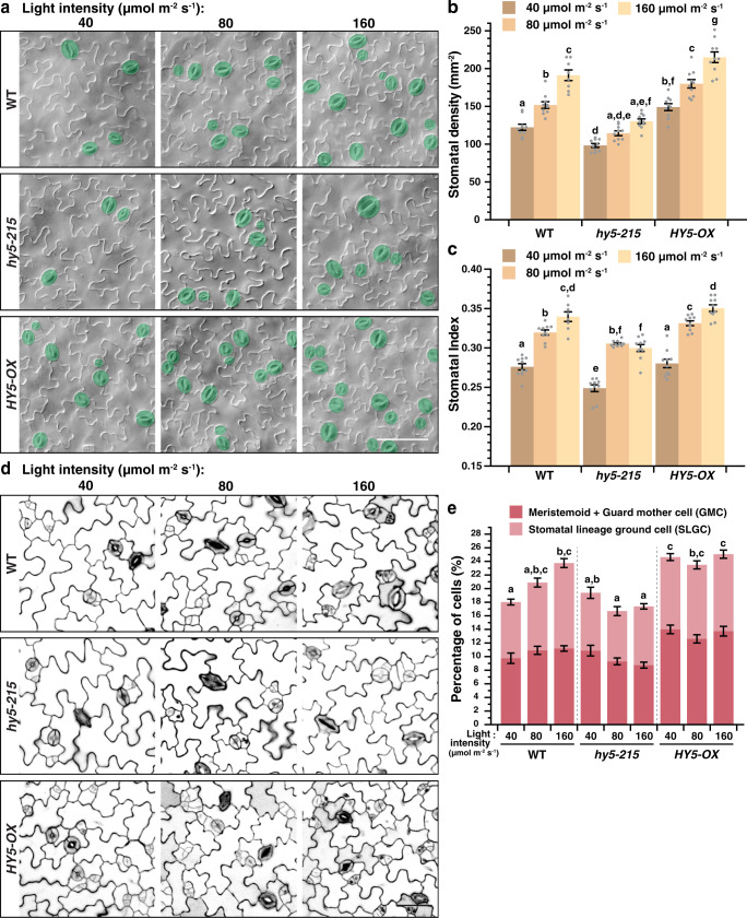
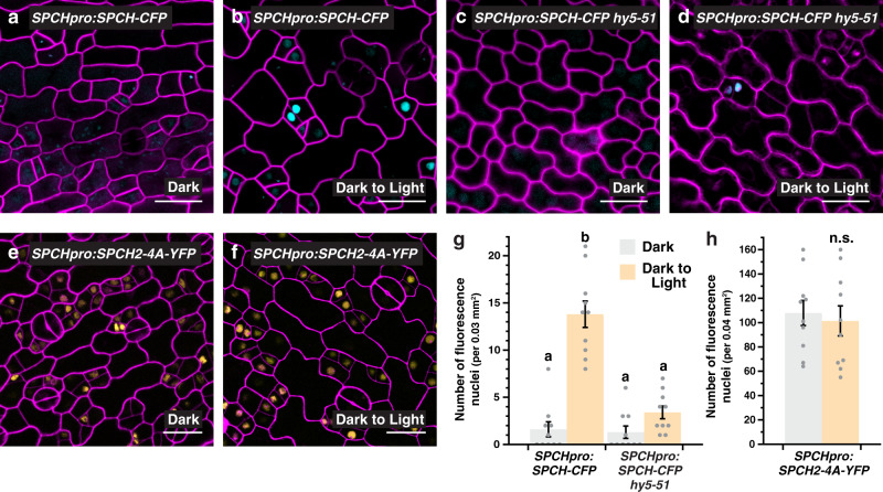
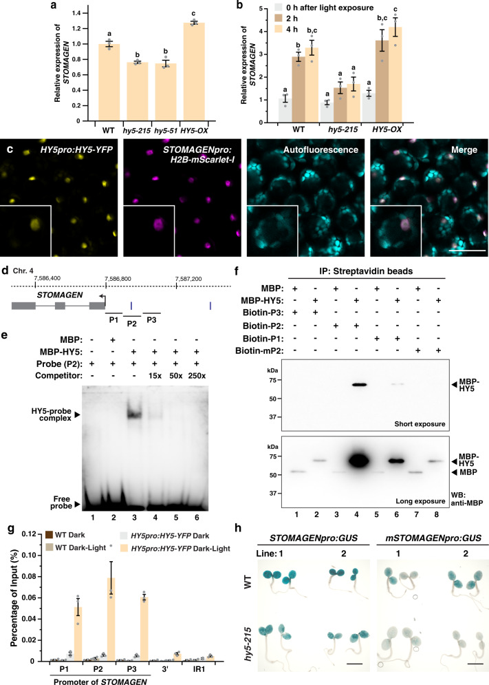
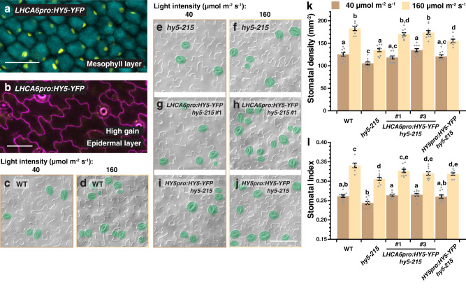
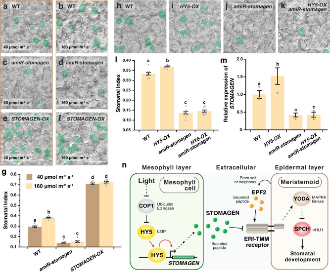
Developmental outcomes are shaped by the interplay between intrinsic and external factors. The production of stomata-essential pores for gas exchange in plants-is extremely plastic and offers an excellent system to study this interplay at the cell lineage level. For plants, light is a key external cue, and it promotes stomatal development and the accumulation of the master stomatal regulator SPEECHLESS (SPCH). However, how light signals are relayed to influence SPCH remains unknown. Here, we show that the light-regulated transcription factor ELONGATED HYPOCOTYL 5 (HY5), a critical regulator for photomorphogenic growth, is present in inner mesophyll cells and directly binds and activates STOMAGEN. STOMAGEN, the mesophyll-derived secreted peptide, in turn stabilizes SPCH in the epidermis, leading to enhanced stomatal production. Our work identifies a molecular link between light signaling and stomatal development that spans two tissue layers and highlights how an environmental signaling factor may coordinate growth across tissue types.


Conflict of interest statement

The authors declare no competing interests.

Figures

Fig. 1
Fig. 1. HY5 is required for the light regulation of stomatal development.
ac Changes in stomatal production in response to light intensities in WT, hy5-215, and HY5-OX (overexpression line of HY5). Arabidopsis seedlings were grown for 10 days at 22 °C under three distinct light intensities (40, 80, and 160 µmol m−2 s−1). Representative images (a), stomatal densities (b), and stomatal indices (c; see text) of the abaxial cotyledons are shown. Stomata are pseudo-colored in green (a). Scale bar, 80 μm. d, e Seedlings of the above genotypes (a) were grown for 3 days to capture the early cell types of the stomatal lineage under the three light intensities (a). Representative confocal images (d) and the percentage of these early cell types, which include meristemoids, guard mother cells (GMCs) and stomatal lineage ground cells (SLGCs) (e; see text and “Methods”) of the abaxial cotyledons are shown. Scale bar, 40 μm. b, c, e Values are mean + /− SEM, n = 10 independent cotyledons. Two-way ANOVA with Tukey’s multiple comparisons test, P < 0.05.
Fig. 2
Fig. 2. HY5 regulates the light-induced accumulation of SPEECHLESS.
ad, g Analysis of a translational reporter of SPEECHLESS (SPCH) in WT (a, b) and hy5-51 (c, d) under light and dark conditions. Confocal images of 3-day-old abaxial cotyledons of SPCHpro:SPCH-CFP, which were grown in darkness and were either kept in the dark (a, c) or transferred to the light for 6 h (b, d) before imaging. Images were taken with the same excitation and acquisition settings. The numbers of CFP-expressing cells (cyan) in an area are quantified (g). e, f, h Analysis of a MAPK-insensitive translational reporter of SPCH. Confocal images of 3-day-old abaxial cotyledons of SPCHpro:SPCH2-4A-YFP grown in darkness (e) or exposed to light (f) (see above). The numbers of YFP-expressing cells (yellow) are quantified (h; color scheme of bar chart as in g). Values are mean + /− SEM, n = 10 independent cotyledons. Two-way ANOVA with Tukey’s multiple comparisons test, P < 0.01 (g) or two-tailed Student’s t test, n.s., not significant (h). Cell outlines were visualized with propidium iodide (magenta). Scale bar, 20 μm.
Fig. 3
Fig. 3. HY5 directly binds and regulates the expression of STOMAGEN.
a, b Gene expression analyses of STOMAGEN in WT, hy5-215, hy5-51 (a only), and HY5-OX by RT-qPCR. In (a), RNA was extracted from 3-day-old seedlings grown under the light. In (b), seedlings were grown in darkness for 4 days and were exposed to light for 0, 2, and 4 h before harvest. Values are mean + /− SEM, n = 3 biological replicates. One-way (a) or two-way (b) ANOVA with Tukey’s multiple comparisons test, P < 0.01. c Co-expression of HY5 and STOMAGEN in the mesophyll layer. Confocal analysis of 3-day-old abaxial cotyledons of a transgenic seedling harboring both HY5pro:HY5-YFP and STOMAGENpro:H2B-mScarlet-I. From left: YFP signals (yellow), mScarlet-I signals (magenta), autofluorescence (cyan) and merged image of all three channels. Scale bar, 50 μm. Three independent cotyledons were examined with similar results (d) Gene structure of STOMAGEN. Arrow indicates the translational start site. Vertical bars mark the position of a Z-box (upstream of TSS only). P1 to 3 represent region(s) tested by EMSA (e), DNA pull down (f) and ChIP-qPCR (g). e EMSA analysis showing the binding of HY5 to a promoter fragment of STOMAGEN (P2). Recombinant MBP (control) and MBP-HY5 were assayed for binding with the biotin-labeled P2 probe. An unlabeled probe (competitor) was used to determine binding specificity (lanes 4–6). f DNA pull-down analysis showing the binding of HY5 to P2 and its dependence on the Z-box. Biotin-labeled probes, including a P2 probe with a mutated Z-box (mP2), were used to pull-down recombinant MBP (control) and MBP-HY5. Results were analyzed by western blotting using an anti-MBP antibody. g ChIP-qPCR assays were performed on WT and HY5pro:HY5-YFP using an anti-GFP antibody. Seedlings were grown for 4 days in darkness before exposed to light for 4 h or kept in the dark. Promoter regions of STOMAGEN (see d) were tested. A genomic region downstream of STOMAGEN and IR1 (see “Methods”) were used as negative controls. Values are mean + /− SEM, n = 3 technical replicates. Assay was repeated with similar results. h GUS reporter assay of two independent lines of STOMAGENpro:GUS and mSTOMAGENpro:GUS, which carries a mutated Z-box in the P2 region, in WT and hy5-215. Seedlings were grown for 3 days under the light. Scale bar, 2 mm. Light intensity used: 200 (a) or 100 µmol m−2 s−1 (b, c, g, h). The experiments in (e) and (f) were carried out two times with similar results.
Fig. 4
Fig. 4. Mesophyll-derived HY5 is capable of driving stomatal development.
a, b Confirmation of the mesophyll-specific expression of LHCA6pro:HY5-YFP. Confocal analysis of 3-day-old abaxial cotyledons of LHCA6pro:HY5-YFP in the mesophyll layer (a) and the epidermis (b). YFP signals (yellow) were detected in (a) but not in (b). Autofluorescence (cyan) of mesophyll cells was captured in (a). In (b), cell outlines were visualized with propidium iodide (magenta). Scale bar, 50 μm. Three independent cotyledons were examined with similar results. cl Genetic complementation of stomatal defects of hy5 by LHCA6pro:HY5-YFP. Arabidopsis seedlings of WT, hy5-215, two independent lines of LHCA6pro:HY5-YFP in hy5-215 and HY5pro:HY5-YFP in hy5-215 were grown for 10 days at 22 °C under low- and high-light intensities (40 and 160 µmol m−2 s−1). Representative images (cj), stomatal densities (k), and stomatal indices (l) of the abaxial cotyledons are shown. Stomata are pseudo-colored in green (cj). Scale bar, 80 μm. Values are mean + /− SEM, n = 10 independent cotyledons. Two-way ANOVA with Tukey’s multiple comparisons test, P < 0.05.
Fig. 5
Fig. 5. STOMAGEN is important for the HY5-mediated promotion of stomatal development.
ag Light-induced stomatal development is impaired in mutants of STOMAGEN. Arabidopsis seedlings of WT, amiR-stomagen (knockdown line of STOMAGEN), and the overexpressor of STOMAGEN (STOMAGEN-OX) were grown for 10 days under either low (40 µmol m−2 s−1) or high (160 µmol m−2 s−1) light intensity. Representative images (af) and stomatal indices (g) of the respective abaxial cotyledons are shown. hl Suppression of the stomatal overproduction phenotype of HY5-OX in a knockdown mutant of stomagen. Arabidopsis seedlings of WT, HY5-OX, amiR-stomagen, and the double-mutant HY5-OX amiR-stomagen were grown for 10 days under 100 µmol m−2 s−1 of light. Representative images (hk) and stomatal indices (l) of the respective abaxial cotyledons are shown. Stomata are pseudo-colored in green (af, hk). Scale bar, 80 μm. (m) Gene expression analysis of STOMAGEN in WT, HY5-OX, amiR-stomagen and HY5-OX amiR-stomagen by RT-qPCR. RNA was extracted from 3-day-old seedlings grown under 100 µmol m−2 s−1 of light. Values are mean + /− SEM, n = 10 independent cotyledons (g, l) or three biological replicates (m). Two-way (g) or one-way (l, m) ANOVA with Tukey’s multiple comparisons test, P < 0.01 (g, l) and P < 0.05 (m). n Model of the HY5-STOMAGEN module in promoting light-mediated stomatal development. Light suppresses COP1 and leads to HY5 accumulation. In mesophyll, HY5 binds and induces STOMAGEN. Increased production of STOMAGEN in turn inhibits the repressive signaling pathway of the epidermal stomatal lineage, leading to the accumulation of SPCH and enhanced stomatal production.

References

    1. Hetherington AM, Woodward FI. The role of stomata in sensing and driving environmental change. Nature. 2003;424:901–908. doi: 10.1038/nature01843. - DOI - PubMed
    1. Vatén A, Bergmann DC. Mechanisms of stomatal development: an evolutionary view. Evodevo. 2012;3:11. doi: 10.1186/2041-9139-3-11. - DOI - PMC - PubMed
    1. Lau OS, Bergmann DC. Stomatal development: a plant’s perspective on cell polarity, cell fate transitions and intercellular communication. Development. 2012;139:3683–3692. doi: 10.1242/dev.080523. - DOI - PMC - PubMed
    1. Han S-K, Torii KU. Lineage-specific stem cells, signals and asymmetries during stomatal development. Development. 2016;143:1259–1270. doi: 10.1242/dev.127712. - DOI - PubMed
    1. MacAlister CA, Ohashi-Ito K, Bergmann DC. Transcription factor control of asymmetric cell divisions that establish the stomatal lineage. Nature. 2007;445:537–540. doi: 10.1038/nature05491. - DOI - PubMed

Publication types

MeSH terms

Substances