. 2021 Jun 7;11(1):12004.
doi: 10.1038/s41598-021-91360-7.

Preclinical immunogenicity testing using anti-drug antibody analysis of GX-G3, Fc-fused recombinant human granulocyte colony-stimulating factor, in rat and monkey models

Affiliations

Preclinical immunogenicity testing using anti-drug antibody analysis of GX-G3, Fc-fused recombinant human granulocyte colony-stimulating factor, in rat and monkey models

Yun Jung Kim et al. Sci Rep. .

Abstract

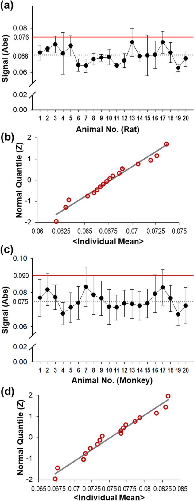
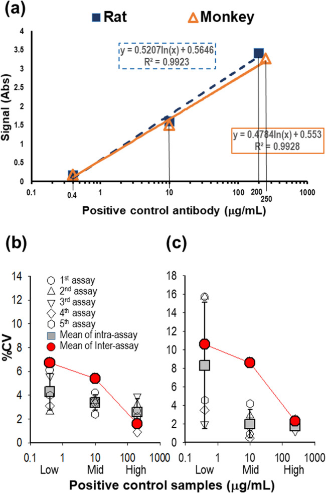
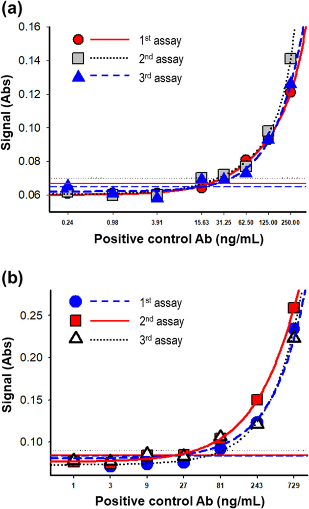
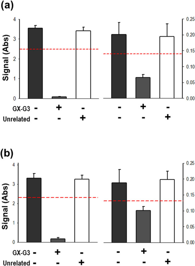
Human granulocyte colony-stimulating factor (G-CSF, this study used Fc-fused recombinant G-CSF; GX-G3) is an important glycoprotein that stimulates the proliferation of granulocytes and white blood cells. Thus, G-CSF treatment has been considered as a crucial regimen to accelerate recovery from chemotherapy-induced neutropenia in cancer patients suffering from non-myeloid malignancy or acute myeloid leukemia. Despite the therapeutic advantages of G-CSF treatment, an assessment of its immunogenicity must be performed to determine whether the production of anti-G-CSF antibodies causes immune-related disorders. We optimized and validated analytical tools by adopting validation parameters for immunogenicity assessment. Using these validated tools, we analyzed serum samples from rats and monkeys injected subcutaneously with GX-G3 (1, 3 or 10 mg/kg once a week for 4 weeks followed by a 4-week recovery period) to determine immunogenicity response and toxicokinetic parameters with serum concentration of GX-G3. Several rats and monkeys were determined to be positive for anti-GX-G3 antibodies. Moreover, the immunogenicity response of GX-G3 was lower in monkeys than in rats, which was relevant to show less inhibition of toxicokinetic profiles in monkeys, at least 1 mg/kg administrated group, compared to rats. These results suggested the establishment and validation for analyzing anti-GX-G3 antibodies and measurement of serum levels of GX-G3 and anti-GX-G3 antibodies, which was related with toxicokinetic profiles. Taken together, this study provides immunogenicity assessment which is closely implicated with toxicokinetic study of GX-G3 in 4-week repeated administrated toxicological studies.


Conflict of interest statement

The authors declare that they have no conflict of interest. The test material studied in this study was supplied by Genexine Inc., Korea, but none of these are in commercial use.

Figures

Figure 1
Figure 1
Negative cut-off (NCO) values were determined using 20 blank serum samples from rats and monkeys. For blank rat sera, (a) determination of the NCO value, (b) normal Q-Q plot of individual data. For blank monkey sera, (c) determination of the NCO value, (d) normal Q-Q plot of individual data.
Figure 2
Figure 2
Evaluation of assay precision. Intra- and inter-assay variability were estimated using the %CV in 5 independent assays. (a) Representative high, middle, and low concentrations of the positive control antibody were selected in the linear range of a sigmoid curve for each set of serum samples. (b) High (200 μg/mL), middle (10 μg/mL), and low (0.4 μg/mL) concentrations of positive control samples (PC) in rat sera were measured for assay precision. (c) High (250 μg/mL), middle (10 μg/mL), and low (0.4 μg/mL) concentrations of PC in monkey sera were measured for assay precision.
Figure 3
Figure 3
Estimation of assay sensitivity. (a) Assay sensitivity was measured in rat sera. PC were spiked into pooled rat serum samples at concentrations ranging from 0.24 to 250 ng/mL. (b) For monkey serum, PC were prepared at concentrations ranging from 1 to 729 ng/mL. The lowest concentration at which the absorbance value was equal to the normalized NCO value in each trial is shown for each sensitivity assay.
Figure 4
Figure 4
Specificity confirmation assay. PC were characterized by testing immune-competition for specificity confirmation. High- and low-concentration PC were pre-incubated for 1 h with the drug GX-G3 or a comparator molecule considered to be unrelated to G-CSF binding, such as rh-GH. (a) High- and low-concentration PC were spiked into pooled rat serum samples at concentrations of 200 μg/mL and 0.4 μg/mL, respectively. (b) High- and low-concentration PC were spiked into pooled monkey serum samples at concentrations of 250 μg/mL and 0.4 μg/mL, respectively. High- and low-concentration PC were pre-incubated with 150 μg/mL GX-G3 for rat serum or 200 μg/mL GX-G3 for monkey serum. rh-GH was used as an unrelated antigen and was spiked into the samples at the same concentrations as GX-G3. High-concentration PC, high-concentration positive control samples; Low-concentration PC, low-concentration positive control samples; Unrelated, unrelated antigen; rh-GH, recombinant human growth hormone.
Figure 5
Figure 5
Estimation of drug interference. Low-concentration PC were pre-incubated with serial dilutions of the drug GX-G3 for 1 h. (a) Low-concentration PC in pooled rat sera were pre-incubated with the drug GX-G3 at a concentration ranging from 250 to 8000 ng/mL. (b) Low-concentration PC in pooled monkey sera were pre-incubated with the drug GX-G3 at a concentration ranging from 250 to 60750 ng/mL. The highest concentration of drug at which the absorbance value was equal to the normalized NCO value is shown in the graphs.
Figure 6
Figure 6
Measurement of ADAs, representing anti-GX-G3 antibodies, in rats subjected to 4-week repeated subcutaneous injection with GX-G3 followed by a 4-week recovery period. Rats were separated into 4 injection subgroups: VC (vehicle control), T1 (1 mg/kg GX-G3), T2 (3 mg/kg GX-G3), and T3 (10 mg/kg GX-G3). ADA levels in rat sera were measured using the method validated in the present study. (a) Immuno-competition assays were performed to discriminate between true and false positive samples. True positivity for GX-G3 specificity was identified based on a greater than 30% difference in absorbance between the non-treated and GX-G3-treated samples. (b) Rat serum samples determined to be true positives were prepared as serial three-fold dilutions beginning from 20-fold. The antibody titer of each sample was expressed as the highest fold-dilution displaying a positive response (absorbance value > normalized NCO value). (c) The mean antibody titer of each group was arithmetically calculated. VC, vehicle control group; P5, pre-dosing day 5; D14 and D28, dosing days 14 and 28, respectively; R29, recovery day 29.
Figure 7
Figure 7
Measurement of ADAs, representing anti-GX-G3 antibodies, in monkeys subjected to 4-week repeated subcutaneous injection with GX-G3 followed by a 4-week recovery period. Monkeys were separated into 4 injection subgroups: VC (vehicle control), T1 (1 mg/kg GX-G3), T2 (3 mg/kg GX-G3), and T3 (10 mg/kg GX-G3). ADA levels in monkey sera were measured using the method validated in the present study. (a) Immuno-competition assays were performed to discriminate between true and false positive samples. True positivity for GX-G3 specificity was identified based on a greater than 30% difference 30% in absorbance between the non-treated and GX-G3-treated samples. (b) Monkey serum samples determined to be true positives were prepared as serial three-fold dilutions beginning from 20-fold. The antibody titer of each sample was expressed as the highest fold-dilution displaying a positive response (absorbance value > normalized NCO value). (c) The mean antibody titer of each group was arithmetically calculated. VC, vehicle control group; D1, D8, D22 and D29, dosing days 1, 8, 22 and 29, respectively; R30, recovery day 30.
Figure 8
Figure 8
Evaluation of the impact of immunogenicity on GX-G3 toxicokinetic analysis, in rats and monkeys subjected to 4-week repeated subcutaneous injection with GX-G3 followed by a 4-week recovery period. Each species was separated into 4 injection subgroups: VC (vehicle control), T1 (1 mg/kg GX-G3), T2 (3 mg/kg GX-G3), and T3 (10 mg/kg GX-G3). (a) Measurement of GX-G3 concentration–time profiles after subcutaneous administration of GX-G3 on Day 1 (Week 0) and Day 22 (Week 3) in rats and monkeys. (b) Representative concentration–time profile of T1 group on Day 1 and Day 22 in rats and monkeys.

Similar articles

Cited by

References

    1. Broxmeyer HE, Vadhan-Raj S. Preclinical and clinical studies with the hematopoietic colony-stimulating factors and related interleukins. Immunol. Res. 1989;8:185–201. doi: 10.1007/BF02918144. - DOI - PubMed
    1. Cohen AM, et al. In vivo stimulation of granulopoiesis by recombinant human granulocyte colony-stimulating factor. Proc. Natl. Acad. Sci. USA. 1987;84:2484–2488. doi: 10.1073/pnas.84.8.2484. - DOI - PMC - PubMed
    1. Welte, K. et al. Recombinant human granulocyte colony-stimulating factor. Effects on hematopoiesis in normal and cyclophosphamide-treated primates. J. Exp. Med.165, 941–948 (1987). - PMC - PubMed
    1. Donadieu J, et al. Analysis of risk factors for myelodysplasias, leukemias and death from infection among patients with congenital neutropenia: experience of the French Severe Chronic Neutropenia Study Group. Haematologica. 2005;90:45–53. - PubMed
    1. Crawford J, et al. Reduction by granulocyte colony-stimulating factor of fever and neutropenia induced by chemotherapy in patients with small-cell lung cancer. New Engl. J. Med. 1991;325:164–170. doi: 10.1056/NEJM199107183250305. - DOI - PubMed

Publication types

MeSH terms