Effect of antioxidant treatment with n-acetylcysteine and swimming on lipid expression of sebaceous glands in diabetic mice
- PMID: 34099835
- PMCID: PMC8184763
- DOI: 10.1038/s41598-021-91459-x
Effect of antioxidant treatment with n-acetylcysteine and swimming on lipid expression of sebaceous glands in diabetic mice
Abstract
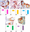
The sebaceous gland (SG) is involved in different inflammatory, infectious and neoplastic processes of the skin and can be related to specific diseases, e.g., diabetes mellitus. Sometimes, the histological diagnosis requires complementary tests due to the ability of diseases to mimic other tumors. We evaluated the sebaceous gland density in Non-obese diabetic mice to analyze the N-acetylcystein effects and swimming exercise treatment in sebaceous glands healing, using specific staining in histochemistry and immunohistochemistry reactions in the identification of the lipid expression in the sebaceous gland. We investigated the intracytoplasmic lipid expression and analysis of gland density from SG in dorsal skin samples from the Non-obese diabetic (NOD mice) and diabetic animals submitted to antioxidant treatment and physical exercise. For histological analysis of the sebaceous glands, specific staining in histochemistry with sudan black and immunohistochemistry reaction with adipophilin were used in the evaluation. Statistical analysis showed significant proximity between the values of the control group and the diabetic group submitted to the swimming exercise (DS group) and similar values between the untreated diabetic group (UD group) and diabetic group treated with the antioxidant N-acetylcysteine (DNa group), which did not prevent possible differences where p < 0.01. Adipophilin (ADPH) immunohistochemistry permitted more intense lipid staining in SGs, the preservation of the SG in the control group, and a morphological deformed appearance in the UD and DNa groups. However, weak morphological recovery of the SG was observed in the DS-Na group, being more expressive in the DS group. In conclusion, the groups submitted to physical exercises showed better results in the recovery of the analyzed tissue, even being in the physiological conditions caused by spontaneous diabetes.
Conflict of interest statement
The authors declare no competing interests.
Figures


References
-
- Gonçalves JF, Mazzanti CM, Becker AG, et al. Oral administration of n-acetylcysteine improves biochemical parameters in diabetic rats. Ciênc Natura. 2012;34(1):81–105.
-
- Santos GR, Ferreira JCR, Corbari TFL, et al. Evaluation of lipid expression in sebaceous glands of diabetic mice by the histochemical method with Sudan III, IV and Black. Rev. Med. Persp. 2019;30(1):5–12.
-
- Paron NG, Lambert PW. Cutaneous manifestations of diabetes mellitus. Dermatology. 2000;27:371–383. - PubMed
Publication types
MeSH terms
Substances
LinkOut - more resources
Full Text Sources
Medical
Research Materials

