. 2021 Jun 7;4(1):688.
doi: 10.1038/s42003-021-02193-z.

REG3A/REG3B promotes acinar to ductal metaplasia through binding to EXTL3 and activating the RAS-RAF-MEK-ERK signaling pathway

Affiliations

REG3A/REG3B promotes acinar to ductal metaplasia through binding to EXTL3 and activating the RAS-RAF-MEK-ERK signaling pathway

Huairong Zhang et al. Commun Biol. .

Abstract

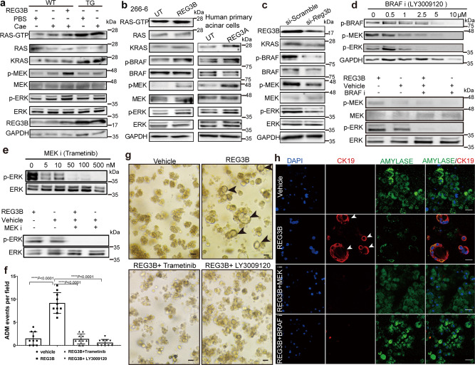
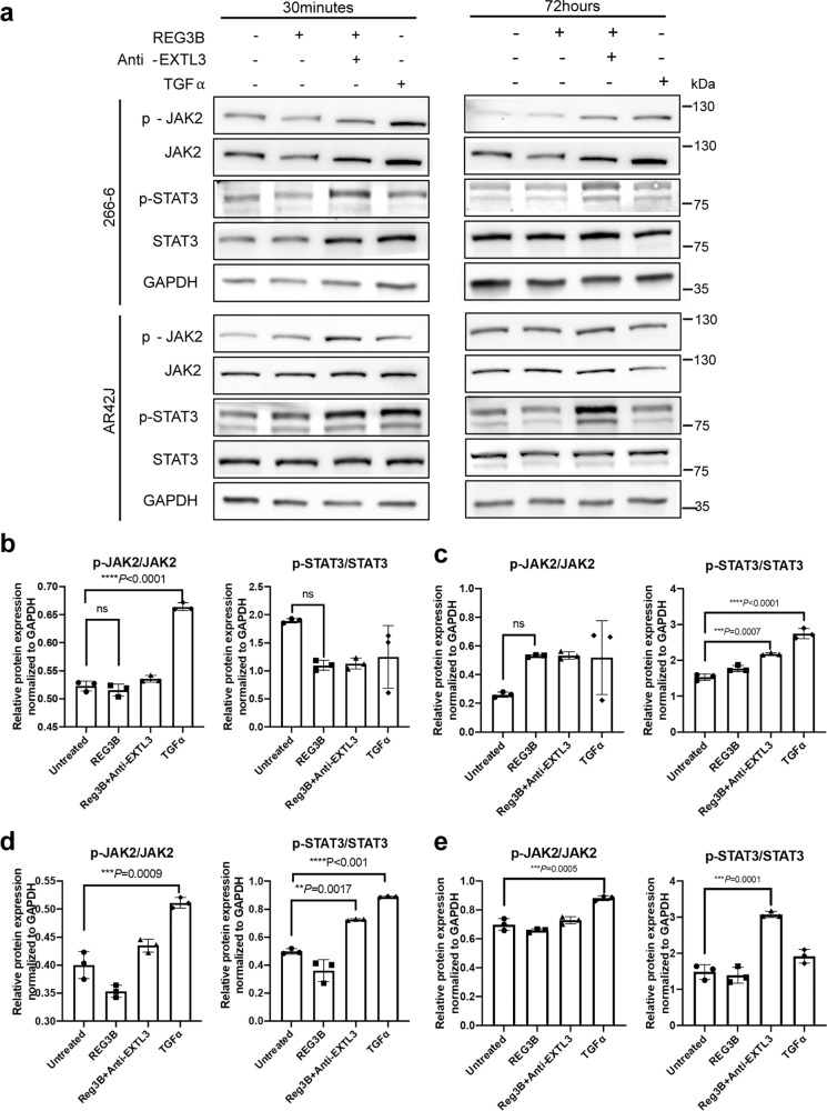
Persistent acinar to ductal metaplasia (ADM) is a recently recognized precursor of pancreatic ductal adenocarcinoma (PDAC). Here we show that the ADM area of human pancreas tissue adjacent to PDAC expresses significantly higher levels of regenerating protein 3A (REG3A). Exogenous REG3A and its mouse homolog REG3B induce ADM in the 3D culture of primary human and murine acinar cells, respectively. Both Reg3b transgenic mice and REG3B-treated mice with caerulein-induced pancreatitis develop and sustain ADM. Two out of five Reg3b transgenic mice with caerulein-induced pancreatitis show progression from ADM to pancreatic intraepithelial neoplasia (PanIN). Both in vitro and in vivo ADM models demonstrate activation of the RAS-RAF-MEK-ERK signaling pathway. Exostosin-like glycosyltransferase 3 (EXTL3) functions as the receptor for REG3B and mediates the activation of downstream signaling proteins. Our data indicates that REG3A/REG3B promotes persistent ADM through binding to EXTL3 and activating the RAS-RAF-MEK-ERK signaling pathway. Targeting REG3A/REG3B, its receptor EXTL3, or other downstream molecules could interrupt the ADM process and prevent early PDAC carcinogenesis.


Conflict of interest statement

The authors declare no competing interests.

Figures

Fig. 1
Fig. 1. REG3A is overexpressed in the ADM area adjacent to human PDAC.
a H&E staining shows histological evidence of transformation from normal acini to ADM and to PDAC. Magnification, ×10; Scale bar: 2 mm. b Enlarged view of ADM area in a. Magnification, ×100; Scale bar: 200 μm. Black arrows indicate typical ADM circular structures. c Single antibody immunohistochemical staining shows intense REG3A expression the ADM zone. Magnification, ×10; Scale bar: 2 mm. d Enlarged view of ADM area in c. Black arrows indicate typical ADM stained strongly with REG3A. Magnification, ×100; Scale bar: 200 μm.
Fig. 2
Fig. 2. REG3B is elevated in caerulein-induced ADM in mice.
a Scheme of caerulein injection to induce acute pancreatitis. Acute pancreatitis was induced by intraperitoneal (i.p.) injection with caerulein dissolved in PBS at a dose of 50 μg/kg at hourly intervals eight times daily, for two consecutive days. PBS injection alone served as control. b The size of pancreas of PBS-treated mice appear smaller than those of caerulein-treated mice (n = 6, student’s t-test). c Percentage of pancreas/body weight of caerulein-treated mice versus PBS-treated mice (n = 6, student’s t-test). d Abundant ADM formation in caerulein-treated mice (H&E, 40×, Scale bar: 500 μm). e Difference in the extent of ADM between PBS-treated mice and caerulein-treated mice, as measured by percentage of ADM area in total pancreatic area on H&E stained tissue sections (n = 6, student’s t-test). f Western blot analysis with quantifications showed higher REG3B protein level and lower AMYLASE protein level in the pancreatic tissue of caerulein-treated mice than that of PBS-treated mice (n = 6, student’s t-test). GAPDH served as a loading control. g RT-qPCR analysis showed higher Ck19 and lower Mist1 mRNA levels in the pancreatic tissue of caerulein-treated mice than that of PBS-treated mice (n = 5, student’s t-test). h H&E staining of caerulein-induced ADM in mice, with corresponding REG3B IHC staining. ADM areas were highlighted by dashed lines, with arrows indicating naïve ADM and asterisks indicating relatively mature ADM. Scale bar: 200 μm. Data are represented as means ± SD; *P < 0.05; **P < 0.01; ***P < 0.001; Non-significant (n.s.) if P > 0.05.
Fig. 3
Fig. 3. Enhanced Reg3B maintains prolonged ADM in mice.
a Scheme of intraperitoneal injection of different reagents in the five experimental groups. b At day 10 (D10), no ADM phenotype was observed in WT mice injected with caerulein. WT mice injected with both recombinant mouse REG3B protein and caerulein, and the REG3B TG mice injected with caerulein showed persistent ADM as indicated by the ductal morphology (black arrow heads) in H&E staining. Focal PanIN (marked by black asterisk) was observed in two out five REG3B TG mice injected with caerulein. Scale bar: 200 μm. c WT mice injected with recombinant REG3B and caerulein and the REG3B TG mice injected with caerulein demonstrate higher levels of Ck19 and lower levels of Mist1 mRNA. (n = 5, one-way ANOVA). d Immunofluorescence staining shows gain of CK19 expression (red) and loss of AMYLASE (green) in the ADM area of WT mice injected with recombinant REG3B and caerulein and in the Reg3b TG mice injected with caerulein. Scale bars: 20 μm. White arrows indicate ADM. e Persistence of ADM in WT mice injected with recombinant REG3B 2, 4, and 10 weeks after caerulein injection. (n = 5, magnification ×200, scale bar: 200 μm). Values in graphs are means ± SD, *P < 0.05; **P < 0.01; ***P < 0.001. Non-significant (n.s.) if P > 0.05. n = 5 per group, WT wild type, TG REG3B transgenic mice, Cae caerulein.
Fig. 4
Fig. 4. Exogenous REG3A/REG3B induces ADM in human and mouse primary acinar cells in vitro.
a Bright field images showing an increase in ADM events (depicted by black arrows) in cultured mouse primary acinar cells after 5 days of REG3B treatment and in cultured human primary acinar cells after 5 days of REG3A treatment (Magnification, ×200). TGFα-treated mouse primary acinar cells served as a positive control. b Bar graph showing increase in ADM quantity in 3D culture of mouse and human primary acinar cells during the 5-day REG3B or REG3A or TGFα treatment (n = 3, 15 fields each group, one-way ANOVA and student’s t-test). c Bright field images in the upper row showing ADM events in cultured mouse primary acinar cells after 5 days of REG3B treatment and in cultured human primary acinar cells after 5 days of REG3A treatment (magnification, ×630). Lower four rows are corresponding confocal immunofluorescence images showing a decrease in AMYLASE protein expression (green) and an increase in CK19 (red) protein in REG3B-induced, REG3A-induced, or TGFα-induced ADM (magnification, ×630. Scale bars: 20 μm). d RT-qPCR analysis showed a decrease in acinar-specific mRNA (Ptf1a, Cpa, and Mist1) and an increase in duct-specific mRNA (Ck19 and Nestin) in mouse and human primary acinar cells after 48 h of REG3B or REG3A treatment. (n = 3 per group, student’s t-test). Values are represented as mean ± standard deviation. *P < 0.05, **P < 0.01, ***P < 0.001. Non-significant (n.s.) if P > 0.05.
Fig. 5
Fig. 5. REG3B induces ADM through the RAS-RAF-MEK-ERK signaling pathway.
a Western blot showing increased expression of p-ERK, p-MEK, p-BRAF, KRAS, and active RAS in the pancreatic tissue of REG3B-treated WT mice with caerulein-induced pancreatitis (WT cae+REG3B) and a moderate increase in TG mice with caerulein-induced pancreatitis (TG cae) (n = 3, one-way ANOVA). b Western blots demonstrating increased expression of p-ERK, p-MEK, p-BRAF, KRAS, and active RAS in cultured human primary acinar cells and mouse acinar cell line 266-6 after REG3A or REG3B treatment respectively, for 48 h (n = 3, student’s t-test). c Western blot showing the reduced expression of phosphorylated ERK, MEK, BRAF, and total KRAS in the 266-6 cell line after Reg3b gene knockdown by siRNA (n = 3, student t-test). d In the upper panel, LY3009120 inhibited ERK, MEK, BRAF phosphorylation in a dose-dependent manner in 266-6 cell line. In the lower panel, LY3009120 (5 μM) blocked REG3B-induced MEK and ERK phosphorylation (n = 3, one-way ANOVA). e Upper panel, Trametinib efficiently attenuated ERK phosphorylation in a dose-dependent manner in the AR42J cell line (n = 3, one-way ANOVA). Lower panel, Trametinib (100 nM) blocked REG3B-induced ERK phosphorylation. f, g Bright field images (g) with quantification (f) show that Trametinib (100 nM) and LY3009120 (5 μM) treatment hindered REG3B-induced ADM in 3D cultures of mouse primary acinar cells (200×, n = 3, one-way ANOVA). h Confocal microscopy shows that Trametinib (100 nM) and LY3009120 (5 μM) treatment reduced CK19 protein expression (red) and increased AMYLASE protein expression (green) in 3D culture of mouse primary acinar cells (Scale bar: 20 μm). Data are represented as means ± SD, n = 3. *P < 0.05, **P < 0.01, ***P < 0.001. Non-significant (n.s.) if P > 0.05.
Fig. 6
Fig. 6. EXTL3 functions as a receptor for REG3B in ADM.
a Co-localization of EXTL3 (green) with human REG3A (red) or rodent REG3B (red) in ADM zones derived from human primary acinar cells in 3D culture, and AR42J and 266-6 cell lines in 2D culture by immunofluorescence microscopy. (magnification: ×630, Scale bars: 10 μm). b Co-immunoprecipitation of REG3B and EXTL3 in mouse primary acinar cells, rat AR42J and mouse 266-6 acinar cell lines. Lysate-bead/antibody conjugate mixture was eluted with sample buffer without DTT for 10 min at 50 °C (elution 1). Sample buffer with DTT (100 mM) was added to the pelleted beads from elution 1 and boiled for 5 min (elution 2). c Confocal immunofluorescence microscopy showed that EXTL3 monoclonal antibody treatment effectively blocks REG3B-induced ADM as indicated by increased expression of the acinar marker AMYLASE and decreased expression of the ductal marker CK19. (magnification: ×630, Scale bar: 20 μm). df Extl3 siRNA or neutralizing antibody treatment reduced the protein expression of KRAS and phosphorylated ERK, MEK, and BRAF in the presence or absence of REG3B for 48 h. d Western blotting analysis of the knockdown of Extl3 by siRNA (20 nM) in 266-6 cell line, e knockdown of Extl3 by siRNA (20 nM) in the context of the 266-6 cell line treated with REG3B for 48 h, f Western blotting analysis of EXTL3 neutralizing antibody treatment (2 μg/ml) in 266-6 and AR42J cell lines and mouse primary acinar cells treated with or without REG3B for 48 h. Human primary acinar cells were treated for 30 min. TGFα treatment serves as a positive control for ADM induction.
Fig. 7
Fig. 7. JAK2/STAT3 is not involved in REG3B-induced ADM.
a Western blot analysis of related protein expression in 266-6 and AR42J cell lines 30 min and 72 h after different stimulations, with quantification data. b, c Expression level of JAK2/STAT3 signaling components in the 266-6 cell line under different conditions for 30 min (b) and 72 h (c). d, e Expression level of JAK2/STAT3 signaling components in the AR42J cell line under different conditions for 30 min (d) and 72 h (e) (n = 3, one-way ANOVA test). REG3B treatment alone did not increase p-JAK nor p-STAT3 expression at two timepoints in two cell lines but did increase their expression once the receptor EXTL3 was blocked.
Fig. 8
Fig. 8. Schematic illustration of the molecular mechanism of REG3B/REG3A-driven ADM.
REG3B/REG3A binds to its receptor, EXTL3 receptor on the acinar cell membrane, and promotes ADM by activating the downstream RAS-RAF-MEK-ERK signaling pathway, in the absence of oncogenic Kras mutation. Targeting REG3B/REG3A, neutralizing its receptor EXTL3, or inhibiting downstream signaling molecules, such as B-RAF (LY3009120) or MEK1/2 (Trametinib), could interrupt the ADM process and potentially prevent early PDAC carcinogenesis.

Similar articles

Cited by

References

    1. Ghaneh P, Costello E, Neoptolemos JP. Biology and management of pancreatic cancer. Gut. 2007;56:1134–1152. doi: 10.1136/gut.2006.113068. - DOI - PMC - PubMed
    1. Distler M, Aust D, Weitz J, Pilarsky C, Grutzmann R. Precursor lesions for sporadic pancreatic cancer: PanIN, IPMN, and MCN. BioMed. Res. Int. 2014;2014:474905. doi: 10.1155/2014/474905. - DOI - PMC - PubMed
    1. Feldmann G, Beaty R, Hruban RH, Maitra A. Molecular genetics of pancreatic intraepithelial neoplasia. J. Hepato Biliary Pancreat. Surg. 2007;14:224–232. doi: 10.1007/s00534-006-1166-5. - DOI - PMC - PubMed
    1. Grippo PJ, Sandgren EP. Acinar-to-ductal metaplasia accompanies c-myc-induced exocrine pancreatic cancer progression in transgenic rodents. Int. J. Cancer. 2012;131:1243–1248. doi: 10.1002/ijc.27322. - DOI - PubMed
    1. Guerra C, et al. Chronic pancreatitis is essential for induction of pancreatic ductal adenocarcinoma by K-Ras oncogenes in adult mice. Cancer Cell. 2007;11:291–302. doi: 10.1016/j.ccr.2007.01.012. - DOI - PubMed

Publication types

MeSH terms