Review
. 2021 Jul;28(7):2029-2044.
doi: 10.1038/s41418-021-00814-y. Epub 2021 Jun 7.

Molecular mechanisms of cell death in neurological diseases

Affiliations
Review

Molecular mechanisms of cell death in neurological diseases

Diane Moujalled et al. Cell Death Differ. 2021 Jul.

Abstract

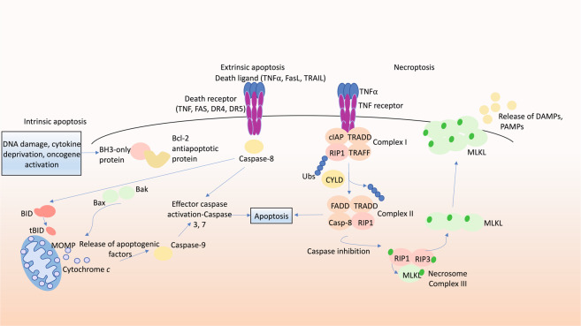
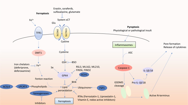
Tightly orchestrated programmed cell death (PCD) signalling events occur during normal neuronal development in a spatially and temporally restricted manner to establish the neural architecture and shaping the CNS. Abnormalities in PCD signalling cascades, such as apoptosis, necroptosis, pyroptosis, ferroptosis, and cell death associated with autophagy as well as in unprogrammed necrosis can be observed in the pathogenesis of various neurological diseases. These cell deaths can be activated in response to various forms of cellular stress (exerted by intracellular or extracellular stimuli) and inflammatory processes. Aberrant activation of PCD pathways is a common feature in neurodegenerative diseases, such as amyotrophic lateral sclerosis (ALS), Alzheimer's disease, Parkinson's disease, and Huntington's disease, resulting in unwanted loss of neuronal cells and function. Conversely, inactivation of PCD is thought to contribute to the development of brain cancers and to impact their response to therapy. For many neurodegenerative diseases and brain cancers current treatment strategies have only modest effect, engendering the need for investigations into the origins of these diseases. With many diseases of the brain displaying aberrations in PCD pathways, it appears that agents that can either inhibit or induce PCD may be critical components of future therapeutic strategies. The development of such therapies will have to be guided by preclinical studies in animal models that faithfully mimic the human disease. In this review, we briefly describe PCD and unprogrammed cell death processes and the roles they play in contributing to neurodegenerative diseases or tumorigenesis in the brain. We also discuss the interplay between distinct cell death signalling cascades and disease pathogenesis and describe pharmacological agents targeting key players in the cell death signalling pathways that have progressed through to clinical trials.


Conflict of interest statement

DM and AS are employees of The Walter and Eliza Hall Institute, which receives milestone payments and royalties from Genentech and AbbVie for the development of ABT-199/venetoclax. AS collaborates with Servier on the development of MCL-1-specific BH3 mimetic drugs for cancer therapy. JRL has no conflict of interest.

Figures

Fig. 1
Fig. 1. Molecular pathways of apoptosis and necroptosis.
Intrinsic apoptosis signalling: in response to growth factor deprivation, DNA damage or oncogene activation, BH3-only pro-apoptotic proteins are induced transcriptionally or post-transcriptionally. The BH3-only proteins bind to and inhibit the anti-apoptotic BCL-2 proteins. This leads to release and activation of the effectors of cell death, BAX and BAK, which then oligomerise and promote mitochondrial outer membrane permeabilisation (MOMP), leading to release of apoptogenic factors, including cytochrome c and Smac/Diablo. This initiates a cascade of caspase activation, cleavage of hundreds of cellular proteins and consequent cell demolition. Death receptor induced (extrinsic) apoptosis signalling: stimulation of death receptors (members of the TNFR family with an intracellular death domain) by their cognate ligands (e.g. stimulation of FAS by FASL) results in adaptor protein (FADD, TRADD) mediated recruitment and activation of the initiator caspase, caspase-8 (in humans also caspase-10), which can then activate downstream effector caspases (caspases-3, -7), resulting in cell demolition (see above). The death receptor induced apoptosis pathway can connect to the intrinsic apoptotic pathway by proteolytic activation of the pro-apoptotic BH3-only protein BID (to generate tBID) by caspase-8. Necroptosis: the best characterised pathway triggering necroptosis is via TNFR1 stimulation. Upon binding of TNFα to TNFR1, cIAPs, RIPK1, TRAFs and TRADD are recruited to the intracellular part of TNFR1, forming the TNFR1 signalling complex I, which activates NFkB and AP1 transcription factors and thereby stimulates cell survival and proliferation. Upon deubiquitylation of RIPK1 by CYLD, RIPK1 can bind to TRADD, FADD and caspase-8 forming complex II which can drive caspase-8 mediated apoptosis (see above). In the absence of caspase-8 (genetic ablation or pharmacological inhibition) and the absence or inhibition of cIAP1/2, the necrosome (complex III) is formed where RIPK3 can phosphorylate MLKL, promoting its translocation to the plasma membrane where MLKL causes cell lysis, resulting in lytic cell death with release of DAMPs and PAMPs.
Fig. 2
Fig. 2. Molecular pathways of ferroptosis and pyroptosis.
Ferroptosis is potentiated by iron which can be imported by TFR1 and DMT1, and requires the presence of susceptible arachidonic acid-containing phospholipids generated by ACSL4 and LPCAT3. Ferroptosis is endogenously inhibited by GPX4 which requires GSH as a substrate, by ubiquinone which is reduced by FSP1, by BH4 which is generated by GCH1, and by other endogenous RTAs such as vitamin E. Ferroptosis is promoted by disruption of cysteine supply via inhibition of the glutamate/cystine antiporter system xCT (by compounds such as erastin, sorafenib, sulfasalazine or glutamate), or impaired GPX4 activity due to insufficient glutathione or selenium or by direct inhibition (e.g. RSL3, ML162, ML210, FIN56, FINO2). Ferroptosis inhibitors include iron chelators (such as deferiprone and deferoxamine), RTAs (including ferrostatin-1 and liproxstatin-1) and lipoxygenase inhibitors. Pyroptosis can be activated in response to physiological or pathological insults which result in activation of inflammasomes, such as the NLRP3 inflammasome, in which the adaptor ASC is recruited, resulting in activation of caspase-1. Caspase-1 proteolytically processes pro-IL-1β and pro-IL-18 into the bio-active forms of these cytokines. Caspase-1 can also cleave GSDMD, and the N‐terminal fragment of GSDMD is recruited to the plasma membrane where it causes pore formation, cell swelling and plasma membrane rupture. BH4 tetrahydrobiopterin, BSO Buthionine sulfoximine, DMT1 divalent metal transporter 1, GCH1 GTP cyclohydrolase-1, GPX4 glutathione peroxidase 4, GSH glutathione, RTAs radical trapping antioxidants, Se selenium, System xCT glutamate/cystine antiporter, TFR1 transferrin receptor 1, NLRP3 NLR family pyrin domain‐containing 3.

Similar articles

Cited by

References

    1. Kerr JF, Wyllie AH, Currie AR. Apoptosis: a basic biological phenomenon with wide-ranging implications in tissue kinetics. Br J Cancer. 1972;26:239–57.. - PMC - PubMed
    1. Kelly GL, Strasser A. Toward targeting antiapoptotic MCL-1 for cancer therapy. Annu Rev Cancer Biol. 2020;4:299–313.
    1. Huang DC, Strasser A. BH3-Only proteins-essential initiators of apoptotic cell death. Cell. 2000;103:839–42. - PubMed
    1. Green DR. The coming decade of cell death research: five riddles. Cell. 2019;177:1094–107.. - PMC - PubMed
    1. Singh R, Letai A, Sarosiek K. Regulation of apoptosis in health and disease: the balancing act of BCL-2 family proteins. Nat Rev Mol Cell Biol. 2019;20:175–93.. - PMC - PubMed

Publication types