[Preprint]. 2021 Jun 4:rs.3.rs-571332.
doi: 10.21203/rs.3.rs-571332/v1.

Rapid Artificial Intelligence Solutions in a Pandemic - The COVID-19-20 Lung CT Lesion Segmentation Challenge

Affiliations

Rapid Artificial Intelligence Solutions in a Pandemic - The COVID-19-20 Lung CT Lesion Segmentation Challenge

Holger R Roth et al. Res Sq. .

Update in

  • Rapid artificial intelligence solutions in a pandemic-The COVID-19-20 Lung CT Lesion Segmentation Challenge.
    Roth HR, Xu Z, Tor-Díez C, Sanchez Jacob R, Zember J, Molto J, Li W, Xu S, Turkbey B, Turkbey E, Yang D, Harouni A, Rieke N, Hu S, Isensee F, Tang C, Yu Q, Sölter J, Zheng T, Liauchuk V, Zhou Z, Moltz JH, Oliveira B, Xia Y, Maier-Hein KH, Li Q, Husch A, Zhang L, Kovalev V, Kang L, Hering A, Vilaça JL, Flores M, Xu D, Wood B, Linguraru MG. Roth HR, et al. Med Image Anal. 2022 Nov;82:102605. doi: 10.1016/j.media.2022.102605. Epub 2022 Sep 6. Med Image Anal. 2022. PMID: 36156419 Free PMC article.

Abstract

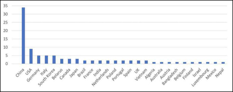
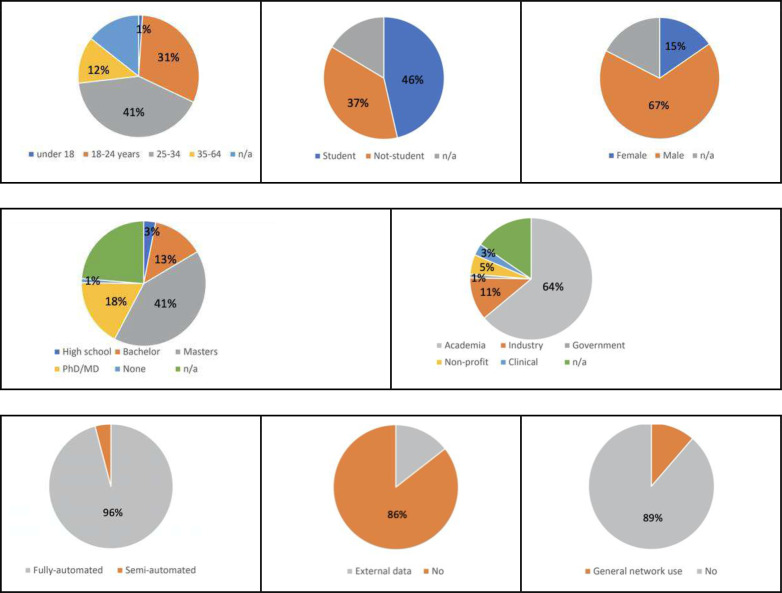
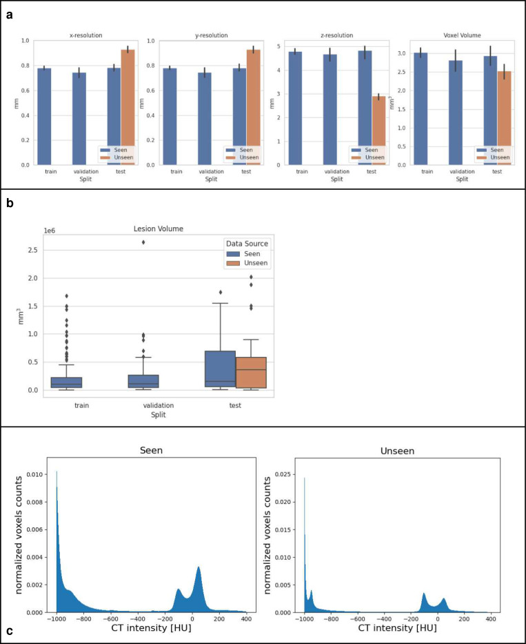
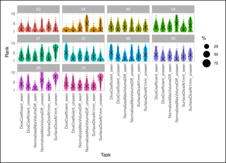
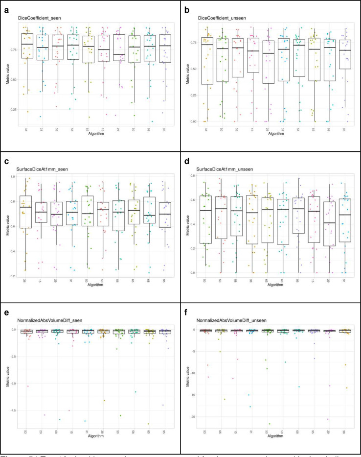
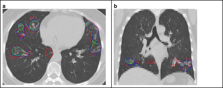
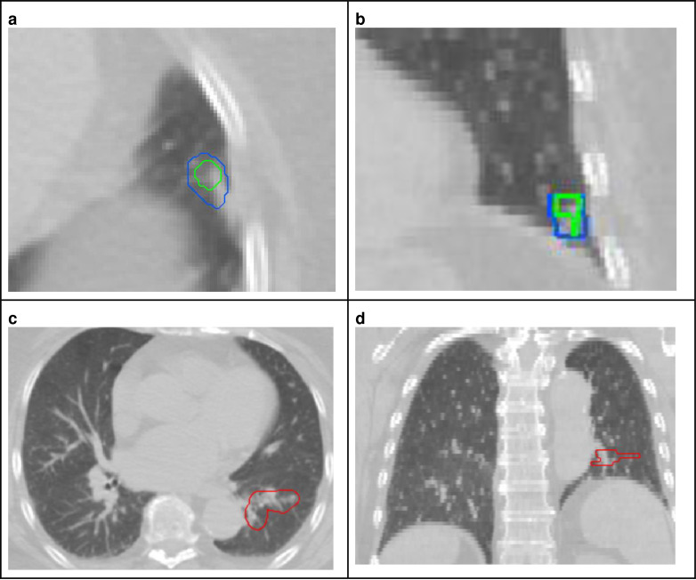
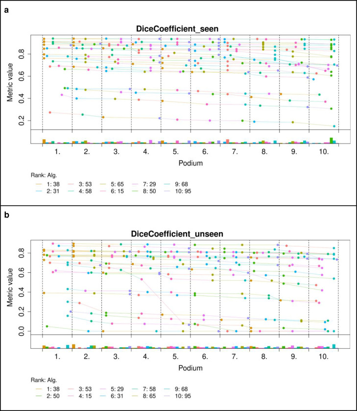
Artificial intelligence (AI) methods for the automatic detection and quantification of COVID-19 lesions in chest computed tomography (CT) might play an important role in the monitoring and management of the disease. We organized an international challenge and competition for the development and comparison of AI algorithms for this task, which we supported with public data and state-of-the-art benchmark methods. Board Certified Radiologists annotated 295 public images from two sources (A and B) for algorithms training (n=199, source A), validation (n=50, source A) and testing (n=23, source A; n=23, source B). There were 1,096 registered teams of which 225 and 98 completed the validation and testing phases, respectively. The challenge showed that AI models could be rapidly designed by diverse teams with the potential to measure disease or facilitate timely and patient-specific interventions. This paper provides an overview and the major outcomes of the COVID-19 Lung CT Lesion Segmentation Challenge - 2020.


Figures

Figure 1 |
Figure 1 |
The countries of origin of the 98 teams that completed the training, validation and test phases of the challenge.
Figure 2 |
Figure 2 |
Demographic information of the leaders of the 98 teams that completed the training, validation and test phases of the challenge. The top row shows the age group (left), student status (middle) and sex (right) of the participant. The middle row shows the highest degree (left) and job category (right). Bottom row shows the algorithm characteristics for the 98 submissions that completed the training, validation and test phases of the challenge. We report if algorithms were fully-automated (left), used external data for training (middle) or used a general pre-trained network for initialization (right).
Figure 3 |
Figure 3 |
Data variability between “seen” and “unseen” sources; a) Illustration of the differences in the image resolution and voxel volume grouped by training, validation, and test sets. b) Differences in COVID-19 lesion volumes across the image data sources. c) Normalized histograms showing the CT intensity distributions of the “seen” and “unseen” data sources in Hounsfield units (HU). Note, −1000 HU corresponds to air, and 750 to cancellous bone .
Figure 4 |
Figure 4 |
Blob plot visualization of the ranking variability via bootstrapping. An algorithm’s ranking stability is shown across the different tasks, illustrating the ranking uncertainty of the algorithm in each task. For more details see .
Figure 5 |
Figure 5 |
Top-10 algorithms performance measured for the m = 6 tasks used in the challenge, namely the Dice coefficient (top row), Normalized Surface Dice (middle row), and Normalized Absolute Volume Error (bottom row) on the “seen” (a, c, e) and “unseen” test datasets (b, d, f), respectively. Algorithms are ranked based on their performance from left to right.
Figure 6 |
Figure 6 |
Example test case from the “seen” data data source (Dataset 1). The performance of the top algorithms #53 and #38 is shown in green and blue, respectively. Ground truth annotations are shown in red (a: axial view, b: coronal view).
Figure 7 |
Figure 7 |
Example test case from the “unseen” data source. (a: axial view, b: coronal view) Top algorithms #53 and #38, shown in green and blue, respectively, both predict a false-positive lesion at the locations of a normal lung vessel. At the same time they missed the real lesion in red (c: axial view, d: coronal view).
Figure 8 |
Figure 8 |
Podium plots for “seen” (a) and “unseen” (b) test data. The participating algorithms are color-coded. Each colored dot shows the Dice coefficient achieved by the respective algorithm. The same test cases are connected by a line. The lower part of the charts displays the relative frequency for a given algorithm to achieve a podium place, i.e. rank achieved by a given algorithm.

References

    1. Johns Hopkins Coronavirus Resource Center. https://coronavirus.jhu.edu/.
    1. Nino G. et al. Pediatric lung imaging features of COVID-19: A systematic review and meta-analysis. Pediatr. Pulmonol. 56, 252–263 (2021). - PMC - PubMed
    1. Zang S.-T. et al. Imaging characteristics of coronavirus disease 2019 (COVID-19) in pediatric cases: a systematic review and meta-analysis. Transl Pediatr 10, 1–16 (2021). - PMC - PubMed
    1. Larici A. R. et al. Multimodality imaging of COVID-19 pneumonia: from diagnosis to follow-up. A comprehensive review. Eur. J. Radiol. 131, 109217 (2020). - PMC - PubMed
    1. Ojha V., Mani A., Pandey N. N., Sharma S. & Kumar S. CT in coronavirus disease 2019 (COVID-19): a systematic review of chest CT findings in 4410 adult patients. Eur. Radiol. 30, 6129–6138 (2020). - PMC - PubMed

Publication types

LinkOut - more resources