. 2021 Jul;231(3-4):73-83.
doi: 10.1007/s00427-021-00677-w. Epub 2021 Jun 7.

Expression patterns of signalling molecules and transcription factors in the early rabbit embryo and their significance for modelling amniote axis formation

Affiliations

Expression patterns of signalling molecules and transcription factors in the early rabbit embryo and their significance for modelling amniote axis formation

Ruben Plöger et al. Dev Genes Evol. 2021 Jul.

Abstract

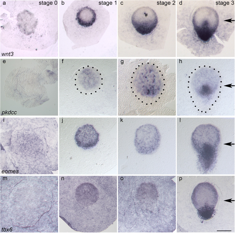
The anterior-posterior axis is a central element of the body plan and, during amniote gastrulation, forms through several transient domains with specific morphogenetic activities. In the chick, experimentally proven activity of signalling molecules and transcription factors lead to the concept of a 'global positioning system' for initial axis formation whereas in the (mammotypical) rabbit embryo, a series of morphological or molecular domains are part of a putative 'three-anchor-point model'. Because circular expression patterns of genes involved in axis formation exist in both amniote groups prior to, and during, gastrulation and may thus be suited to reconcile these models, the expression patterns of selected genes known in the chick, namely the ones coding for the transcription factors eomes and tbx6, the signalling molecule wnt3 and the wnt inhibitor pkdcc, were analysed in the rabbit embryonic disc using in situ hybridisation and placing emphasis on their germ layer location. Peripheral wnt3 and eomes expression in all layers is found initially to be complementary to central pkdcc expression in the hypoblast during early axis formation. Pkdcc then appears - together with a posterior-anterior gradient in wnt3 and eomes domains - in the epiblast posteriorly before the emerging primitive streak is marked by pkdcc and tbx6 at its anterior and posterior extremities, respectively. Conserved circular expression patterns deduced from some of this data may point to shared mechanisms in amniote axis formation while the reshaping of localised gene expression patterns is discussed as part of the 'three-anchor-point model' for establishing the mammalian body plan.

Keywords: Germ layers; Primitive streak; Rabbit; T-box transcription factors; Twinning; Wnt-signalling.


Conflict of interest statement

Not applicable

Figures

Fig. 1
Fig. 1
Expression patterns of wnt3 (ad), pkdcc (eh), eomes (il) and tbx6 (mp) in en face views at stages 0 (a, e, i, m), 1 (b, f, j, n), 2 (c, g, k, o) and 3 (d, h, l, p) using bright-field illumination. Embryonic discs are oriented with the anterior pole to the top. Stages are indicated by Arabic numbers; genes names are printed in italics. Dotted lines (f, g, h) mark the border of the embryonic disc as determined by cell density in dark-field microscopy (see suppl. Fig. 1). Black arrows mark the tip of the primitive streak. The hash symbol (c) indicates a staining artefact. The scale bar corresponds to 25 μm in panels showing stage 0 embryonic discs (a, e, i, m), to 450 μm (g), 550 μm (o), and 500 μm (bd, f, h, jl, n, p) in panels showing  later stages, respectively
Fig. 2
Fig. 2
Near-median sagittal Technovit(R) sections of wnt3 (ac), pkdcc (df), eomes (gi), and tbx6 (jl) expression domains in the anterior margin at stages 1 (a, d, g, j), 2 (b, e, h, k) and 3 (c, f, i, l). Anterior is to the left, asterisks indicate the border of the embryonic disc  as determined by the most anterior epiblast cell. Arrow heads point to Rauber cells (ad, g, j). e: epiblast, h: hypoblast . Scale bar : 200 μm
Fig. 3
Fig. 3
Near-median sagittal Technovit(R) sections of wnt3 (a, b, i), pkdcc (c, d, j), eomes (e, f, k) and tbx6 (g, h, l) expression domains in the posterior margin at stages 1 (a, c, e, g), 2 (b, d, f, h) and 3 (i, j, k, l). Orientation of sections and inscriptions are as in Fig. 2; asterisks, however, indicate the posterior border of the embryonic disc. The ring symbol indicates the border between the anterior gastrula plate (AGP) and the posterior gastrula extension (PGE). The plus symbol marks bottle cells.  m: mesoderm cells . Scale bar (l): 200 μm
Fig. 4
Fig. 4
Schematic representation of mammalian initial a.-p. axis formation stages in dorsal views (ae) and median sagittal sections (g and h) using the rabbit as a model and compass needle symbols (f) to illustrate the increasing restriction of possible a.-p. axis orientation in the TAP model, in which  AMC, ASD and EN are the  putative anchors 1, 2, and 3, respectively. T: trophoblast (grey), Y: yolk sac epithelium (orche), E: epiblast (red, boxed in upper layer), H: hypoblast (boxed in lower layer), MZ: marginal zone (camel), CZ: central zone (light blue), AMC: anterior marginal crescent (light green), ASD: anterior streak domain (dark blue), red dashed line: border between anterior gastrula plate (AGP) and posterior gastrula extension (PGE), EN: end node (yellow), PS: primitive streak (purple), arrows: expansion of CZ, stick figure superimposed on PS in f, right hand panel: contribution of PS to adult body parts. Scale bar: roughly 500 μm

References

    1. Arias CF, Herrero MA, Stern CD, Bertocchini F. A molecular mechanism of symmetry breaking in the early chick embryo. Sci Rep. 2017;7:15776. doi: 10.1038/s41598-017-15883-8. - DOI - PMC - PubMed
    1. Arnold SJ, Hofmann UK, Bikoff EK, Robertson EJ. Pivotal roles for eomesodermin during axis formation, epithelium-to-mesenchyme transition and endoderm specification in the mouse. Development. 2008;135:501–511. doi: 10.1242/dev.014357. - DOI - PMC - PubMed
    1. Bachvarova RF, Skromne I, Stern CD. Induction of primitive streak and Hensen’s node by the posterior marginal zone in the early chick embryo. Development. 1998;125:3521–3534. doi: 10.1242/dev.125.17.3521. - DOI - PubMed
    1. Behringer RR, Wakamiya M, Tsang TE, Tam PP. A flattened mouse embryo: leveling the playing field. Genesis. 2000;28:23–30. doi: 10.1002/1526-968X(200009)28:1<23::AID-GENE30>3.0.CO;2-G. - DOI - PubMed
    1. Bellairs R. The primitive streak. Anat Embryol. 1986;174:1–14. doi: 10.1007/BF00318331. - DOI - PubMed

Publication types

LinkOut - more resources