. 2021 Aug;12(4):454-465.
doi: 10.1007/s13239-021-00535-w. Epub 2021 Jun 7.

VICTORIA: VIrtual neck Curve and True Ostium Reconstruction of Intracranial Aneurysms

Affiliations

VICTORIA: VIrtual neck Curve and True Ostium Reconstruction of Intracranial Aneurysms

Philipp Berg et al. Cardiovasc Eng Technol. 2021 Aug.

Abstract

Purpose: For the status evaluation of intracranial aneurysms (IAs), morphological and hemodynamic parameters can provide valuable information. For their extraction, a separation of the aneurysm sac from its parent vessel is required that yields the neck curve and the ostium. However, manual and subjective neck curve and ostium definitions might lead to inaccurate IA assessments.

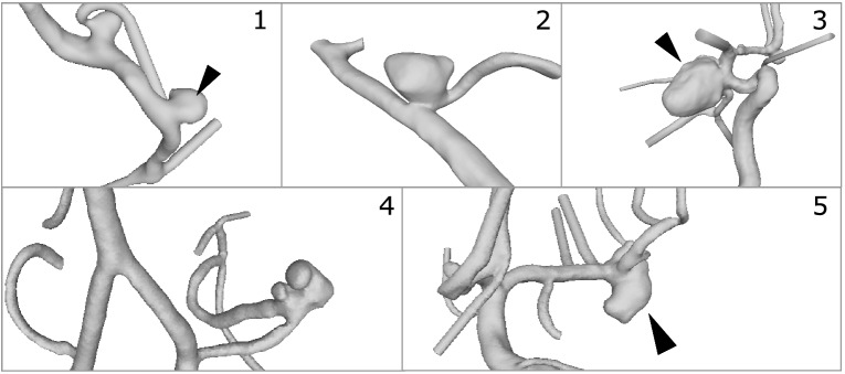
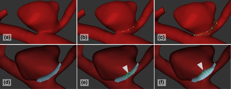
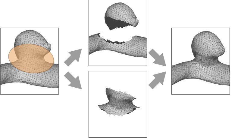
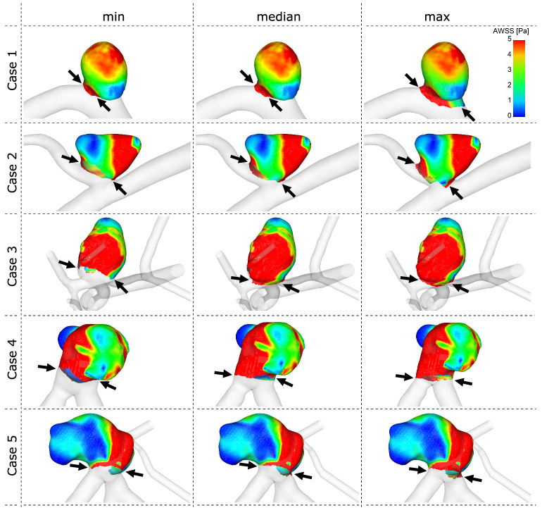
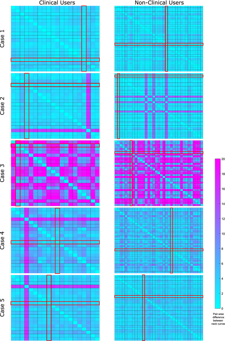
Methods: The research project VICTORIA was initiated, allowing users to interactively define the neck curve of five segmented IA models using a web application. The submitted results were qualitatively and quantitatively compared to identify the minimum, median and maximum aneurysm surface area. Finally, image-based blood flow simulations were carried out to assess the effect of variable neck curve definitions on relevant flow- and shear-related parameters.

Results: In total, 55 participants (20 physicians) from 18 countries participated in VICTORIA. For relatively simple aneurysms, a good agreement with respect to the neck curve definition was found. However, differences among the participants increased with increasing complexity of the aneurysm. Furthermore, it was observed that the majority of participants excluded any small arteries occurring in the vicinity of an aneurysm. This can lead to non-negligible deviations among the flow- and shear-related parameters, which need to be carefully evaluated, if quantitative analysis is desired. Finally, no differences between participants with medical and non-medical background could be observed.

Conclusions: VICTORIAs findings reveal the complexity of aneurysm neck curve definition, especially for bifurcation aneurysms. Standardization appears to be mandatory for future sac-vessel-separations. For hemodynamic simulations a careful neck curve definition is crucial to avoid inaccuracies during the quantitative flow analysis.

Keywords: Hemodynamics; Intracranial aneurysm; Neck curve; Rupture risk assessment; VICTORIA.


Figures

Figure 1
Figure 1
Depiction of the five patient-specific IAs used in the VICTORIA study. The aneurysms were located at the middle cerebral artery (Case 1 and 5), posterior inferior cerebellar artery (Case 2 and 4) and posterior communicating artery (Case 3), respectively. In the presence of multiple IAs, the selected one is highlighted (see arrow heads).
Figure 2
Figure 2
Illustration of the different steps involved in the neck curve and ostium definition: (a) 3D visualization of the surface model is shown; (b) The user can interactively select points on the aneurysm surface; (c) If the points are close to each other, the neck curve is automatically closed; (d) In the second step, an automatic ostium triangulation is provided; (e) The user can hover over the points and the active point is highlighted in blue (see arrowhead); (f) The point can be moved (including a reduced movement of its neighbors) until the user is satisfied with the ostium shape.
Figure 3
Figure 3
Illustration of the subdivision of triangles within the neck region: for each model, we cut the neck region and subdivided it such that the influence of the triangle size of the neck curve course is drastically reduced. Afterwards, the subdivided neck and the rest of the model were merged into a single model.
Figure 4
Figure 4
Illustration of the median neck curves for all five aneurysms of all users (green), the clinical users (cyan) and the non-clinical users (violet) showing a high agreement. The small inlays with arrows provide context information, which part of the aneurysm is shown. Furthermore, for each view, the most similar neck curve to the median neck curve is color-coded in white, the least similar neck curve is color-coded in gray and linear interpolation of colors-value is carried out in between based on all users.
Figure 5
Figure 5
Qualitative comparison of the time-averaged wall shear stress (AWSS) prediction depending on the neck curve definition. Notice the differences occurring at the transition between each aneurysm and the corresponding parent vessel (marked by black arrows).
Figure 6
Figure 6
Heatmap-based illustration of the pairwise comparison of neck curves. Each matrix (i.e. the heatmap) color-codes the difference between the users. On the left column, only clinical users are listed, on the right, the non-clinical users are presented for each aneurysm case. The color-coding is kept constant for all cases to allow for a visual comparison. Each cell ci,j of a heatmap depicts the distance between the neck curve from user i and the neck curve from user j. The neck curves with smallest distances to all other neck curves are selected as median neck curves (highlighted in red). The overview shows strong agreement for aneurysm Case 1 and very poor agreement for Case 3, the most complex case. Also, there is one outlier for Case 2, Case 4 and Case 5 among the clinical users and three outliers for Case 2 among the non-clinical users. A detailed listing of all users for all cases including quantitative results is provided in the supplemental material.

References

    1. Antiga L, Piccinelli M, Botti L, Ene-Iordache B, Remuzzi A, Steinman DA. An image-based modeling framework for patient-specific computational hemodynamics. Med. Biol. Eng. Comput. 2008;46(11):1097–1112. doi: 10.1007/s11517-008-0420-1. - DOI - PubMed
    1. Behrendt, B., S. Voß, B. Preim, P. Berg, and S. Saalfeld. VICTORIA—an interactive online tool for the VIrtual neck Curve and True Ostium Reconstruction of Intracranial Aneurysms. In: Proceedings of the Workshop on Image Processing (BVM), pp. 1–6, 2020
    1. Berg P, Saalfeld S, Voß S, Beuing O, Janiga G. A review on the reliability of hemodynamic modeling in intracranial aneurysms: why computational fluid dynamics alone cannot solve the equation. Neurosurg. Focus. 2019;47(1):E15. doi: 10.3171/2019.4.FOCUS19181. - DOI - PubMed
    1. Berg P, Saalfeld S, Voß S, Redel T, Preim B, Janiga G, Beuing O. Does the DSA reconstruction kernel affect hemodynamic predictions in intracranial aneurysms? an analysis of geometry and blood flow variations. J. Neurointerv. Surg. 2018;10(3):290–296. doi: 10.1136/neurintsurg-2017-012996. - DOI - PubMed
    1. Berg P, Stucht D, Janiga G, Beuing O, Speck O, Thévenin D. Cerebral blood flow in a healthy circle of Willis and two intracranial aneurysms: computational fluid dynamics versus four-dimensional phase-contrast magnetic resonance imaging. J. Biomech. Eng. 2014 doi: 10.1115/1.4026108. - DOI - PubMed

Publication types