. 2021 Oct;12(5):530-538.
doi: 10.1007/s13239-021-00549-4. Epub 2021 Jun 7.

Transcatheter Mitral Valve Repair Simulator Equipped with Eye Tracking Based Performance Assessment Capabilities: A Pilot Study

Affiliations

Transcatheter Mitral Valve Repair Simulator Equipped with Eye Tracking Based Performance Assessment Capabilities: A Pilot Study

Jan Michael Zimmermann et al. Cardiovasc Eng Technol. 2021 Oct.

Abstract

Background: The increase in cardiovascular disease cases that require minimally invasive treatment is inducing a new need to train physicians to perform them safely and effectively. Nevertheless, adaptation to simulation-based training has been slow, especially for complex procedures.

Objectives: We describe a newly developed mitral valve repair (MVR) simulator, equipped with new objective performance assessment methods, with an emphasis on its use for training the MitraClip™ procedure.

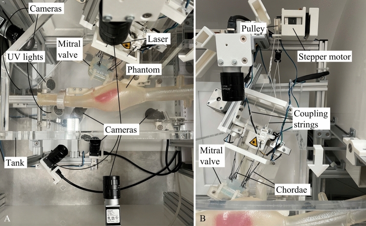
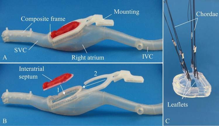
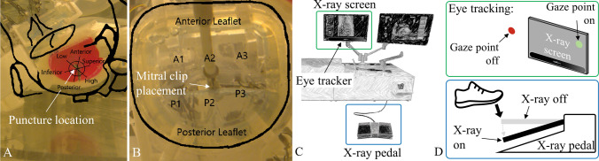
Methods: The MVR contains phantoms of all anatomical structures encountered during mitral valve repair with a transvenous, transseptal approach. In addition, several cameras, line lasers, and ultraviolet lights are used to mimic echocardiographic and fluoroscopic imaging and with a remote eye tracker the cognitive behaviour of the operator is recorded. A pilot study with a total of 9 interventional cardiologists, cardiac surgeons and technical experts was conducted. All participants performed the MitraClip procedure on the MVR simulator using standard interventional tools. Subsequently, each participant completed a structured questionnaire to assess the simulator.

Results: The simulator functioned well, and the implemented objective performance assessment methods worked reliably. Key performance metrics such as x-ray usage were comparable with results from studies assessing these metrics in real interventions. Fluoroscopy imaging is realistic for the transseptal puncture but reaches its limits during the final steps of the procedure.

Conclusion: The functionality and objective performance assessment of the MVR simulator were demonstrated. Especially for complex procedures such as the MitraClip procedure, this simulator offers a suitable platform for risk-free training and education.

Keywords: Education; MitraClip; Mitral valve; Objective assessment; Simulation training; Transseptal puncture.


Figures

Figure 1
Figure 1
Mitral valve repair simulator. The housing containing the anatomical phantoms, cameras, line lasers and UV lights for the medical imaging simulation (details see Fig. 2). Hip module containing a femoral access catheterization pad. The screen displaying the simulated fluoroscopic images. The detailed view shows an eye tracker attached at the bottom of the screen. The screen displaying the graphical user interface and the simulated transesophageal echocardiographic images during training. Controls to switch, zoom and orient fluoroscopy view. The left foot pedal activates fluoroscopy and the right foot pedal activates a direct camera image of the anatomy.
Figure 2
Figure 2
Inside views of the simulator. (a) Top view of the inside of the Mitral valve repair simulator. The simulator contains anatomical silicone phantoms of the inferior vena cava, right atrium with an exchangeable interatrial septum, superior vena cava and the mitral valve. A line laser, ultraviolet (UV) lights and 8 cameras are used to generate the necessary medical images (fluoroscopy and ultrasound) in various views for the entire intervention. (b) Front view of the inside of the Mitral valve repair simulator. Depicted are the actuation system of the mitral valve. The leaflets are connected via the chordae to a pulley by coupling springs. The pulley is actuated using a stepper motor.
Figure 3
Figure 3
Overview of the multi-material phantoms used in the mitral valve repair simulator. (a) Silicone phantom of the inferior vena cava, right atrium, exchangeable interatrial septum, superior vena cava (SVC) and integrated rigid mountings and frames, (b) The interatrial septum can be exchanged in a two-step process. First, the mounting (2) is removed and second, the interatrial septum can be exchanged (1). (c) The mitral valve consists of a silicone annulus and leaflets which are actuated through the chordae. The chordae are realized using strings that are on one end attached to the leaflets and on the other to a stepper motor for an artificial actuation.
Figure 4
Figure 4
Simulated transesophageal echocardiography (TEE) workflow. (a) Illustration of the overall concept (b) Line laser creates “ultrasound like” cut through the mitral valve. Ultraviolet (UV) lights highlight the MitraClip delivery system which is coated with a fluorescent dye. (c) Recorded images of the mitral valve leaflets and the MitraClip after image colour threshold processing. (d) Overlay of simulated tools and anatomical parts shown in (c) onto a real patient TEE template to create the final simulated TEE image.
Figure 5
Figure 5
Overview of feedback capabilities of the mitral valve repair simulator. (a) Feedback image of the transseptal puncture location. The rough dimension of the left atrium, interatrial septum and mitral valve are sketched and overlaid in black. Additionally, a position grid is overlaid. (b) Feedback image of the MitraClip placement in the mitral valve. (c) Illustration of the remote eye tracker implemented on the simulator and used to compute the percentage of “correctly” used fluoroscopy. (d) Correctly used fluoroscopy means that the x-ray pedal is pressed (“X-ray on”) and at the same time the users gaze point is on the x-ray screen (“gaze point on”). If the x-ray pedal is pressed (“X-ray on”) and the gaze point is not on the x-ray screen (“gaze point off”), it is classified as unnecessary x-ray usage.
Figure 6
Figure 6
Comparison between the MVR simulator simulated and real echocardiography (echo) and fluoroscopy (fluoro) images. (a–b) Simulated and real echo image of the transseptal puncture. (c–d) Simulated and real echo image of the MitraClip right above the mitral valve. (e–f) Simulated and real 3D echo view looking down onto the mitral valve. (g–h) Simulated and real fluoro image of a guidewire inside the right atrium. (k–l) Simulated and real fluoro image of the TSP sheath right before puncture. (m–n) Simulated and real image of the MitraClip inside the left atrium and ventricle. The simulation fails to emulate the clip inside the left atrium.

References

    1. Agha RA, Fowler AJ. The role and validity of surgical simulation. Int. Surg. 2015;100:350–357. doi: 10.9738/INTSURG-D-14-00004.1. - DOI - PMC - PubMed
    1. Alsalamah A, Campo R, Tanos V, Grimbizis G, Van Belle Y, Hood K, Pugh N, Amso N. Face and content validity of the virtual reality simulator ‘ScanTrainer®’. Gynecol. Surg. 2017;14:18. doi: 10.1186/s10397-017-1020-6. - DOI - PMC - PubMed
    1. Boulet JR, Durning SJ. What we measure … and what we should measure in medical education. Med. Educ. 2019;53:86–94. doi: 10.1111/medu.13652. - DOI - PubMed
    1. Chhatriwalla AK, Vemulapalli S, Holmes DR, Dai D, Li Z, Ailawadi G, Glower D, Kar S, Mack MJ, Rymer J, Kosinski AS, Sorajja P. Institutional experience with transcatheter mitral valve repair and clinical outcomes. JACC Cardiovasc. Interv. 2019;12:1342–1352. doi: 10.1016/j.jcin.2019.02.039. - DOI - PubMed
    1. Gong, R.H., B. Jenkins, R.W. Sze, Z. Yaniv. A cost effective and high fidelity fluoroscopy simulator using the Image-Guided Surgery Toolkit (IGSTK), 2014, vol. 903618, p. 903618.

Publication types

MeSH terms