. 2021 Jun 15;10(6):bio056457.
doi: 10.1242/bio.056457. Epub 2021 Jun 8.

Postnatal expression profiles of atypical cadherin FAT1 suggest its role in autism

Affiliations

Postnatal expression profiles of atypical cadherin FAT1 suggest its role in autism

Jeannine A Frei et al. Biol Open. .

Abstract

Genetic studies have linked FAT1 (FAT atypical cadherin 1) with autism spectrum disorder (ASD); however, the role that FAT1 plays in ASD remains unknown. In mice, the function of Fat1 has been primarily implicated in embryonic nervous system development with less known about its role in postnatal development. We show for the first time that FAT1 protein is expressed in mouse postnatal brains and is enriched in the cerebellum, where it localizes to granule neurons and Golgi cells in the granule layer, as well as inhibitory neurons in the molecular layer. Furthermore, subcellular characterization revealed FAT1 localization in neurites and soma of granule neurons, as well as being present in the synaptic plasma membrane and postsynaptic densities. Interestingly, FAT1 expression was decreased in induced pluripotent stem cell (iPSC)-derived neural precursor cells (NPCs) from individuals with ASD. These findings suggest a novel role for FAT1 in postnatal development and may be particularly important for cerebellum function. As the cerebellum is one of the vulnerable brain regions in ASD, our study warrants further investigation of FAT1 in the disease etiology.

Keywords: Autism; Cadherin; Cerebellum; FAT1; Granule cells; Neural precursor cells.


Conflict of interest statement

Competing interests The authors declare no competing or financial interests.

Figures
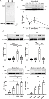
Fig. 1.
Fig. 1.
The postnatal cerebellum exhibits high levels of FAT1 protein. (A) The specificity of FAT1 antibody was tested by Western blot. Lysates from vascular smooth muscle cells isolated from aortas of wild-type (WT) and FAT1 knockout (KO) mice were probed with anti-FAT1 antibody. The antibody recognizes one band around 500 kDa, the predicted size of FAT1, in the WT but not KO lysate. (B) Temporal expression profile of FAT1 in mouse whole brain collected at E14, P1, P7, P14, P21 and 5-month-old adults. N=6 whole brains per age from three independent litters. (C) Spatial expression profile of FAT1 in cortex, hippocampus, cerebellum and thalamus/striatum at P7 and P14 of mouse brain development. P7: ***p=0.0002, ****p<0.0001; P14: ***p=0.0002 CX versus CB, p=0.00023 HC versus CB, p=0.0005 CB versus Th/St; one-way ANOVA with Tukey's multiple comparison test. N=4 samples per brain area with 2–3 pooled brain areas per sample. (D) Temporal expression of FAT1 in the cerebellum at P1, P7, P14 and P21. *p=0.0116, **p=0.0014, ***p=0.0002; one-way ANOVA with Tukey's multiple comparison test. N=4 samples per time point with 2–3 pooled cerebellums per sample. (E) Temporal expression profile of FAT1 in cultured cerebellar granule neurons harvested at 1, 3, 7, and 14 DIV. **p=0.0094; one-way ANOVA with Tukey's multiple comparison test. N=3 independent cultures.
Fig. 2.
Fig. 2.
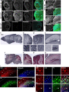
FAT1 is expressed in granule cells and interneurons in the cerebellum and hippocampus. (A) The specificity of FAT1 antibody was tested on sagittal sections of embryonic eyes from E13.5 FAT1 KO and WT mice. Scale bars: 100 μm. (B) Magnification of boxed areas depicted in A. FAT1 (green) colocalizes with phalloidin-stained actin (red) in WT but not KO eyes. Sections were counter-stained with DAPI (blue). Scale bars: 50 μm. (C–H) DAB immunostaining of FAT1 and (F–H) FAT1-DAB and neutral red co-staining on P14 mouse brain sagittal sections. Magnifications of cerebellum are depicted in (D), (D′), (G) and (G′) and magnifications of hippocampus are depicted in (E) and (H). FAT1 is expressed in the cerebellum (D,D′) in the surrounding but not in the soma of Purkinje cells (box 1, arrowheads), in the granule cell layer (box 2) in granule cells (asterisk) and Golgi cells (arrows), and in interneurons in the molecular layer (box 3, open arrowheads). FAT1 is also expressed in the hippocampus (E) in interneurons (box 1, open arrowheads) and in granule cells of the dentate gyrus (box 2, asterisk). Note that FAT1 is enriched in cell dense structures of cerebellar granule layer (G,G′) and hippocampal dentate gyrus (H). Scale bars: 1 mm (C,D,F,G), 0.5 mm (E,H), 100 μm (D′,G′), 50 μm (box 1–3 in D′, box 1,2 in E). (I) Sagittal sections of P14 cerebellum co-immunostained for FAT1 (red), calbindin (blue), and parvalbumin (PV; green). Dashed lines indicate layers: GCL, granule cell layer; PCL, Purkinje cell layer; ML, molecular layer. Scale bar: 100 μm. (J) Magnification of GCL, PCL, and ML depicted in merged image in I. Scale bar: 50 μm. FAT1 is expressed in granule cells (asterisk) and Golgi cells (arrows) in the GCL, around calbindin- and PV-positive Purkinje cells (open arrowheads) in the PCL, and PV-positive interneurons (arrowheads) in the ML.
Fig. 3.
Fig. 3.
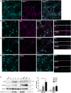
FAT1 localizes to neurites and soma of cerebellar granule cells. Confocal images of cerebellar granule cell cultures fixed at 14 DIV and immunostained with FAT1 (cyan) together with different neuronal and synaptic markers (magenta), including (A) MAP2, (B) Tau, (C) vGlut1, and (D) PSD-95. FAT1 is expressed in dendrites, axons, and soma and localizes to vGlut1 and PSD-95 puncta. Scale bars: 10 μm, 2 μm (magnifications in B–D). (E) Synaptic fractionation of P21 cerebellar tissue was performed to determine the subcellular localization of FAT1. Western blots were probed, stripped, and reprobed with markers PSD-95, syntaxin-1 and β-actin to confirm successful separation and purity of different fractions. Total, total protein input; P1, nuclear; S1, cytosol/membranes; P2, crude synaptosome; S2, cytosol/light membranes; P3, synaptosome; SPM, synaptic plasma membrane; PSD, postsynaptic density; S3, synaptic vesicles. *p=0.0422; one-way ANOVA with Tukey's multiple comparison test. N=6 mice, two pooled cerebellums per sample.
Fig. 4.
Fig. 4.
FAT1 levels are reduced in iPSC-derived cortical neural progenitors from autistic individuals. (A) Western blot of FAT1 expression in 19 DIV iPSC-derived cortical NPCs from typically-developing control and autistic individuals. (B) Quantification of FAT1 expression represented as a bar graph and as box and whisker plot. ****p<0.0001; unpaired two-tailed t-test. N=4 control and eight autistic individuals, duplicates of each line.

Similar articles

Cited by

References

    1. Abou Jamra, R., Becker, T., Georgi, A., Feulner, T., Schumacher, J., Stromaier, J., Schirmbeck, F., Schulze, T. G., Propping, P., Rietschel, M.et al. (2008). Genetic variation of the FAT gene at 4q35 is associated with bipolar affective disorder. Mol. Psychiatry 13, 277-284. 10.1038/sj.mp.4002111 - DOI - PubMed
    1. American Psychiatric Association (2013). Diagnostic and Statistical Manual of Mental Disorders, 5th edn. Arlington, VA: American Psychiatric Publishing.
    1. Avilés, E. C. and Goodrich, L. V. (2017). Configuring a robust nervous system with Fat cadherins. Semin. Cell Dev. Biol. 69, 91-101. 10.1016/j.semcdb.2017.06.001 - DOI - PMC - PubMed
    1. Badouel, C., Zander, M. A., Liscio, N., Bagherie-Lachidan, M., Sopko, R., Coyaud, E., Raught, B., Miller, F. D. and McNeill, H. (2015). Fat1 interacts with Fat4 to regulate neural tube closure, neural progenitor proliferation and apical constriction during mouse brain development. Development 142, 2781-2791. 10.1242/dev.123539 - DOI - PMC - PubMed
    1. Baron, O., Grieshober, D., Dias, C. and Fanto, M. (2019). Fat cadherins in mouse models of degenerative ataxias. Sci. Rep. 9, 16155. 10.1038/s41598-019-52684-7 - DOI - PMC - PubMed

Publication types