. 2021 Jun 29;12(3):e0117921.
doi: 10.1128/mBio.01179-21. Epub 2021 Jun 8.

A Chlamydial Plasmid-Dependent Secretion System for the Delivery of Virulence Factors to the Host Cytosol

Affiliations

A Chlamydial Plasmid-Dependent Secretion System for the Delivery of Virulence Factors to the Host Cytosol

Lei Lei et al. mBio. .

Abstract

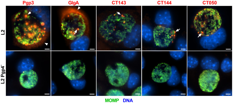
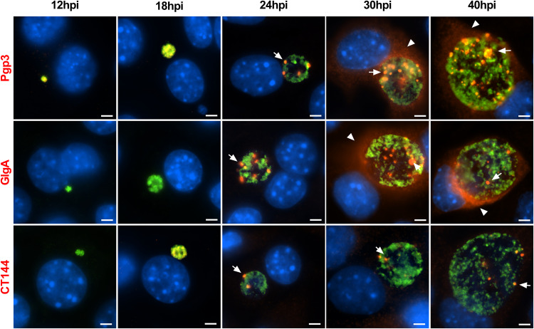
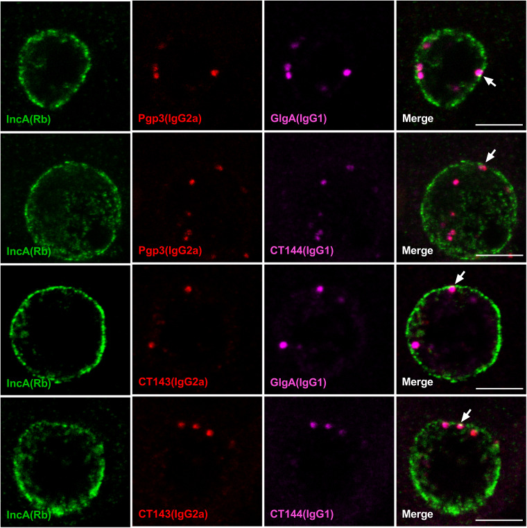
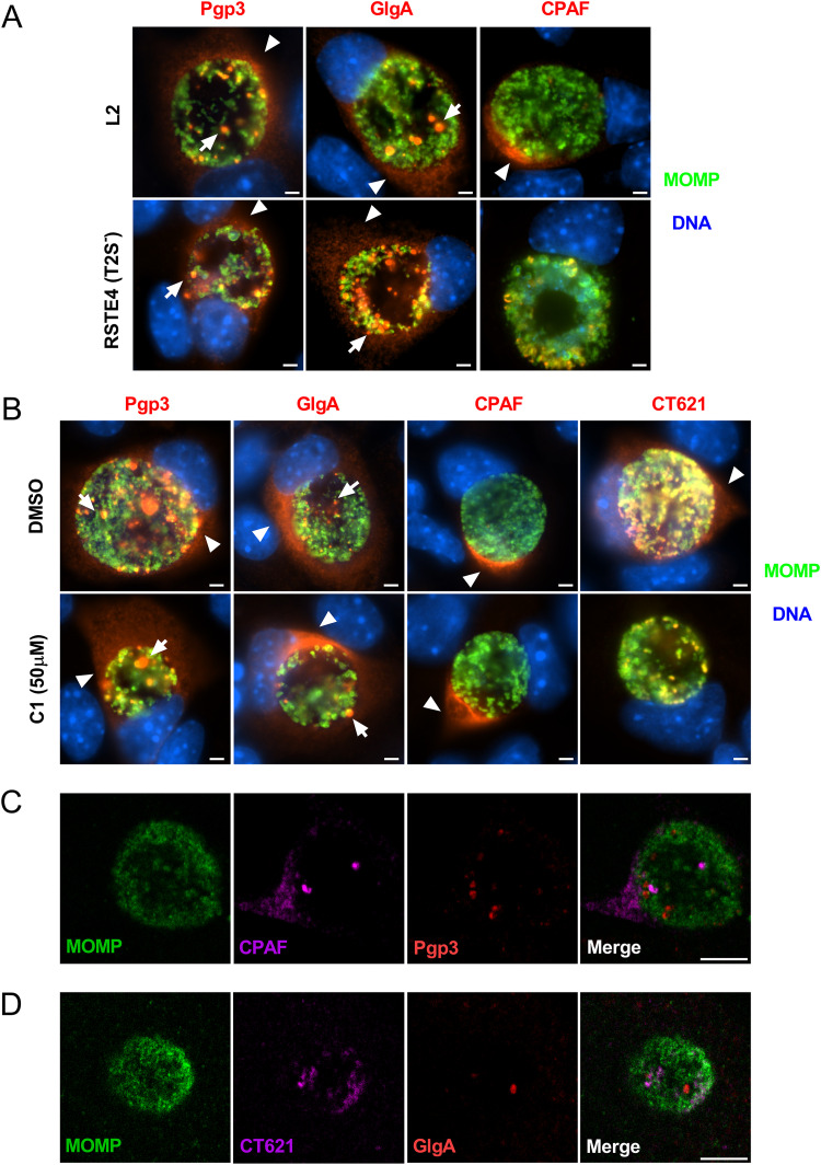
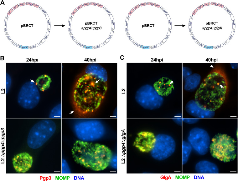
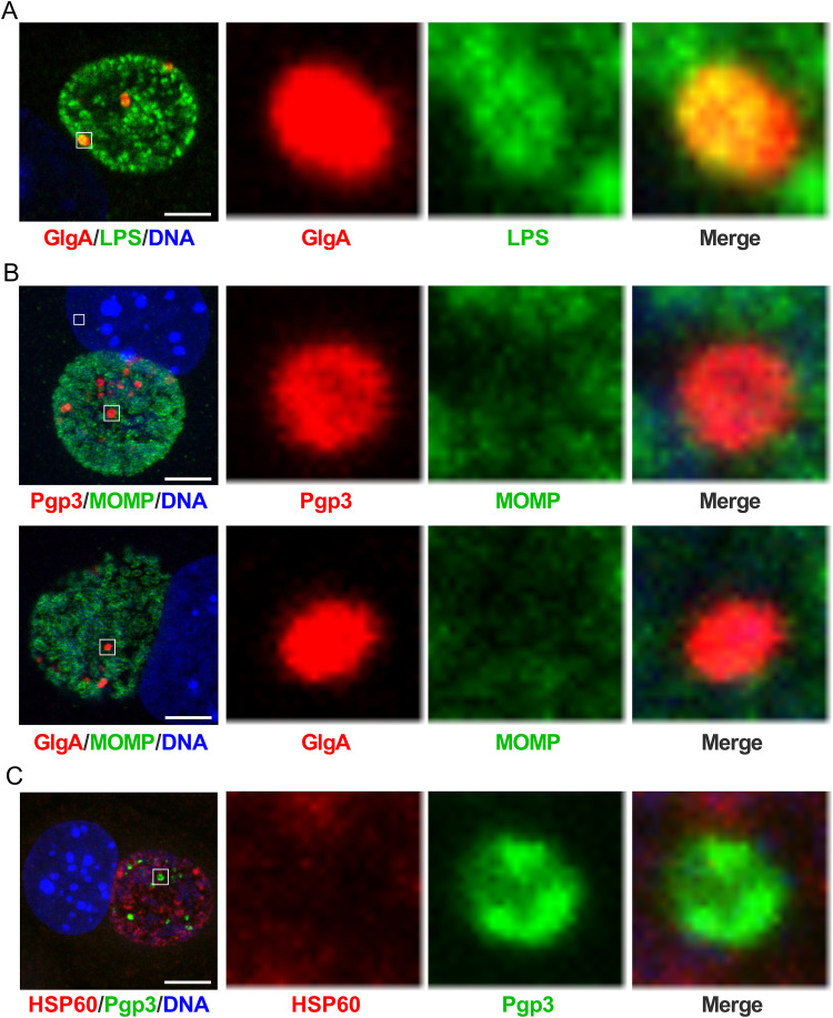
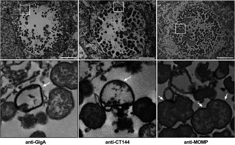
Chlamydia are obligate intracellular Gram-negative bacteria distinguished by a unique developmental biology confined within a parasitophorous vacuole termed an inclusion. The chlamydial plasmid is a central virulence factor in the pathogenesis of infection. Plasmid gene protein 4 (Pgp4) regulates the expression of plasmid gene protein 3 (Pgp3) and chromosomal glycogen synthase (GlgA), virulence factors secreted from the inclusion to the host cytosol by an unknown mechanism. Here, we identified a plasmid-dependent secretion system for the cytosolic delivery of Pgp3 and GlgA. The secretion system consisted of a segregated population of globular structures originating from midcycle reticulate bodies. Globular structures contained the Pgp4-regulated proteins CT143, CT144, and CT050 in addition to Pgp3 and GlgA. Genetic replacement of Pgp4 with Pgp3 or GlgA negated the formation of globular structures, resulting in retention of Pgp3 and GlgA in chlamydial organisms. The generation of globular structures and secretion of virulence factors occurred independently of type 2 and type 3 secretion systems. Globular structures were enriched with lipopolysaccharide but lacked detectable major outer membrane protein and heat shock protein 60, implicating them as outer membrane vesicles. Thus, we have discovered a novel chlamydial plasmid-dependent secretion system that transports virulence factor cargo from the chlamydial inclusion to the host cytosol. IMPORTANCE The Chlamydia trachomatis plasmid regulates the expression and secretion of immune evasion virulence factors to the host cytosol by an unknown mechanism. In this study, we identified a novel plasmid gene protein 4 (Pgp4)-dependent secretion system. The system consists of globular structures distinct from typical chlamydial developmental forms that export Pgp3 and GlgA to the host cytosol. Globular structures emerged at mid-chlamydial growth cycle from distinct populations of reticulate bodies. The formation of globular structures occurred independently of known chlamydial secretion systems. These results identify a Pgp4-dependent secretory system required for exporting plasmid regulated virulence factors to the host cytosol.

Keywords: Chlamydia; plasmid; secretion; virulence factors.


Figures

FIG 1
FIG 1
Plasmid-regulated proteins exhibit a globular staining pattern localized to the inclusion lumen of infected cells. McCoy cells infected with C. trachomatis wild-type L2 strain or an L2 pgp4-null strain were fixed with PFA at 40 hpi and stained with antibodies against Pgp3, GlgA, CT143, CT144, or CT050, together with the chlamydial major outer membrane protein (MOMP). All 5 proteins displayed a globular staining pattern found in the lumen of the chlamydial inclusion (arrows). Pgp3 and GlgA were detected in the host cell cytosol (arrowheads). Bar, 10 μm.
FIG 2
FIG 2
Temporal kinetics of Pgp3, GlgA, and CT144 expression in infected McCoy cells. McCoy cells infected with the C. trachomatis wild-type L2 strain were harvested at different times postinfection, as indicated, and processed for IFA assay. Pgp3 was detected as early as 12 hpi. The globular staining pattern (arrowheads) of Pgp3, GlgA, and CT144 was evident at 24 hpi. At 30 hpi, Pgp3 and GlgA staining was markedly stronger and more abundant than CT144 staining, which did not significantly change from the 24-h time point. The increase expression of Pgp3 and GlgA at 30 and 40 hpi coincided with protein secretion into the host cell cytosol (arrows). Bar, 10 μm.
FIG 3
FIG 3
Pgp3, GlgA, CT143, and CT144 colocalize to the same globular structure. WT L2-infected McCoy cells were fixed with PFA at 24 hpi, stained with different combination of anti-Pgp3, GlgA, CT143, CT144, and IncA, and analyzed by confocal microscopy. Anti-IncA was used to identify the boundary of the chlamydial inclusion membrane. Bar, 5 μm. Note that each of the plasmid-regulated proteins is contained within the same globular structure. Bar, 5 μm.
FIG 4
FIG 4
The formation of globular structures and GlgA and Pgp3 secretion to the host cytosol are independent of T2SS and T3SS. (A) WT-L2- or T2SS-deficient-L2 (RSTE4)-infected McCoy cells were fixed with PFA at 40 hpi and immunolabeled with anti-Pgp3, anti-GlgA, or anti-CPAF, together with anti-MOMP. The globular (arrows) and host cell cytosol (arrowheads) staining of Pgp3 and GlgA was detected in both L2- and RSTE4-infected cells. In contrast, the secretion of CPAF into the host cell cytosol was detected only in cells infected with WT L2, not in cells infected with RSTE4. Bar, 10 μm. (B) McCoy cells were infected with WT L2, and at 18 hpi, the T3SS inhibitor C1 was added to the culture medium. Cells were fixed at 40 hpi and immunolabeled with anti-Pgp3, anti-GlgA, anti-CPAF, or anti-CT621, together with anti-MOMP. Bar, 10 μm. (C) McCoy cells infected with WT L2 were fixed at 24 hpi and immunolabeled with anti-Pgp3 MAb and anti-CPAF MAb together with anti-MOMP. Staining of Pgp3 did not overlap staining of CPAF. Bar, 5 μm. (D) McCoy cells infected with WT L2 were fixed at 24 hpi and immunolabeled with anti-CT621 serum and anti-MOMP, with relevant secondary antibodies. Then cells were immunolabeled with Alexa Fluor 568-conjugated anti-GlgA MAb. Staining of CT621 did not overlap staining of GlgA. Bar, 5 μm.
FIG 5
FIG 5
The formation of globular structures and GlgA and Pgp3 secretion to the host cytosol are dependent on Pgp4. (A) The Pgp4 ORF was replaced by the Pgp3 or GlgA ORF. (B) The Pgp4 promoter drove the expression of Pgp3. In the absence of Pgp4, Pgp3 was highly expressed but there was no cytosolic secretion observed. (C) The expression of GlgA was driven by the Pgp4 promoter. In the absence of Pgp4, GlgA was highly expressed but expression was also restricted to chlamydial organisms. Bar, 10 μm.
FIG 6
FIG 6
The globular structures contain chlamydial LPS but do not costain with chlamydial MOMP and HSP60. (A) WT L2-infected McCoy cells were fixed at 24 hpi, stained with anti-GlgA together with anti-LPS, and analyzed by confocal microscopy. GlgA colocalized with the LPS. Bar, 5 μm. (B) WT L2-infected McCoy cells were fixed at 24 hpi and costained with anti-Pgp3 and anti-MOMP or with anti-GlgA and anti-MOMP. Pgp3 and GlgA did not colocalize with MOMP. (C) WT L2-infected McCoy cells were fixed at 24 hpi and costained with anti-Pgp3 and anti-HSP60. Pgp3 did not colocalize with HSP60. Bar, 5 μm. In each row, the area highlighted by the white box in the leftmost panel is magnified in the second, third, and fourth panels.
FIG 7
FIG 7
Transmission immunoelectron microscopy shows globular structures in the inclusion lumen. WT L2-infected McCoy cells were fixed at 24 hpi, immunolabeled with anti-GlgA, anti-CT144, or anti-MOMP, and prepared for TEM. Anti-GlgA and -CT144 antibodies specifically recognize globular structures in the inclusion lumen (arrows). Anti-MOMP antibodies labeled chlamydial EB and RB developmental forms (arrows). The globular structures did not react with anti-MOMP antibodies. Bars, 5 μm. The bottom row shows magnifications of the areas highlighted by the white boxes in the top row.

References

    1. Burton MJ, Mabey DC. 2009. The global burden of trachoma: a review. PLoS Negl Trop Dis 3:e460. doi:10.1371/journal.pntd.0000460. - DOI - PMC - PubMed
    1. World Health Organization. 2016. WHO Guidelines for the Treatment of Chlamydia trachomatis. World Health Organization, Geneva, Switzerland. - PubMed
    1. Moulder JW. 1991. Interaction of chlamydiae and host cells in vitro. Microbiol Rev 55:143–190. doi:10.1128/MR.55.1.143-190.1991. - DOI - PMC - PubMed
    1. Ouellette SP, Lee J, Cox JV. 2020. Division without binary fission: cell division in the FtsZ-less Chlamydia. J Bacteriol 202:e00252-20. doi:10.1128/JB.00252-20. - DOI - PMC - PubMed
    1. Elwell C, Mirrashidi K, Engel J. 2016. Chlamydia cell biology and pathogenesis. Nat Rev Microbiol 14:385–400. doi:10.1038/nrmicro.2016.30. - DOI - PMC - PubMed

Publication types