. 2021 Jul 1;108(7):1330-1341.
doi: 10.1016/j.ajhg.2021.05.007. Epub 2021 Jun 7.

De novo and bi-allelic variants in AP1G1 cause neurodevelopmental disorder with developmental delay, intellectual disability, and epilepsy

Affiliations

De novo and bi-allelic variants in AP1G1 cause neurodevelopmental disorder with developmental delay, intellectual disability, and epilepsy

Muhammad A Usmani et al. Am J Hum Genet. .

Abstract

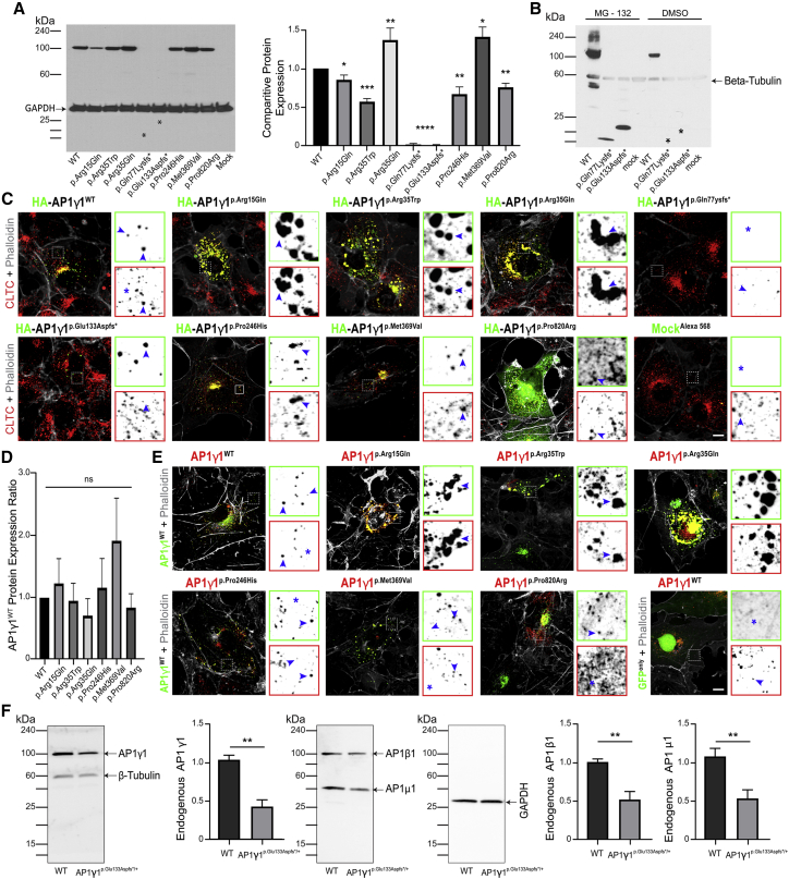
Adaptor protein (AP) complexes mediate selective intracellular vesicular trafficking and polarized localization of somatodendritic proteins in neurons. Disease-causing alleles of various subunits of AP complexes have been implicated in several heritable human disorders, including intellectual disabilities (IDs). Here, we report two bi-allelic (c.737C>A [p.Pro246His] and c.1105A>G [p.Met369Val]) and eight de novo heterozygous variants (c.44G>A [p.Arg15Gln], c.103C>T [p.Arg35Trp], c.104G>A [p.Arg35Gln], c.229delC [p.Gln77Lys11], c.399_400del [p.Glu133Aspfs37], c.747G>T [p.Gln249His], c.928-2A>C [p.?], and c.2459C>G [p.Pro820Arg]) in AP1G1, encoding gamma-1 subunit of adaptor-related protein complex 1 (AP1γ1), associated with a neurodevelopmental disorder (NDD) characterized by mild to severe ID, epilepsy, and developmental delay in eleven families from different ethnicities. The AP1γ1-mediated adaptor complex is essential for the formation of clathrin-coated intracellular vesicles. In silico analysis and 3D protein modeling simulation predicted alteration of AP1γ1 protein folding for missense variants, which was consistent with the observed altered AP1γ1 levels in heterologous cells. Functional studies of the recessively inherited missense variants revealed no apparent impact on the interaction of AP1γ1 with other subunits of the AP-1 complex but rather showed to affect the endosome recycling pathway. Knocking out ap1g1 in zebrafish leads to severe morphological defect and lethality, which was significantly rescued by injection of wild-type AP1G1 mRNA and not by transcripts encoding the missense variants. Furthermore, microinjection of mRNAs with de novo missense variants in wild-type zebrafish resulted in severe developmental abnormalities and increased lethality. We conclude that de novo and bi-allelic variants in AP1G1 are associated with neurodevelopmental disorder in diverse populations.

Keywords: AP-1 complex; AP1G1; Pakistani families; developmental delay; epilepsy; exome sequencing; genetic heterogeneity; intellectual disabilities; neurodevelopment disorder.


Conflict of interest statement

The authors declare no competing interests.

Figures

Figure 1
Figure 1
Homozygous as well as de novo variants in AP1G1 cause neurodevelopment disorder (NDD) with intellectual disability and dysmorphic features (A) Pedigree of Italian CPBO family segregating intellectual disability associated with a c.737C>A (p.Pro246His) variant in AP1G1. The filled symbols represent affected individuals, and a double horizontal line connecting parents represents a consanguineous marriage. (B) Pedigree of Pakistani family PKMR328 segregating intellectual disabilities with dysmorphic features associated with a homozygous c.1105A>G (p.Met369Val) allele AP1G1 variant. (C) Pedigrees of nine outboard families segregating with de novo missense, frameshift, as well as splice variants. (D) Schematic representation of human AP1G1 gene and protein structures along with bi-allelic (above) and de novo (below) variants identified in families with NDD. Yellow hexagon represents Alpha adaptin C2 domain, while blue rectangles marked the exons encoding transmembrane domains. (E) Clustal W alignment revealed high evolutionary conservation of residues mutated in families with NDD.
Figure 2
Figure 2
Impact of NDD-associated variants on AP1γ1 steady state level and targeting in heterologous cells (A) Immunoblot revealed statistically significant differences in AP1γ1 steady state levels when overexpressed levels in HEK293T cells. AP1γ1 harboring missense variants p.Arg15Glu, p.Arg35Trp, p.Pro246His, and p.Pro820Arg had significantly decreased levels (p < 0.04, ∗∗p < 0.007, ∗∗∗∗p < 0.0001), while slightly increased protein levels were observed for AP1γ1 with p.Arg35Gln and p.Met369Val variants (p < 0.01 and ∗∗p < 0.007, respectively) when compared with HA-AP1γ1WT. In contrast, no detectable protein was observed for AP1γ1 harboring p.Gln77Lysfs11 and p.Glu133Aspfs37 frameshift variants. (B) Protease inhibitor (MG132) assay. Levels of AP1γ1-truncated proteins due to p.Gln77Lysfs11 and p.Glu133Aspfs37 were significantly increased in HEK293T cells treated with MG132 (20 mM) for 12 h as compared to vehicle (0.5% DMSO)-treated cells. (C) Confocal images of COS7 cells transfected with HA-AP1γ1 constructs and immunolabeled with CLTC (clathrin heavy chain marker) and phalloidin (actin marker). HA-tagged WT AP1γ1 (HA-AP1γ1WT) colocalizes with clathrin-coated pits predominantly at perinuclear area. Similarly, no apparent differences in the distribution of HA-AP1γ1p.Pro246His or HA-AP1γ1p.Met369Val proteins were noted. However, AP1γ1 proteins harboring de novo missense variants (HA-AP1γ1p.Arg15Glu, HA-AP1γ1p.Arg35Trp, and HA-AP1γ1p.Arg35Gln) form aggregates in the transfected cells, and HA-AP1γ1p.Pro820Arg had diffuse cytoplasmic localization. Finally, as anticipated from immunoblot analysis, no to very little protein was observed for AP1γ1 with frameshift variants (p.Gln77Lysfs11 and p.Glu133Aspfs37). The regions magnified with split channels next to each image are boxed. Scale bars: 10 µm and 2 µm (inset). (D) Immunoblot revealed no statistically significant differences in steady state levels of GFP-AP1γ1WT when co-transfected with HA-AP1γ1 harboring either p.Arg15Glu, p.Arg35Trp, p.Arg35Gln, p.Pro246His, p.Met369Val, or p.Pro820Arg missense variants in HEK293T cells. (E) Confocal images of COS7 cells co-transfected with GFP-AP1γ1WT and HA- AP1γ1 constructs harboring NDD-associated missense variants. Only the HA- AP1γ1 with de novo missense variants, HA-AP1γ1p.Arg15Glu, HA-AP1γ1p.Arg35Trp and HA-AP1γ1p.Arg35Gln, impaired the distribution of GFP-AP1γ1WT proteins and formed large protein aggregates in the cytoplasm. (F) Immunoblot revealed statistically significant reduced AP1γ1, AP1β1, and AP1μ1 steady state levels in the AP1γ1p.Glu133Aspfs∗37 variant-harboring affected individual-derived primary fibroblast cells as compared WT control cells (∗∗p < 0.007).
Figure 3
Figure 3
AP1γ1 NDD-associated variants impact the endosomal recycling pathway, evaluated via transferrin recycling (A) COS7 cells transiently transfected with GFP-AP1γ1WT, GFP-AP1γ1p.Pro246His, or GFP-AP1γ1p.Met369Val and immunostained with RAB11 to mark the recycling endosomes. Cells were pulsed with transferrin and chased for time points shown in (B). Single channels are shown as inverted grayscale images with their respective channel color boxes. Colocalization is indicated with arrow heads and asterisks are indicating the absence of the respective marker. Representative images at time point 0 min (initiation of transferrin chase), 45 min, and 60 min are shown. Images for other studies’ time points are given in Figure S4. White rectangles marked the area enlarged from transfected (T) or neighboring untransfected (U) cells. (B) Quantification of colocalization pattern with ImageJ software at different time points revealed significant difference for both variants compared to WT protein. In cells transfected with AP1γ1WT, most of the transferrin enters in recycling endosomes, as early as 15 min, and is recycled out after 30 min, while AP1γ1p.Met369Val-transfected cells were unable to recycle transferrin through recycling endosomes, as colocalization of AP1γ1 with transferrin and RAB11 was observed even at 45 min (∗∗∗∗p < 0.00001, Student’s t test) and 60 min post chase. In contrast, transferrin was recycled out before 30 min (∗∗∗∗p < 0.00001) through fast recycling route from cells transfected with AP1γ1p.Pro246His, and very few transferrin-positive recycling endosomes were observed at later time points. Scale bars, 10 μm and 2 μm (inset).
Figure 4
Figure 4
Ap1g1 is essential for zebrafish growth and survival (A) Representative images of ap1g1+/+, ap1g1+/−, and ap1g1−/− larvae at 5 days post fertilization (dpf). Severe morphological defects, including edema, were observed in ap1g1−/− as early as 4 dpf. (B) Kaplan-Meier curve showed only 16% of ap1g1−/− larvae survived until 10 dpf (∗∗∗∗p < 0.0003, Kaplan-Meier estimator, Student’s t test). No significant survival deficits were observed for ap1g1+/− larvae when compared with ap1g1+/+, indicating that the Ap1g1 encoded by the single copy of ap1g1 is sufficient for zebrafish survival. (C) Human WT (AP1γ1WT), but not the NDD-associated missense variants (p.Arg15Gln, p.Arg35Trp, p.Arg35Gln, p.Pro246His, and p.Met366Val) encoding mRNAs, rescued the edema phenotype and increased the survival of ap1g1−/− larvae (∗∗∗∗p < 0.0001, Kaplan-Meier estimator, Student’s t test). Blue asterisks marked statistically significant poor survival of the de novo variants injected larvae at 4 dpf when compared with uninjected ap1g1−/−. Please see Figure S7 for additional experimental data graphs, including the microinjection of human AP1G1 mRNA in all three genotypes (ap1g1+/+, ap1g1+/−, and ap1g1−/−). Scale bar, 400 μm. (D) In vivo microinjection of human AP1G1 WT or mutant mRNAs in AB/TU wild-type zebrafish. Representative images of resulting phenotypes in developing larvae at 2 dpf. Larvae were categorized into five classes on the basis of their developmental morphology, edema, and survival. (E) Only AP1γ1-harboring de novo missense variants, p.Arg15Glu, p.Arg35Trp, and Arg35Gln, showed dominant-negative impact and impaired the zebrafish development (∗∗∗p < 0.001 and ∗∗∗∗p < 0.0001, Student’s t test) when compared with AP1γ1WT-injected fish.

References

    1. Hirst J., Robinson M.S. Clathrin and adaptors. Biochim. Biophys. Acta. 1998;1404:173–193. - PubMed
    1. Robinson M.S. Forty Years of Clathrin-coated Vesicles. Traffic. 2015;16:1210–1238. - PubMed
    1. Ohno H. Clathrin-associated adaptor protein complexes. J. Cell Sci. 2006;119:3719–3721. - PubMed
    1. Popova N.V., Deyev I.E., Petrenko A.G. Clathrin-mediated endocytosis and adaptor proteins. Acta Naturae. 2013;5:62–73. - PMC - PubMed
    1. Traub L.M., Kornfeld S., Ungewickell E. Different Domains of AP1 Adaptor complex are required for Golgi membrane binding and clathrine recruitment. J. Biol. Chem. 1995;270:4933–4942. - PubMed

Publication types

Substances