. 2021 Jul 15;81(14):3018-3030.e5.
doi: 10.1016/j.molcel.2021.05.009. Epub 2021 Jun 7.

XRCC1 prevents toxic PARP1 trapping during DNA base excision repair

Affiliations

XRCC1 prevents toxic PARP1 trapping during DNA base excision repair

Annie A Demin et al. Mol Cell. .

Abstract

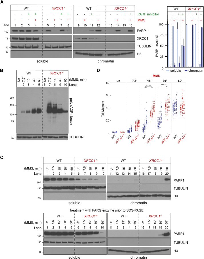
Mammalian DNA base excision repair (BER) is accelerated by poly(ADP-ribose) polymerases (PARPs) and the scaffold protein XRCC1. PARPs are sensors that detect single-strand break intermediates, but the critical role of XRCC1 during BER is unknown. Here, we show that protein complexes containing DNA polymerase β and DNA ligase III that are assembled by XRCC1 prevent excessive engagement and activity of PARP1 during BER. As a result, PARP1 becomes "trapped" on BER intermediates in XRCC1-deficient cells in a manner similar to that induced by PARP inhibitors, including in patient fibroblasts from XRCC1-mutated disease. This excessive PARP1 engagement and trapping renders BER intermediates inaccessible to enzymes such as DNA polymerase β and impedes their repair. Consequently, PARP1 deletion rescues BER and resistance to base damage in XRCC1-/- cells. These data reveal excessive PARP1 engagement during BER as a threat to genome integrity and identify XRCC1 as an "anti-trapper" that prevents toxic PARP1 activity.

Keywords: PARP inhibitors; PARP trapping; PARP1; XRCC1 protein complexes; base excision repair; single-strand breaks.


Conflict of interest statement

Declaration of interests The authors declare no competing interests.

Figures

None
Graphical abstract
Figure 1
Figure 1
XRCC1 suppresses PARP1-dependent SSB accumulation and toxicity during BER (A) Clonogenic survival of wild-type (WT) and gene-edited RPE-1 cells after treatment with the indicated concentrations of MMS for 30 min, followed by incubation in drug-free medium for 10–14 days. Data are the mean (±SEM) of 3 independent experiments. The level of the indicated proteins in the indicated gene-edited cell lines is shown (right). Statistical significance was assessed by two-way ANOVA with Tukey’s multiple comparisons test. All cell lines were significantly different from the WT (p ≤ 0.01), and other relevant comparisons are shown on the graph (ns, not significant; p ≤ 0.05, ∗∗p ≤ 0.01, ∗∗∗p ≤ 0.001, ∗∗∗∗p ≤ 0.0001). See also Figures S1A and S1B. (B), Survival of WT and the indicated gene-targeted TK6 cells after treatment with the indicated concentration of MMS for 1 h, followed by incubation in complete medium for 72 h. Cell viability was assessed by ATP assays. Data are the mean (±SEM) of 3 independent experiments, and statistics are as in (A). (C) Clonogenic survival of WT and gene-edited RPE-1 cells following continuous treatment with the indicated concentrations of camptothecin (CPT) for 10–14 days. Data are the mean (±SEM) of 3 independent experiments, and statistics are as in (A). (D) DNA strand breaks quantified by alkaline comet assays in the WT and the indicated gene-edited RPE-1 cell lines following treatment or no treatment with 0.1 mg/mL MMS for 15 min. Data plotted are the individual comet tail moments (an arbitrary measure of DNA strand breakage) of 100 cells per sample per experiment for 3 independent experiments, with individual cell tail moments for each experiment plotted vertically and each experiment plotted side by side. Statistical significance was ascertained by one-way ANOVA of the mean tail moments from 3 experiments with Sidak’s multiple comparisons test (∗∗p ≤ 0.01, ∗∗∗p ≤ 0.001, ∗∗∗∗p ≤ 0.0001). (E) DNA strand breaks quantified as above in the indicated RPE-1 cell lines transfected with non-targeting or PARP1 siRNA following treatment or no treatment with 0.05 mg/mL MMS for 15 min. A western blot illustrating the efficiency of PARP1 depletion is shown in (A). Data and statistics are as in (D). See also Figure S1A. (F) DNA strand breaks quantified as above in the indicated RPE-1 cell lines following treatment or no treatment with 0.1 mg/mL MMS for 15 min in the presence or absence of 20 μM Ara-A, as indicated. A western blot shows the efficiency of PARP1 depletion (inset). Data and statistics are as in (D). (G) DNA strand breaks quantified as above in the indicated RPE-1 cell lines following treatment or no treatment with 0.1 mg/mL MMS for 15 min in the presence of DMSO vehicle (−PARP inhibitor) or PARP inhibitor (10 μM) as indicated. Data and statistics are as in (D).
Figure 2
Figure 2
XRCC1 suppresses endogenous PARP1 trapping during BER (A) PARP1 levels in cell-equivalent aliquots of soluble and chromatin-containing fractions of WT and XRCC1−/− RPE-1 cells, measured by western blotting. Cells were incubated or not with 10 μM PARP inhibitor (KU0058948) and/or MMS (0.1 mg/mL) for 1 h, as indicated, prior to subcellular fractionation. Representative immunoblots are shown on the left and quantification on the right. See also Figures S1C and S1D. (B) Levels of PARP1 auto-ribosylation in WT and XRCC1−/− RPE-1 cells during treatment with 0.1 mg/mL MMS, detected by the poly(ADP-ribose)-specific detection reagent MABE1031. (C) Top: PARP1 levels in cell-equivalent aliquots of soluble and chromatin-containing fractions from WT and XRCC1−/− RPE-1 cells treated for the indicated times with 0.1 mg/mL MMS. Bottom: as above, but the cell extracts were treated with recombinant PARG to remove all poly(ADP-ribose) immediately prior to SDS-PAGE. (D) DNA strand breaks quantified by alkaline comet assays in WT and XRCC1−/− RPE-1 cells during treatment with 0.1 mg/mL MMS. Data plotted are the individual comet tail moments (an arbitrary measure of DNA strand breakage) of 50 cells per sample per experiment, with tail moments for each experiment plotted vertically and three independent experiments plotted side by side. Statistical significance was ascertained by one-way ANOVA of the mean tail moments from 3 experiments with Sidak’s multiple comparisons test (∗∗∗∗p ≤ 0.0001).
Figure 3
Figure 3
XRCC1 regulates PARP1 activity during BER (A) Levels of PARP1 auto-ribosylation detected by poly(ADP-ribose)-specific detection reagent in WT and XRCC1−/− RPE-1 cells following treatment or not (untreated [Un]) with 0.1 mg/mL MMS for 1 h. Where indicated, the PARG inhibitor (PARGi) was present during the final 5 min of MMS treatment. (B) Levels of PARP1 auto-ribosylation detected as above in total extracts prepared from Un or MMS-treated (as in A) WT and XRCC1−/− cells and following incubation of the cell extracts in the absence or presence of recombinant PARG enzyme and/or PARGi, as indicated. The PARP inhibitor was present in all cell extracts to prevent further ADP-ribosylation. (C) Levels of PARP1 auto-ribosylation detected as above in WT and XRCC1−/− RPE-1 cells that were Un or treated with 0.1 mg/mL MMS for 1 h, with 2 mM H2O2 for 10 min, or sequentially with MMS and then H2O2. (D) XRCC1 protein complexes regulate PARP1 activity during BER. Left: aliquots of the purified recombinant human PARP1, XRCC1-His, His-LIG3, and POLβ proteins employed here were fractionated by SDS-PAGE and stained with Coomassie brilliant blue. Center: PARP1 (0.3 μM) was incubated with or without 0.15 μM of duplex hairpin substrate harboring a site-specific uracil residue following mock treatment or pre-treatment with uracil-DNA glycosylase (UDG)/APE1 to create the SSB in the presence or absence of 10 μM NAD+. Reaction products were fractionated by SDS-PAGE and immunoblotted with anti-poly(ADP-ribose) antibodies to detect auto-ribosylated PARP1. Note that generation of the SSB intermediate of BER (a cleaved abasic site) was required for efficient PARP1 activation. Right: PARP1 (0.3 μM) was incubated with UDG/APE1-treated substrate as above in the presence of 10 μM NAD+ and 0.3 μM of each of the indicated recombinant proteins for 5 min at room temperature, and reaction products were processed as above.
Figure 4
Figure 4
XRCC1 assembles protein complexes that regulate PARP1 activity, NAD+ consumption, and trapping during BER (A) Levels of PARP1 auto-ribosylation detected as above in XRCC1−/− U2OS cell lines stably transfected with empty vector or with an expression vector encoding full-length recombinant Myc-His-XRCC1 or truncated Myc-His-XRCC1161–406 during incubation or not (Un) for the indicated times with 0.1 mg/mL MMS. The expression level of the recombinant XRCC1 proteins is shown (right). (B) Levels of PARP1, XRCC1, LIG3, and POL β in cell-equivalent aliquots of soluble and chromatin-containing fractions from the indicated U2OS cell lines following treatment for the indicated times with 0.1 mg/mL MMS. The fractionated cell extracts were treated with recombinant PARG immediately prior to SDS-PAGE to ensure that auto-ribosylation did not obscure detection of PARP1. (C) DNA strand breaks quantified by alkaline comet assays in the indicated U2OS cell lines following treatment with the indicated concentrations of MMS for 15 min. Data plotted are the individual comet tail moments of 50 cells per sample per experiment, with tail moments plotted vertically and each of three independent experiments plotted side by side. Statistical significance was ascertained by one-way ANOVA of the mean tail moments from 3 independent experiments with Sidak’s post hoc multiple comparisons test (p ≤ 0.05, ∗∗p ≤ 0.01, ∗∗∗∗p ≤ 0.0001). (D) Cell extracts prepared from Un or MMS-treated (0.1 mg/mL, 60 min) WT and XRCC1−/− RPE-1 cells were incubated for 45 min in the absence or presence of 1 mM NAD+, as indicated. See also Figure S3.
Figure 5
Figure 5
Endogenous PARP1 trapping impedes POLβ recruitment into chromatin during BER (A) PARP1, XRCC1, and POLβ levels in the soluble and chromatin-containing fractions (1:4 cell equivalents, respectively) of WT and the indicated RPE-1 cell lines, measured by western blotting. Cells were pre-treated or not with the PARP inhibitor (10 μM) and/or MMS (0.1 mg/mL) for 1 h, as indicated. A western blot showing total PARP1, XRCC1, and POLβ levels in the cell lines is shown (right). (B) A model for endogenous PARP1 trapping during BER. Blue box: in WT cells, XRCC1 protein complexes limit PARP1 engagement and activity during BER by promoting efficient hand-off of SSB intermediates to POLβ and LIG3, preventing PARP1 from impeding repair. Orange box: in XRCC1−/− cells, the absence of XRCC1 protein complexes results in excessive cycles of PARP1 association/activation at SSB intermediates, which impedes access by other BER enzymes and blocks their repair, resulting in SSB accumulation. If this scenario is sufficiently prolonged, such as at high levels of base damage, then this increased PARP1 engagement leads progressively to NAD+ depletion, declining PARP1 auto-ribosylation and dissociation, and accumulation of PARP1 in chromatin. PARP1 trapping in this scenario thus reflects both increased PARP1 association at SSB intermediates and subsequently decreased PARP1 dissociation, both of which impede BER in a manner reminiscent of chemical PARP inhibitors (pink box shown for comparison). Green box: additional deletion of PARP1 in XRCC1−/− cells allows access of BER intermediates by POLβ, LIG3, and/or alternative DNA repair enzymes, restoring normal rates of BER.

References

    1. Ahel D., Horejsí Z., Wiechens N., Polo S.E., Garcia-Wilson E., Ahel I., Flynn H., Skehel M., West S.C., Jackson S.P. Poly(ADP-ribose)-dependent regulation of DNA repair by the chromatin remodeling enzyme ALC1. Science. 2009;325:1240–1243. - PMC - PubMed
    1. Amé J.-C., Rolli V., Schreiber V., Niedergang C., Apiou F., Decker P., Muller S., Höger T., Ménissier-de Murcia J., de Murcia G. PARP-2, A novel mammalian DNA damage-dependent poly(ADP-ribose) polymerase. J. Biol. Chem. 1999;274:17860–17868. - PubMed
    1. Andrabi S.A., Kim N.S., Yu S.-W., Wang H., Koh D.W., Sasaki M., Klaus J.A., Otsuka T., Zhang Z., Koehler R.C. Poly(ADP-ribose) (PAR) polymer is a death signal. Proc. Natl. Acad. Sci. USA. 2006;103:18308–18313. - PMC - PubMed
    1. Baker J.L., Faustoferri R.C., Quivey R.G., Jr A Modified Chromogenic Assay for Determination of the Ratio of Free Intracellular NAD+/NADH in Streptococcus mutans. Bio. Protoc. 2016;6:e1902. - PMC - PubMed
    1. Beard W.A., Horton J.K., Prasad R., Wilson S.H. Eukaryotic Base Excision Repair: New Approaches Shine Light on Mechanism. Annu. Rev. Biochem. 2019;88:137–162. - PMC - PubMed

Publication types

MeSH terms