Review
. 2021 Jun 7;31(11):R741-R762.
doi: 10.1016/j.cub.2021.04.001.

The tectum/superior colliculus as the vertebrate solution for spatial sensory integration and action

Affiliations
Review

The tectum/superior colliculus as the vertebrate solution for spatial sensory integration and action

Tadashi Isa et al. Curr Biol. .

Abstract

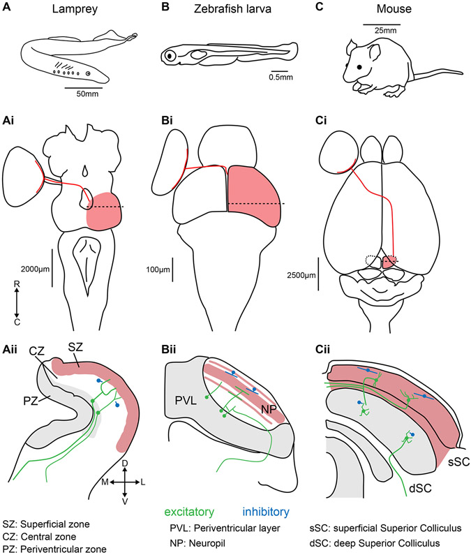
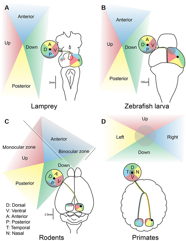
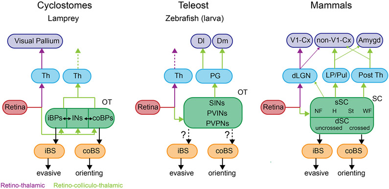
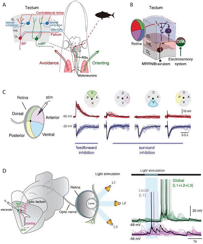
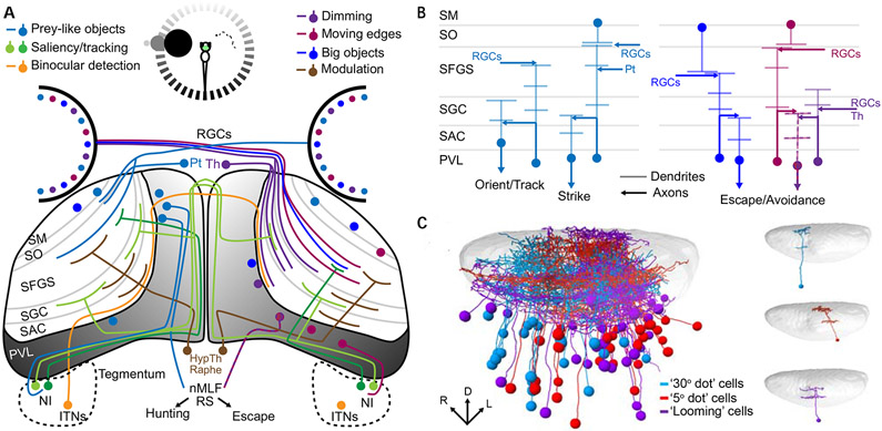
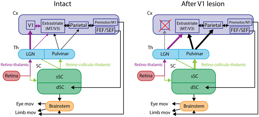
The superior colliculus, or tectum in the case of non-mammalian vertebrates, is a part of the brain that registers events in the surrounding space, often through vision and hearing, but also through electrosensation, infrared detection, and other sensory modalities in diverse vertebrate lineages. This information is used to form maps of the surrounding space and the positions of different salient stimuli in relation to the individual. The sensory maps are arranged in layers with visual input in the uppermost layer, other senses in deeper positions, and a spatially aligned motor map in the deepest layer. Here, we will review the organization and intrinsic function of the tectum/superior colliculus and the information that is processed within tectal circuits. We will also discuss tectal/superior colliculus outputs that are conveyed directly to downstream motor circuits or via the thalamus to cortical areas to control various aspects of behavior. The tectum/superior colliculus is evolutionarily conserved among all vertebrates, but tailored to the sensory specialties of each lineage, and its roles have shifted with the emergence of the cerebral cortex in mammals. We will illustrate both the conserved and divergent properties of the tectum/superior colliculus through vertebrate evolution by comparing tectal processing in lampreys belonging to the oldest group of extant vertebrates, larval zebrafish, rodents, and other vertebrates including primates.


Conflict of interest statement

Declaration of interests The authors declare no competing interests.

Figures

Figure 1:
Figure 1:. Positions, sizes, and retinal inputs to the tectum/SC in lampreys, zebrafish larvae, and mice.
(A) The lamprey (Lampetra fluviatilis). (Ai) Dorsal view of a lamprey brain. Retinal ganglion cell projections (red line) from the left eye cross the optic chiasm to enter the right tectum (red shading). The dashed line represents the position of the coronal section displayed in (Aii). Major divisions are based on Butler and Hodos, 2005 . The red shaded region represents the layers that receive retinal input. The grey shaded area represents regions with a high density of neuronal somae. Local inhibitory interneurons are represented in blue. Ipsilateral and contralateral glutamatergic projecting neurons are represented in green. The same features are shown for larval zebrafish (B-Bii) and mouse (C-Cii). R: rostral, C: caudal, D: dorsal, V: ventral, M: medial, L: lateral.
Figure 2:
Figure 2:. Eye position and retinotectal space across lineages.
Visual topography of the retinotectal projections in the lamprey (A), zebrafish larva (B), rodents (C) and primates (D). Projections from various aspects of the visual field (Up, Down, Anterior and Posterior in A-C, and Right and Left in D) to the retina and tectum/SC are shown in corresponding colors. The center of retina is represented with black dots. The binocular zone for rodents (C) and the foveal region for primates (D) are shaded in gray.
Figure 3:
Figure 3:. Phylogenetic conservation of brain-wide visual pathways.
Retino-thalamic (violet lines) and retino-collicullo-thalamic (green) lines in the lamprey, zebrafish larva and mammals. Abbreviations: Amygd, amygdala; iBPs, ipsilateral brainstem projecting neurons; coBPs, contralateral brainstem projecting neurons; Cx, cortex ; Dl, dorsolateral subdivision of pallium; Dm, dorsomedial subdivision of pallium; iBS, ipsilateral brainstem; coBS, contralateral brainstem; INs, inhibitory interneurons; Th, thalamus; SINs, GABAergic superficial inhibitory neurons; PVINs, periventricular interneurons; PVPNs, periventricular projection neurons; NF, narrow field vertical cells, H, horizontal cells ; St, stellate cells; WF, wide field vertical cells; OT, optic tract; PG, periglomerular nucleus.
Figure 4:
Figure 4:. Structure and circuitry of the lamprey tectum.
(A) Drawing of the lamprey head – brain with the circuitry leading to orienting and avoidance responses. In the upper left the tectal circuitry is represented, with the ipsilateral projecting neurons (iBP) underlying avoidance reaction and the contra-laterally projecting neurons(coBP) orienting responses. These neurons have monosynaptic input from retinal afferents and GABAergic interneurons. The output layer has input from both the pallium (cortex) and the substantia nigra pars reticulata (SNr). (B) Diagram illustrating convergence between the retinal input and the electro-sensory input activated from the same point in space. At the distal dendrites the retinal afferents form synapses, while the electro-sensory afferents target the same dendrite but closer to the cell soma. The tectal output neuron targets middle rhombencephalic reticular nucleus (MRRN), located in the brainstem. Abbreviations: SL, superficial layer; IntL, intermediate layer; DL, deep layer; OLA, octavolateral area. (C) Stimulation of retina in one quadrant leads to excitation in tectal neurons in the specific retinotopic projection area in tectum, while stimulation in all other parts of retina instead leads to a strong inhibition mediated by the tectal GABAergic interneurons. The red traces are recorded with a holding potential of −65 mV, and the blue traces at −20 mV, when the inhibition can be seen clearly. There is thus a powerful lateral inhibition. (D) Shows recordings from a lamprey eye-brain preparation in which the tectal output neurons can be patched, while the eye is illuminated with local brief light pulses (blue area) or globally with light on in all parts of retina. The neuron illustrated is excited by the local light (L1), while there is no response when the entire retina is illuminated (global). This illustrates the strong surround inhibition also shown in B. There is, however, a post-inhibitory rebound.
Figure 5:
Figure 5:. Integral circuitry of the larval zebrafish tectum.
A schematic representation of the larval zebrafish’s tectal inputs and outputs (A) illustrates its role in receiving and integrating information from diverse sources. The inputs are strongly spatial (shading of circle at the top of (A) and of the PVL in the tectum), and differ depending on whether a small stimulus (prey item seen by the right eye) or a large looming stimulus (left eye) is presented. The processing of prey-like stimuli (left tectum, since all retinal ganglion cell axons cross the midline) results in hunting behavior, while looming stimuli (right tectum) trigger escape responses. Inputs from various cell types and brain regions are color coded to indicate the stimulus properties that they encode (legend at the top of A). Different types of information are delivered selectively to different laminae of the tectal neuropil (B), and this dictates the response properties of PVL neurons with dendrites in a specific lamima or laminae. The spatial registration (left, C) of many individual PVL neurons’ morphologies (right, C), taking into account their visual response profiles (colors), reveals the spatial and functional architecture of visual processing in the tectum (Panel C is adapted from Förster et al, 2020 ). Abbreviations: HypTh: Hypothalamus; ITNs: Intertectal neurons; NI: Nucleus Isthmi; nMLF: nucleus of the medial longitudinal fasciculus; Pt: Pretectum; PVL: Periventricular cell layer; RGCs: Retinal ganglion cells; RS: Reticulospinal neurons; SAC: stratum album centrale; SFGS: stratum fibrosum et griseum superficiale; SGC: stratum griseum centrale; SM: stratum marginale; SO: stratum opticum; Th: Thalamus
Figure 6:
Figure 6:. Canonical SC circuits in mammals.
(A) Interlaminar connection. (A1) From the sSC to the dSC. When an electrical stimulus is delivered to narrow field vertical (NF) cells in the sSC (red lightning bolt), excitation is transmitted to brainstem-projecting neurons in the dSC (open circles) through excitatory synapses with AMPA and NMDA receptors. In parallel, GABAergic feedforward inhibition (black circle cells) curtails the excitation as shown in the inset (see the upper inset trace showing intracellular electrophysiological recordings, “w/o disinhibition”) and prevents successive spiking responses upon stimulation of the NF cells. However, if the feedforward GABAergic inhibition is removed, long lasting excitatory responses are induced in the dSC neurons (lower inset trace, “w disinhibition”) through bidirectional reverberating excitatory circuits existing in the dSC (see B2) . (A2) Interlaminar connection from the dSC to sSC. The brainstem projection neurons in the dSC (open circle) send collaterals to the GABAergic dSC neurons (black circle), which send axons to the sSC and inhibit WF cells that project to the LP. (Adapted from Phongphanphanee et al. 2014). (B) Intralaminar connections of the sSC and dSC. (B1) In the sSC, when an electrical stimulus is delivered to the NF cells (red lightening bolt), they excite nearby horizontal cells (H, black circles) which are GABAergic and send horizontal neurites to inhibit remote NF cells. Here, the extent of horizontal excitatory connections (red trace in the upper inset) is narrower than the inhibitory connections (blue trace in the upper inset) and the net effect (green trace in the lower inset) becomes ”Mexican-hat”-like center excitation and surround inhibition in NF cells . Here, “distance” in the inset indicates the medial-lateral distance of the NF cells relative to the stimulated cell. (B2) In the dSC, when an electrical stimulus is delivered to the brainstem-projecting neurons (open circle) the lateral excitation (red trace in the upper inset) is wider than the lateral inhibition in other brainstem-projecting neurons (blue trace in the upper inset) mediated by local GABAergic neurons (black circles), and the net effect (green trace in the lower inset) is an “Excitatory hill”. (Adapted from Phongphanphanee et al. 2014 ).
Figure 7:
Figure 7:. Output pathways from the dSC.
(A) SC output circuits controlling horizontal saccades. (A1) Firing patterns of individual neuron types belonging to the circuits involved in the onset of saccades. The presence of the visual target and movements of the eye are indicated (bottom). (A2) dSC neurons are connected to long-lead burst neurons (LLBNs) and medium-lead excitatory burst neurons (EBNs) in the paramedian pontine reticular formation (PPRF), which transmit the velocity-related high frequency firing activity to abducens motoneuron (VIn). EBN signals are integrated by the tonic neurons (TNs), which relay eye position-related tonic activity to VIn. EBNs are connected to inhibitory burst neurons (IBNs) which further inhibit the VIn on the contralateral side. Panel A2 is adapted from Sparks, 2002 and Takahashi and Shinoda, 2018 . (B) dSC output circuits controlling eye and head movements. dSC neurons send bifurcating axons to the ipsilateral mesodiencephalic junction (MDJ) and contralateral pontomedullary reticular formation (PMRF). The former includes Forel’s field H (FFH) (or rostral interstitial nucleus of MLF (riMLF)) and interstitial nucleus of Cajal (INC). Both of these structures are connected to the oculomotor (IIIn) and trochlear motor (IVn) nuclei to control eye movements and dorsal neck motoneurons (MNs) either directly or indirectly via the nucleus reticularis gigantocellularis (NRGc) to control movements of the head. The latter descending axons are connected to the nucleus reticularis pontis caudalis (NRPc) which is connected to VIn and lateral neck MNs either directly or indirectly via NRGc (horizontal system). Some dSC neurons descend to the spinal cord and connected to MNs via spinal cord interneurons (Spc INs). Panel B is adapted from Isa and Sasaki, 2002 .
Figure 8.
Figure 8.. Circuits controlling innate visual responses in the mouse.
(A) Projections of the crossed pathway, principally from the dSC to the contralateral PMRF (green), drive orienting movements. In addition to the PMRF, descending collaterals are projected to the contralateral dSC (co-dSC), the precerebellar nuclei such as the nucleus reticularis tegmenti pontis (NRTP) and inferior olive (IO), the raphe, and further down to the spinal cord (Spc). In addition, collaterals are also projected ipsilaterally to the pedunculopontine tegmental nucleus (PPN) and partly to the PRMF in the pons, mesencephalic reticular formation (mRt) and substantia nigra pars compacta (SNc) in the midbrain. Furthermore, ascending axons are projected to several thalamic and midbrain nuclei on the ipsilateral side, some of which might function an efference copy signal. (B) Projections forming the uncrossed pathway (magenta), from the dSC to the ipsilateral cuneiform nucleus (CnF), control evasive movements. Here, collaterals of the descending branch are projected to the PPN, inferior colliculus (IC), PMRF, raphe and IO. In the midbrain, collaterals are projected to the dorsal periaqueductal gray matter (dPAG), SNc and mRt, some of which might be related to the emotional and reward-related aspects of the behavior. Furthermore, ascending axons are projected to several thalamic and midbrain nuclei on the ipsilateral side. Adapted from Dean et al, 1989 ; Sahibzada et al, 1986 and Isa et al, 2020 .
Box legend:
Box legend:. Schematic of visual pathways before and after V1 damage.
Visuo-motor pathway for the control of eye or limb movements with the intact V1 (left) and following damage to the V1 (right). The thickness of the arrows indicates the strength of connectivity.

References

    1. Kardamakis AA, Perez-Fernandez J, and Grillner S (2016). Spatiotemporal interplay between multisensory excitation and recruited inhibition in the lamprey optic tectum. Elife 5. - PMC - PubMed
    1. Kardamakis AA, Saitoh K, and Grillner S (2015). Tectal microcircuit generating visual selection commands on gaze-controlling neurons. Proc. Natl. Acad. Sci. U. S. A 112, E1956–E1965. - PMC - PubMed
    1. Suzuki DG, Perez-Fernandez J, Wibble T, Kardamakis AA, and Grillner S (2019). The role of the optic tectum for visually evoked orienting and evasive movements. Proc. Natl. Acad. Sci. U. S. A 116, 15272–15281. - PMC - PubMed
    1. Sparks DL (1986). Translation of sensory signals into commands for control of saccadic eye movements: role of primate superior colliculus. Physiol. Rev 66, 118–171. - PubMed
    1. Dean P, Redgrave P, and Westby GW (1989). Event or emergency? Two response systems in the mammalian superior colliculus. Trends Neurosci. 12, 137–147. - PubMed

Publication types