. 2021 Jun:68:103418.
doi: 10.1016/j.ebiom.2021.103418. Epub 2021 Jun 5.

Gain-of-function Tibetan PHD2D4E;C127S variant suppresses monocyte function: A lesson in inflammatory response to inspired hypoxia

Affiliations

Gain-of-function Tibetan PHD2D4E;C127S variant suppresses monocyte function: A lesson in inflammatory response to inspired hypoxia

Sulagna Bhattacharya et al. EBioMedicine. 2021 Jun.

Abstract

Background: We have previously described an evolutionarily selected Tibetan prolyl hydroxylase-2 (PHD2D4E;C127S) variant that degrades the hypoxia-inducible factor (HIFα) more efficiently and protects these highlanders from hypoxia-triggered elevation in haemoglobin concentration. High altitude is known to cause acute mountain sickness (AMS) and high-altitude pulmonary edema (HAPE) in a section of rapidly ascending non-acclimatised lowlanders. These morbidities are often accompanied by inflammatory response and exposure to hypobaric hypoxia is presumed to be the principal causative agent. We have investigated whether PHD2D4E;C127S variant is associated with prevention of hypoxia-mediated inflammatory milieu in Tibetan highlanders and therefore identify a potential target to regulate inflammation.

Methods: We genotyped the Tibetans using DNA isolated from whole blood. Thereafter immunophenotying was performed on PBMCs from homozygous PHD2D4E;C127S and PHD2WT individuals using flow cytometry. RNA isolated from these individuals was used to evaluate the peripheral level of important transcripts associated with immune as well as hypoxia response employing the nCounter technology. The ex-vivo findings were validated by generating monocytic cell lines (U937 cell line) expressing PHD2D4E;C127S and PHD2WT variants post depletion of endogenous PHD2. We had also collected whole blood samples from healthy travellers and travellers afflicted with AMS and HAPE to evaluate the significance of our ex-vivo and in vitro findings. Hereafter, we also attempted to resolve hypoxia-triggered inflammation in vitro as well as in vivo by augmenting the function of PHD2 using alpha-ketoglutarate (αKG), a co-factor of PHD2.

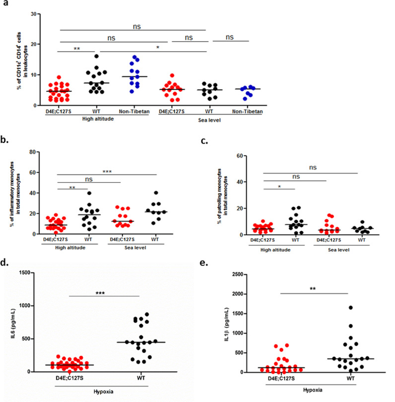
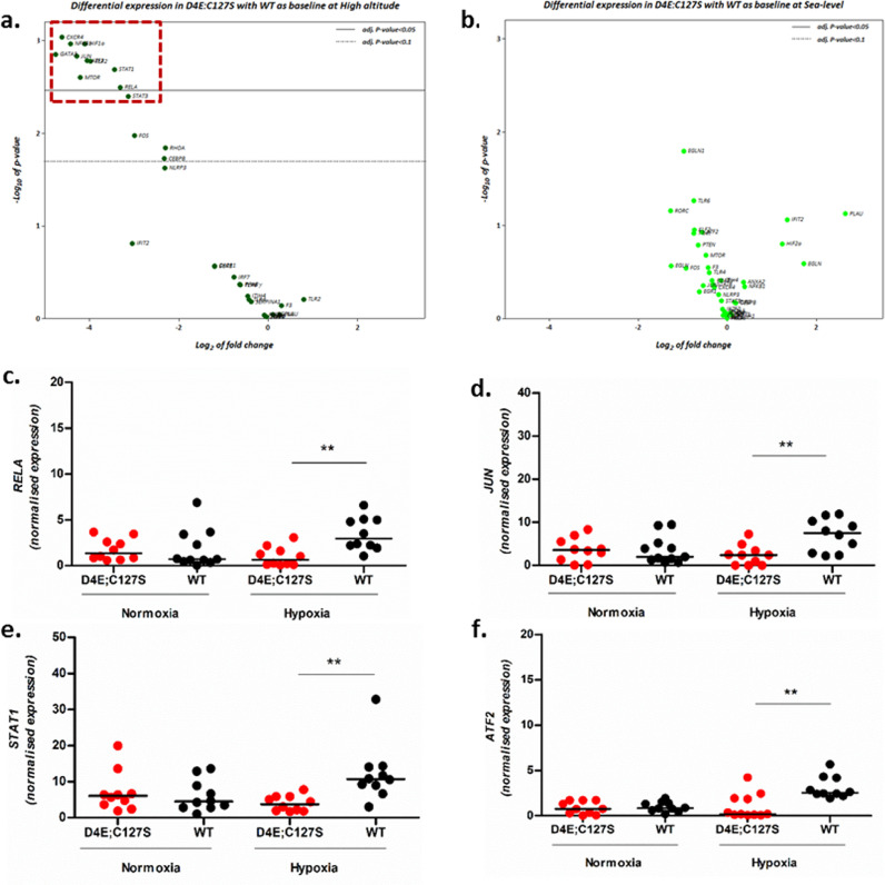
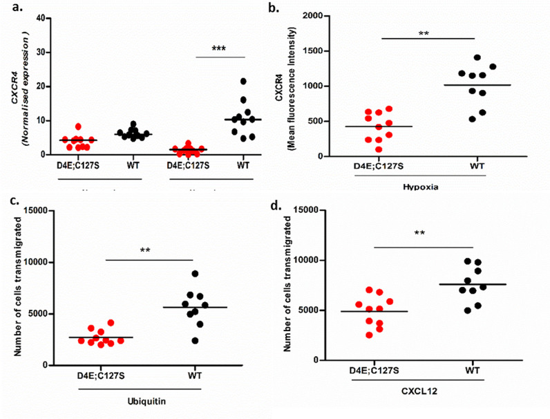
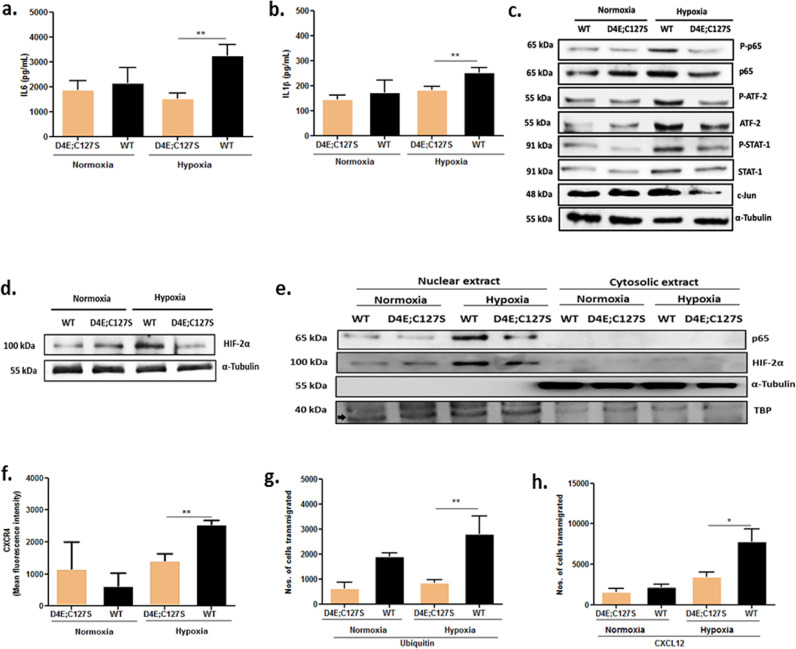
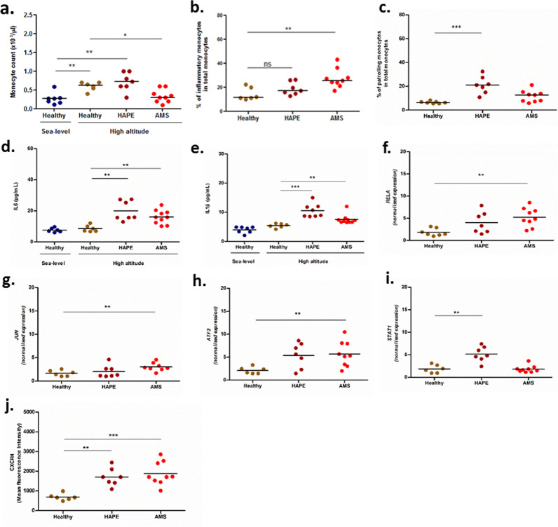
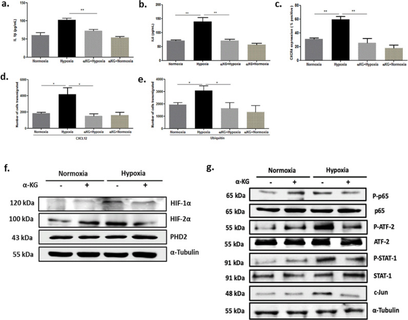
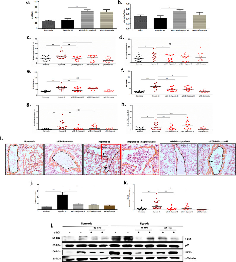
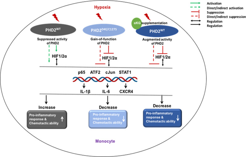
Findings: We report that homozygous PHD2D4E;C127S highlanders harbour less inflammatory and patrolling monocytes in circulation as compared to Tibetan PHD2WT highlanders. In response to in vitro hypoxia, secretion of IL6 and IL1β from PHD2D4E;C127S monocytes, and their chemotactic response compared to the PHD2WT are compromised, corresponding to the down-modulated expression of related signalling molecules RELA, JUN, STAT1, ATF2 and CXCR4. We verified these functional outcomes in monocytic U937 cell line engineered to express PHD2D4E;C127S and confirmed the down-modulation of the signalling molecules at protein level under hypoxia. In contrast, non-Tibetan sojourners with AMS and HAPE at high altitude (3,600 m above sea level) displayed significant increase in these inflammatory parameters. Our data henceforth underline the role of gain-of-function of PHD2 as the rate limiting factor to harness hyper-activation of monocytes in hypoxic environment. Therefore upon pre-treatment with αKG, we observed diminished inflammatory response of monocytes in vitro and reduction in leukocyte infiltration to the lungs in mice exposed to normobaric hypoxia.

Interpretation: Our report suggests that gain-of-function PHD2 D4E;C127S variant can therefore protect against inflammation elicited by hypobaric hypoxia. Augmentation of PHD2 activity therefore may be an important method to alleviate inflammatory response to inspired hypoxia.

Funding: This study is supported by the Department of Biotechnology, Government of India.

Keywords: HAPE; High altitude hypoxia; Inflammation; Monocyte; PHD2 variant; Sojourners; Tibetan; α-ketoglutarate.


Conflict of interest statement

Declaration of Competing Interest The authors declare no competing interests.

Figures

Fig 1
Fig. 1
Elevated monocyte counts in PHD2WT Tibetans than PHD2D4E;C127S at high altitude. Monocyte count was measured in PBMCs of healthy Tibetans carrying either homozygous PHD2D4E;C127S or PHD2WT from high altitude and sea level using flow cytometry. (a) Percentage of monocytes in total leukocyte, (b) inflammatory and (c) patrolling subsets in total monocytes were measured. The detail immunophenotyping gating strategy is described in Fig. S1. Each dot represents the percentage of cells in each individual. Kruskal Wallis test was used on median values for comparison between genotypes followed by Dunn's multiple comparison post test, ***p<0.001, **p<0.01, *p<0.05 and ns=non-significant. Classical monocyte subtype count is mentioned in Fig. S2d. Also the monocyte count was analysed from whole blood using haematology analyser; these data show similar trend, depicted in Fig. S2a. A similar count of monocytes was observed in Tibetans between PHD2 heterozygous and wild-type, is shown in Fig. S2a. Reduced secretion of pro-inflammatory cytokines by PHD2D4E;C127S monocytes than PHD2WT. The CD14+ monocytes isolated from PBMCs were exposed to 1% oxygen for 24 hrs. CBA assay was used to assess supernatants for the levels of (d) IL6 and (e) IL1β, to compare between PHD2D4E;C127S and PHD2WT Tibetans. Unpaired t-test was used for comparison between genotypes, ***p<0.001 and **p<0.01. Levels of other cytokines as well as comparison of values amongst homozygous, heterozygous and wild type are mentioned in Fig. S3.
Fig 2
Fig. 2
Differential expression of hypoxia and immune response genes in Tibetans. Total RNA isolated from PBMCs was probed for expression of 47 genes at the interface of hypoxia and immune response. The differential expression of the genes is shown as volcano plot for (a) high altitude and (b) sea level Tibetans. The nCounter technology was used to measure gene expression. Differential expression between PHD2D4E;C127S and PHD2WT (n=10 each) Tibetans was calculated using the advanced analysis module of the nSolver software. It uses the Wald test with adjusted p-value. Data in detail are described in Table S2. The differentially expressed genes in nCounter data were validated in CD14+ monocytes of PHD2D4E;C127S and PHD2WT individuals residing at sea level. The monocytes were exposed to 21% or 1% oxygen for 24 hrs and gene expression of (c-f)RELA, JUN, ATF2 and STAT1 was evaluated using real-time PCR. Each dot represents the relative quantification for each individual. Unpaired t-test was used for comparison between genotypes, **p<0.01. The expression of above genes between homozygous, heterozygous and wild type is mentioned in Fig. S6.
Fig 3
Fig. 3
Reduced chemotactic ability of PHD2D4E;C127S monocytes. CXCR4 level was measured in CD14+ monocytes isolated from PBMCs of PHD2D4E;C127S or PHD2WT individuals residing at sea level. The monocytes were exposed to 21% O2 or 1% O2 and evaluated for (a) gene expression and (b) cell surface expression of CXCR4. Unpaired t-test was used for comparison between genotypes, ***p<0.001 and **p<0.01. Chemotactic ability of these cells was measured using (c) ubiquitin and (d) CXCL12 in the lower chamber of the transwell plate. Each dot represents data from a single individual. Unpaired t-test was used for comparison between genotypes, **p<0.01. The comparative expression of CXCR4 amongst homozygous, heterozygous and wild type is depicted in Fig. S7.
Fig 4
Fig. 4
U937 cell line expressing PHD2D4E;C127S displays reduced cytokine secretion. Endogenous PHD2 was depleted using shRNA targeting the 3′ UTR of endogenous EGLN1. Then, cells were transduced with constructs expressing either PHD2D4E;C127S or PHD2WT using lentiviral particles, detail in Fig. S8a-b. U937 cells expressing either PHD2D4E;C127S or PHD2WT were exposed to 21% or 1% oxygen for 24 hrs and cytometry based assay was used to assess supernatants for the level of (a) IL6 and (b) IL1β. Each bar represents mean ± SEM from three independent experiments in duplicates. Unpaired t-test was used for comparison, **p<0.01. Other cytokine data are mentioned in Fig. S8c-d. Down-modulated expression of signalling molecules in PHD2D4E;C127S cells. The cells from above experiments were assessed for genes, RELA, JUN, ATF2 and STAT1 (Fig. S8f-i) and (c) proteins- P-p65, P-ATF2, P-STAT1 and c-Jun, and (d) HIF2α. (e) Nuclear localisation of p65 and HIF2α was evaluated with respect to TBP. Protein expression was evaluated by western blot from three independent experiments. Densitometry data are shown in Fig. S15a-h. Reduced chemotactic ability of PHD2D4E;C127S cells. The above cells assessed for gene (Fig. S8j) and (e) cell surface expression of CXCR4. Chemotactic ability of these cells was assessed using (f) ubiquitin and (g) CXCL12 in the lower chamber of the transwell plate. Data represented as mean ± SEM from three independent experiments. Unpaired t-test was used for comparison, ***p<0.001, **p<0.01 and *p<0.05.
Fig 5
Fig. 5
Elevated inflammatory response of monocytes in AMS and HAPE patients. PBMCs were isolated from whole blood of non-Tibetan sojourners with AMS/HAPE or healthy individuals from high altitude. Healthy individuals from sea level were used as reference. (a) Total monocytes were counted using haematology analyser. (b) Inflammatory and (c) patrolling monocyte subsets were measured using flow cytometry as described in Fig. 1. Each dot represents a single individual. Kruskal Wallis test was used on median values for comparison between patients and healthy individuals followed by Dunn's multiple comparison post test, ***p<0.001 and **p<0.01. Plasma level of cytokines (d) IL6 and (e) IL1β was measured, alongside TNFα and TGF β (Fig. S9e-f) using CBA assay. Data were recorded in duplicates and calculated as mentioned above, ***p<0.001, **p<0.01 and *p<0.05. (f-i) The gene expression of RELA, JUN, ATF2 and STAT1 was measured using real-time PCR and data were normalized using expression of housekeeping gene TUBB. Data were calculated as mentioned above, **p<0.01. The CXCR4 gene expression (Fig. S9g) was measured in these individuals as mentioned above. (j): Alongside the surface expression of CXCR4 on CD14+ monocytes were assessed as mentioned in Fig. 3. Data were calculated as mentioned above, **p<0.01 and *p<0.05.
Fig 6
Fig. 6
αKG-mediated augmentation of PHD2 and inhibition of hypoxia-mediated inflammatory response in U937 cell line. U937 cells were pre-treated with either octyl α-ketoglutarate (1 mM) or vehicle for 4 hrs and kept under either 21% or 1% oxygen for 24 hrs. Cytokines were measured in the supernatant using CBA assay as described in Fig. 1. (a) IL1β and (b) IL6 levels were elevated in hypoxia but reduced after αKG treatment. Data are mean ± SEM of three independent experiments. Unpaired t-test was used for comparison, **p<0.01. Other cytokines levels are mentioned in Fig. S10a-c. Cell surface expression of (c) CXCR4 and chemotactic potential of the cells in response to (d) CXCL12 and (e) Ubiquitin was evaluated and data analyzed from three independent experiments, **p<0.01, and *p<0.05. Expression of (f) HIF1α, HIF2α and PHD2, and (g) P-p65, P-ATF2, P-STAT1 and c-Jun was evaluated by western blot to ascertain the effect of enhanced enzymatic function of PHD2 in monocytes after octyl αKG treatment under normoxia and hypoxia. Detail densitometry data are described in Fig. S15i-t.
Fig 7
Fig. 7
Effect of dietary αKG on hypoxia-immune response in mice. The C57/BL6 mice (5-6 weeks old) were administered with 1% αKG via drinking water, 12 hrs prior to hypoxia exposure (11% oxygen for 48 hrs), experimental design is presented as a schematic in Fig. S14a. (a-b) After treatment, plasma and PBMC-granulocyte cell pellet were used for measuring αKG in (a) plasma and (b) PBMCs (1 × 106 cells) lysate using colorimetry based assay. Unpaired t-test was used for comparison between groups (n=7 in each group), **p<0.01, *p<0.05 and ns=non-significant. (c-d) Monocytes and neutrophils counts were measured from whole blood using hematology analyzer. (e-f) Plasma level of cytokines IL1β and IL6 were assessed using CBA assay. (g-h) Monocyte and neutrophil counts were measured from bronchoalveolar lavage fluid (BALF) using hematology analyzer. The counts of both cell types in whole blood and BALF were confirmed using staining with CD45, CD11b, Ly6C and Ly6G and analyzed using flow cytometry analysis, mentioned in Fig. S13. Each dot represents a mouse from three independent experiments. Unpaired t-test was used for comparison between the groups as in Fig. 7c-h, ***p<0.001 **p<0.01, and *p<0.05. (i) The H & E staining of lung section of mice showed the infiltration of immune cells into airway space under hypoxia treatment (magnification view shows large numbers of leukocytes inside airway space), which was rescued by 48 hrs αKG supplementation; arrows indicate the cell infiltration, bars in all images ~ 50 μm. (J) The infiltration score was calculated in case of each treatment group based on H & E lung sections from independent experiment. Additional images for quantification has been depicted in Fig. S14g. (k) The expression of Cxcr4 gene in leukocytes was assessed using real-time PCR and normalized with housekeeping gene Hprt. Unpaired t-test was used for comparison between groups, **p<0.01, and *p<0.05.(l) Expression of P-p65 and HIF2α in leukocytes pellet was evaluated by western blot to ascertain the enzymatic function of PHD2 after αKG administration under hypoxia. Representative blot shows data of 2 mice from each group. Densitometry showed data from 5 mice for P-p65/p65 and 8 mice for HIF2α from independent experiments as mentioned in Fig. S15u-w. The αKG treatment for 48 hrs rescued more effectively than 24 hrs treatment for all above parameters in mice exposed for 48 hrs under 11% hypoxia.
Fig 8
Fig. 8
PHD2 regulates inflammatory response of monocyte. (Left panel) The monocytes of PHD2D4E;C127S Tibetans highlanders exhibit protection against hypoxia-triggered elevation of inflammatory cytokines including IL6 and IL1β, as well as CXCR4-mediated chemotactic ability by down-modulating transcription factors such as p65, ATF2, c-Jun, STAT1 and HIFα compared to their wild type counterparts, suggesting the gain-of-function for PHD2D4E;C127S variant. (Mid panel) The non-Tibetans sojourners who traveled to high altitudes and developed AMS and HAPE display high level of pro-inflammatory cytokines alongside elevation of above-mentioned transcription factors.(Right panel) The dietary supplementation of α-ketoglutarate (αKG, co-factor of PHD2) to mice exposed to hypoxia (11% oxygen) suppressed the hypoxia-induced elevation of inflammatory cytokines and transmigration of immune cells in lungs in conjunction with augmented PHD2 activity.

References

    1. Imray C, Wright A, Subudhi A, Roach R. Acute mountain sickness: pathophysiology, prevention and treatment. Prog Cardiovasc Dis. 2010;5:467–484. - PubMed
    1. Luks AM, Swenson ER, Bartsch P. Acute high altitude sickness. Eur Respir Rev. 2016;26 - PMC - PubMed
    1. Schoene RB, Swenson ER, Pizzo CJ, Hackett PH, Roach RC, Mills Jr WJ. The lung at high altitude: bronchoalveolar lavage in acute mountain sickness and pulmonary edema. J Appl Physiol. 1988;6:2605–2613. - PubMed
    1. Wang C, Jiang H, Duan J, Chen J, Wang Q, Liu X, Wang C. Exploration of acute phase proteins and inflammatory cytokines in early stage of diagnosis of acute mountain sickness. High Altitude Med Biol. 2018;19:170–177. - PubMed
    1. Bartsch P, Swenson ER. Acute high-altitude illness. New Eng J Medicine. 2013;368:2294–2302. - PubMed

MeSH terms

Substances

Supplementary concepts