

3D magnetic resonance spectroscopic imaging reveals links between brain metabolites and multidimensional pain features in fibromyalgia
- PMID: 34102707
- PMCID: PMC9176690
- DOI: 10.1002/ejp.1820
3D magnetic resonance spectroscopic imaging reveals links between brain metabolites and multidimensional pain features in fibromyalgia
Abstract
Background: Fibromyalgia is a centralized multidimensional chronic pain syndrome, but its pathophysiology is not fully understood.
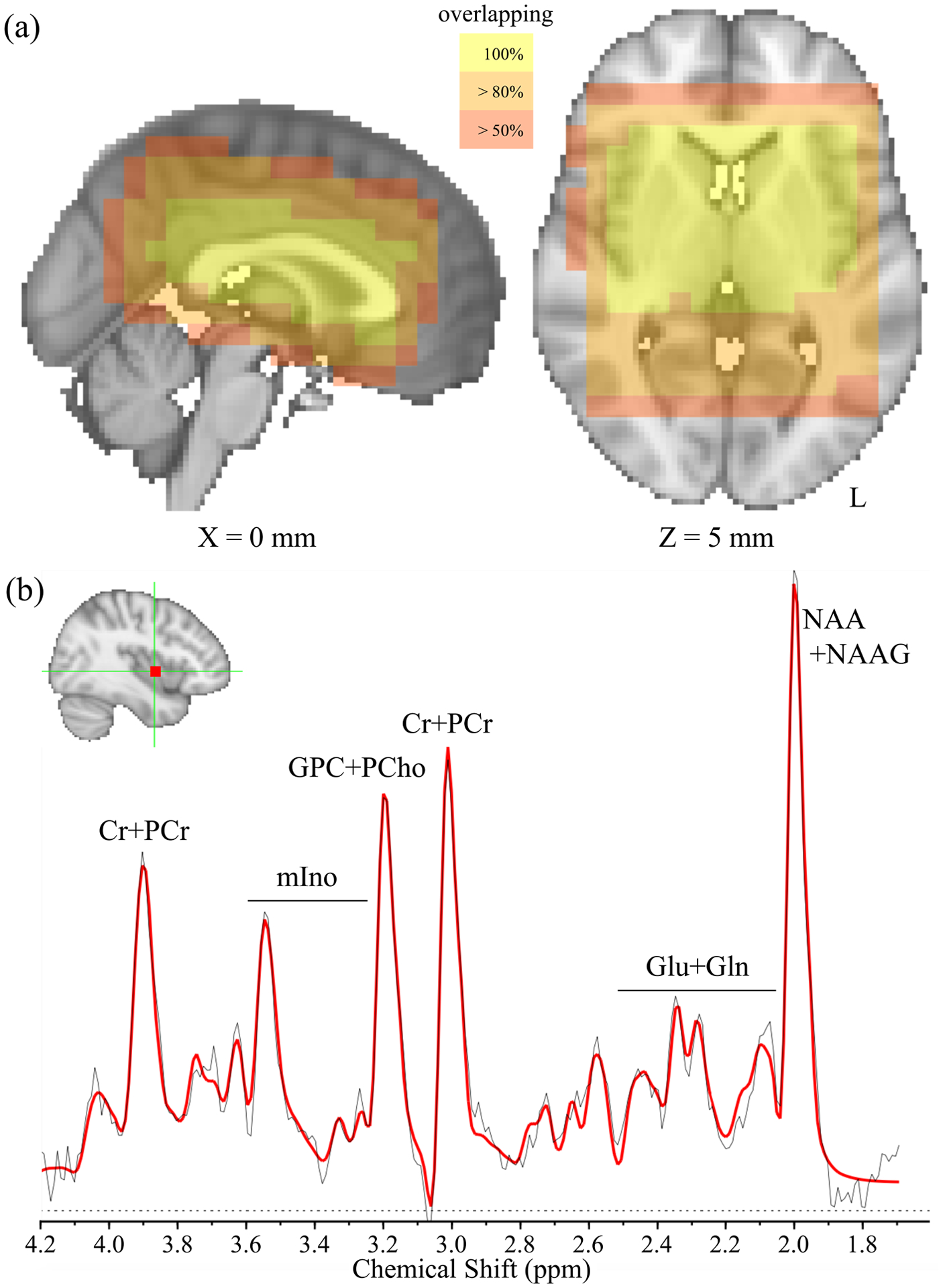
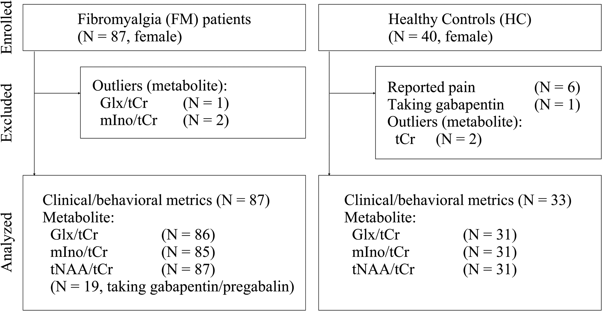
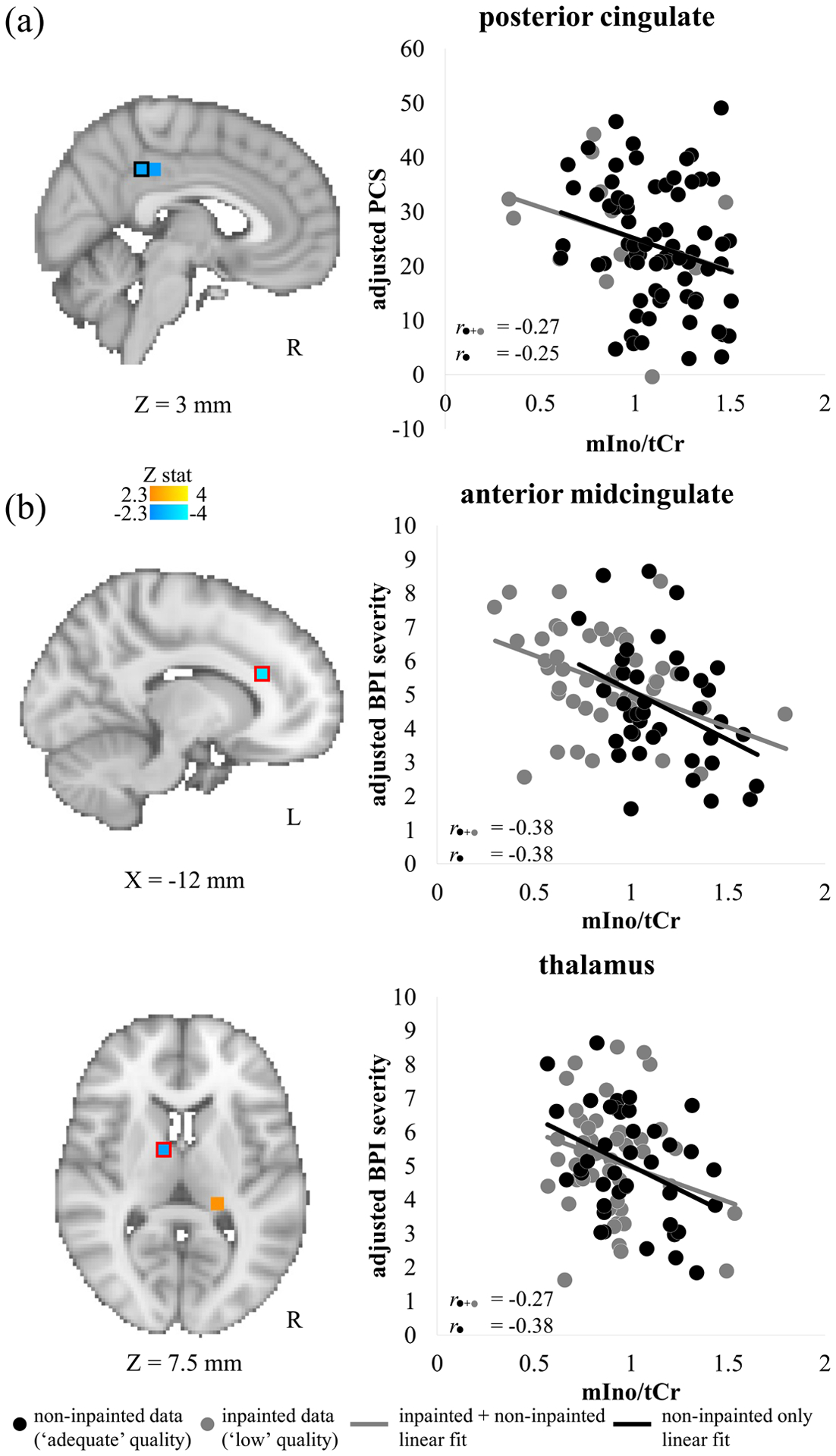
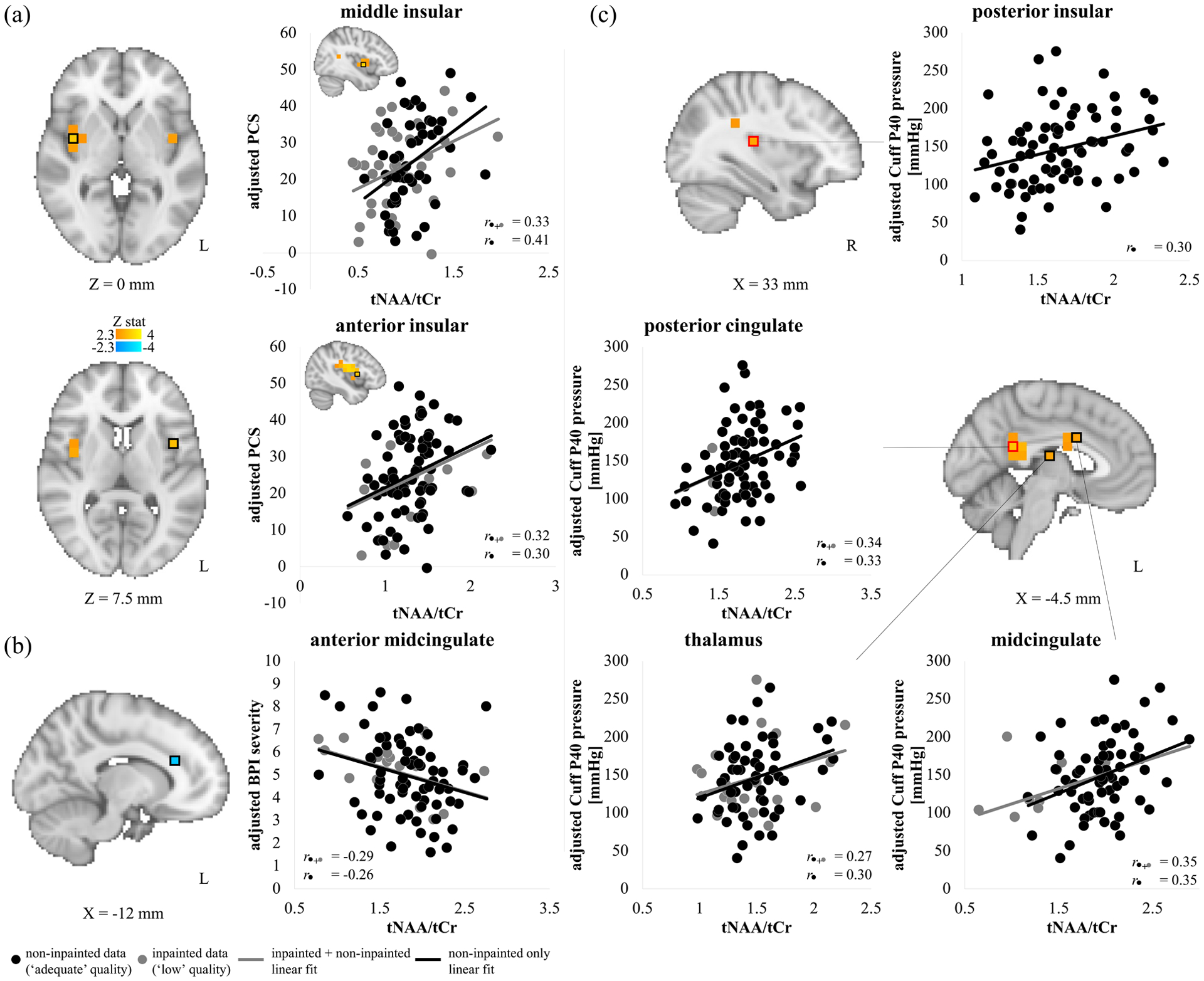
Methods: We applied 3D magnetic resonance spectroscopic imaging (MRSI), covering multiple cortical and subcortical brain regions, to investigate the association between neuro-metabolite (e.g. combined glutamate and glutamine, Glx; myo-inositol, mIno; and combined (total) N-acetylaspartate and N-acetylaspartylglutamate, tNAA) levels and multidimensional clinical/behavioural variables (e.g. pain catastrophizing, clinical pain severity and evoked pain sensitivity) in women with fibromyalgia (N = 87).
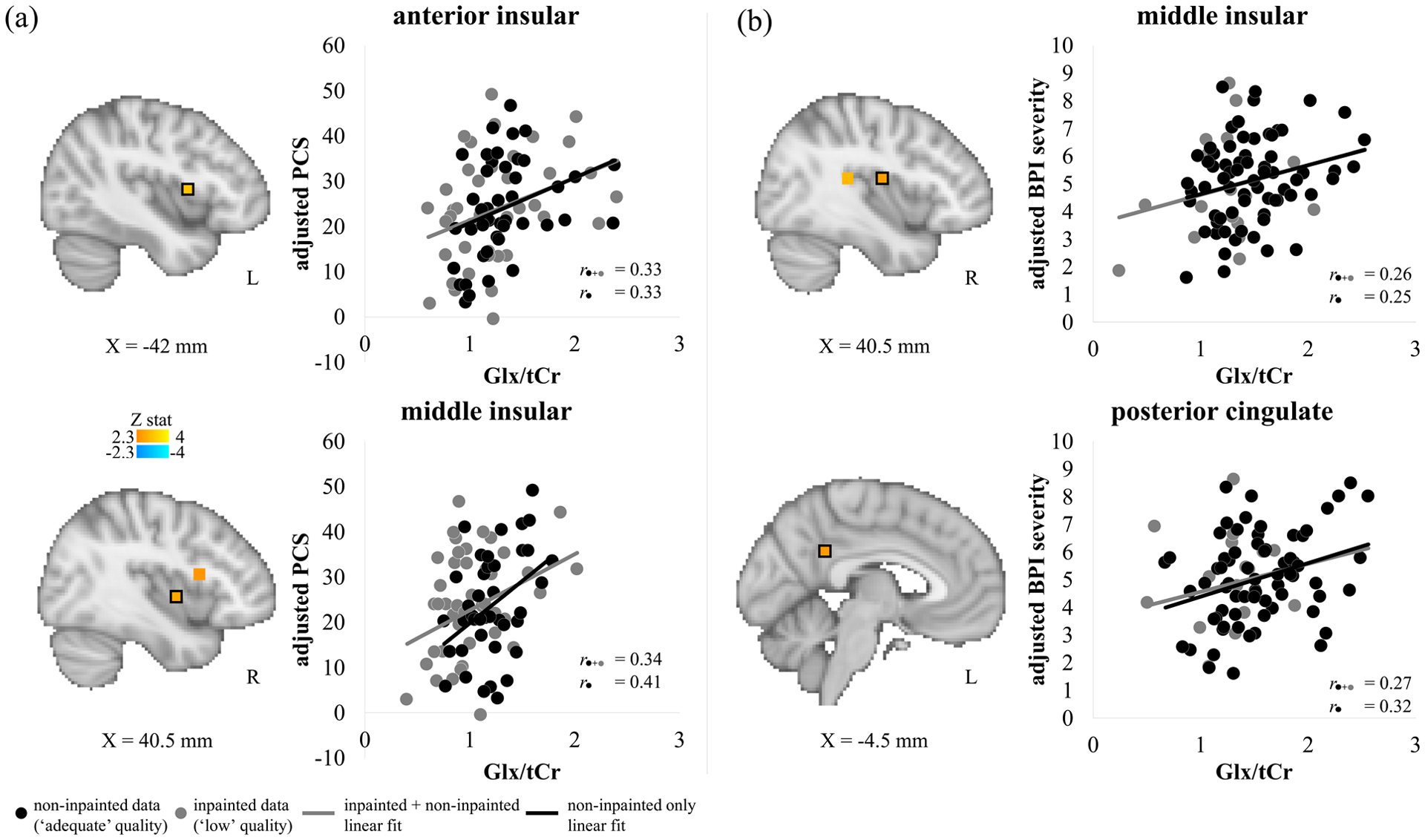
Results: Pain catastrophizing scores were positively correlated with Glx and tNAA levels in insular cortex, and negatively correlated with mIno levels in posterior cingulate cortex (PCC). Clinical pain severity was positively correlated with Glx levels in insula and PCC, and with tNAA levels in anterior midcingulate cortex (aMCC), but negatively correlated with mIno levels in aMCC and thalamus. Evoked pain sensitivity was negatively correlated with levels of tNAA in insular cortex, MCC, PCC and thalamus.
Conclusions: These findings support single voxel placement targeting nociceptive processing areas in prior 1 H-MRS studies, but also highlight other areas not as commonly targeted, such as PCC, as important for chronic pain pathophysiology. Identifying target brain regions linked to multidimensional symptoms of fibromyalgia (e.g. negative cognitive/affective response to pain, clinical pain, evoked pain sensitivity) may aid the development of neuromodulatory and individualized therapies. Furthermore, efficient multi-region sampling with 3D MRSI could reduce the burden of lengthy scan time for clinical research applications of molecular brain-based mechanisms supporting multidimensional aspects of fibromyalgia.
Significance: This large N study linked brain metabolites and pain features in fibromyalgia patients, with a better spatial resolution and brain coverage, to understand a molecular mechanism underlying pain catastrophizing and other aspects of pain transmission. Metabolite levels in self-referential cognitive processing area as well as pain-processing regions were associated with pain outcomes. These results could help the understanding of its pathophysiology and treatment strategies for clinicians.
© 2021 European Pain Federation - EFIC®.
Conflict of interest statement
Figures
Similar articles
-
Encoding of Self-Referential Pain Catastrophizing in the Posterior Cingulate Cortex in Fibromyalgia.Arthritis Rheumatol. 2018 Aug;70(8):1308-1318. doi: 10.1002/art.40507. Epub 2018 Jun 22. Arthritis Rheumatol. 2018. PMID: 29579370 Free PMC article.
-
Effects of intensive cognitive-behavioral therapy on cingulate neurochemistry in obsessive-compulsive disorder.J Psychiatr Res. 2013 Apr;47(4):494-504. doi: 10.1016/j.jpsychires.2012.11.010. Epub 2013 Jan 4. J Psychiatr Res. 2013. PMID: 23290560 Free PMC article.
-
Spectroscopic differences in posterior insula in patients with chronic temporomandibular pain.Scand J Pain. 2018 Jul 26;18(3):351-361. doi: 10.1515/sjpain-2017-0159. Scand J Pain. 2018. PMID: 29794260
-
Cerebral magnetic resonance changes associated with fibromyalgia syndrome.Med Clin (Barc). 2017 Jun 7;148(11):511-516. doi: 10.1016/j.medcli.2017.01.034. Epub 2017 Apr 25. Med Clin (Barc). 2017. PMID: 28450073 Review. English, Spanish.
-
Alterations of brain activity in fibromyalgia patients.J Clin Neurosci. 2017 Apr;38:13-22. doi: 10.1016/j.jocn.2016.12.014. Epub 2017 Jan 10. J Clin Neurosci. 2017. PMID: 28087191 Review.
Cited by
-
Neuropathic Pain and Spinal Cord Injury: Management, Phenotypes, and Biomarkers.Drugs. 2023 Jul;83(11):1001-1025. doi: 10.1007/s40265-023-01903-7. Epub 2023 Jun 16. Drugs. 2023. PMID: 37326804 Review.
-
Molecular Mechanisms of Chronic Pain and Therapeutic Interventions.MedComm (2020). 2025 Aug 7;6(8):e70325. doi: 10.1002/mco2.70325. eCollection 2025 Aug. MedComm (2020). 2025. PMID: 40787071 Free PMC article. Review.
-
Deciphering nociplastic pain: clinical features, risk factors and potential mechanisms.Nat Rev Neurol. 2024 Jun;20(6):347-363. doi: 10.1038/s41582-024-00966-8. Epub 2024 May 16. Nat Rev Neurol. 2024. PMID: 38755449 Review.
-
Reevaluating fibromyalgia diagnosis: a proposal to integrate deep tendon reflex responses into current criteria.Rheumatol Int. 2025 Apr 2;45(4):84. doi: 10.1007/s00296-025-05846-y. Rheumatol Int. 2025. PMID: 40172661 Free PMC article. Review.
-
Topology of pain networks in patients with temporomandibular disorder and pain-free controls with and without concurrent experimental pain: A pilot study.Front Pain Res (Lausanne). 2022 Oct 17;3:966398. doi: 10.3389/fpain.2022.966398. eCollection 2022. Front Pain Res (Lausanne). 2022. PMID: 36324873 Free PMC article.
References
-
- Al-Iedani O, Ribbons K, Gholizadeh N, Lechner-Scott J, Quadrelli S, Lea R, … Ramadan S (2020). Spiral MRSI and tissue segmentation of normal-appearing white matter and white matter lesions in relapsing remitting multiple sclerosis patients(). Magn Reson Imaging, 74, 21–30. doi:10.1016/j.mri.2020.09.001 - DOI - PubMed
Publication types
MeSH terms
Substances
Grants and funding
LinkOut - more resources
Full Text Sources
Medical
