. 2021 Aug 31;144(9):712-727.
doi: 10.1161/CIRCULATIONAHA.121.054885. Epub 2021 Jun 9.

PKM1 Exerts Critical Roles in Cardiac Remodeling Under Pressure Overload in the Heart

Affiliations

PKM1 Exerts Critical Roles in Cardiac Remodeling Under Pressure Overload in the Heart

Qinfeng Li et al. Circulation. .

Abstract

Background: Metabolic remodeling precedes most alterations during cardiac hypertrophic growth under hemodynamic stress. The elevation of glucose utilization has been recognized as a hallmark of metabolic remodeling. However, its role in cardiac hypertrophic growth and heart failure in response to pressure overload remains to be fully illustrated. Here, we aimed to dissect the role of cardiac PKM1 (pyruvate kinase muscle isozyme 1) in glucose metabolic regulation and cardiac response under pressure overload.

Methods: Cardiac-specific deletion of PKM1 was achieved by crossing the floxed PKM1 mouse model with the cardiomyocyte-specific Cre transgenic mouse. PKM1 transgenic mice were generated under the control of tetracycline response elements, and cardiac-specific overexpression of PKM1 was induced by doxycycline administration in adult mice. Pressure overload was triggered by transverse aortic constriction. Primary neonatal rat ventricular myocytes were used to dissect molecular mechanisms. Moreover, metabolomics and nuclear magnetic resonance spectroscopy analyses were conducted to determine cardiac metabolic flux in response to pressure overload.

Results: We found that PKM1 expression is reduced in failing human and mouse hearts. It is important to note that cardiomyocyte-specific deletion of PKM1 exacerbates cardiac dysfunction and fibrosis in response to pressure overload. Inducible overexpression of PKM1 in cardiomyocytes protects the heart against transverse aortic constriction-induced cardiomyopathy and heart failure. At the mechanistic level, PKM1 is required for the augmentation of glycolytic flux, mitochondrial respiration, and ATP production under pressure overload. Furthermore, deficiency of PKM1 causes a defect in cardiomyocyte growth and a decrease in pyruvate dehydrogenase complex activity at both in vitro and in vivo levels.

Conclusions: These findings suggest that PKM1 plays an essential role in maintaining a homeostatic response in the heart under hemodynamic stress.

Keywords: cardiomegaly; glycolysis; heart failure; pyruvate dehydrogenase complex; pyruvate kinase.


Figures

Figure 1.
Figure 1.. PKM1 expression and pyruvate kinase activity are decreased in failing mouse hearts.
A. Heart failure was triggered by pressure overload (transverse aortic constriction, TAC) with wild-type mice. Cardiac tissues were harvested for metabolomics analysis. PCA (principal component analysis) is shown. n=4. B. Out of 133 metabolites, 13 showed significant changes between normal and heart failure groups (P<0.05). C. Pathway analysis showed that glycolysis was among the most significantly changed signaling pathways. D. Cardiac tissue lysates were extracted and used for Western blotting. Note that the induction of ANF is a molecular signature of heart failure in vivo. n=5 to 7. E. PKM1 mRNA level was decreased under heart failure. n=6 to 7. F. Pyruvate kinase activity was reduced in failing mouse hearts. n=6 to 7. Unpaired Student’s t test was conducted (D-F). Data are represented as mean±SEM.
Figure 2.
Figure 2.. Cardiac specific deletion of PKM1 exacerbates heart failure progression under pressure overload.
A. Schematic of the cardiac specific PKM1 knockout mouse model (cKO). Triangles represent loxP sites. B. Representative photos of H&E (hematoxylin & eosin) staining and Masson’s trichrome staining 1 week after TAC. Scale: 1 mm. C. WGA (Wheat germ agglutinin) staining was conducted to calculate cardiomyocyte cross-sectional area. Pint<0.05. Cardiomyocytes quantified: n=85 for sham/FF; n=96 for TAC/FF; n=95 for sham/cKO; n=105 for TAC/cKO. Scale: 50 μm. D. Representative M-mode echocardiography images of control and cKO mice after either sham or TAC. E. Cardiac specific loss of PKM1 led to aggravated cardiac dysfunction, as revealed by reduced ejection fraction and fractional shortening. All Pint<0.05. n=8-9. F. The wet lung weight/body weight ratio was elevated in cKO mice after TAC, compared to controls. Pint<0.05. n=8 to 9. G. Western blotting analysis showed an increase in heart failure markers in PKM1 cKO hearts after pressure overload in comparison to controls. H. Representative Masson’s trichrome staining images of control and cKO mouse hearts. Fibrotic area was calculated at right. Pint<0.05. n=6 to 9. Scale: 100 μm. Two-way ANOVA was conducted, followed by Tukey’s multiple comparisons test (C, E, F, H). Data are represented as mean±SEM.
Figure 3.
Figure 3.. Cardiac specific overexpression of PKM1 attenuates the progression of cardiac dysfunction under pressure overload.
A. Schematic illustration of the cardiac specific inducible PKM1 overexpression mouse model. Doxycycline-containing water was used to suppress PKM1 expression in control (either TRE-PKM1 only or αMHC-tTA only) and TG (TRE-PKM1;αMHC-tTA double transgenics) mice. Removal of doxycycline from drinking water stimulated PKM1 expression in TG hearts. B. PKM1 expression was turned on 1 week after TAC by replacing doxycycline-containing water with regular drinking water. Cardiac function was examined by echocardiography for 5 consecutive weeks. Representative echocardiography images are shown. C. Serial echocardiographic measurements showed that ejection fraction was maintained in transgenic mice after PKM1 turn-on, and it was gradually but significantly decreased in control littermates. All Pint<0.05. n=7 to 12. D. The decline of fractional shortening by TAC was attenuated under PKM1 overexpression. All Pint<0.05 except week 5. n=7 to 12. Two-way ANOVA was conducted, followed by Tukey’s multiple comparisons test (C, D). Data are represented as mean±SEM.
Figure 4.
Figure 4.. PKM1 is required for cardiomyocyte hypertrophic growth in vitro.
A. Representative images of cultured cardiomyocytes stained for α-actinin (red) and nuclei (blue). PKM1 was silenced in neonatal rat ventricular myocytes (NRVMs). The cells were then stimulated with phenylephrine (PE) for 24 hours and harvested for immunofluorescence staining. Pint<0.05. Cardiomyocytes quantified: n=149 for veh/ctrl si; n=189 for PE/ctrl si; n=153 for veh/PKM1 si; n=196 for PE/PKM1 si. Scale: 50 μm. B. Leucine incorporation assay showed significant suppression of cardiac hypertrophic growth by PKM1 silencing. Pint>0.05. n=6 to 9. C. Molecular markers of hypertrophic growth in vitro were reduced by PKM1 knockdown at the protein level. Rcan1.1 was used as a negative control. All Pint<0.05. n=5 to 6. D. PKM1 silencing led to a decrease of the mRNA level of hypertrophic marker genes. For ANF and BNP, Pint<0.05; for SMA, Pint>0.05. n=5 to 6. Two-way ANOVA was conducted, followed by Tukey’s multiple comparisons test (A-D). Data are represented as mean±SEM.
Figure 5.
Figure 5.. PKM1 deficiency leads to decreases in glycolysis and anabolism in vitro.
A. Seahorse analysis of extracellular acidification rates (ECARs) in NRVMs treated with either vehicle or PE (50 μM) for 24 hours. B. PKM1 knockdown led to inhibition of both basal glycolysis and glycolytic capacity. For basal glycolysis, Pint<0.05; for glycolytic capacity, Pint>0.05. n=3 to 5. C. Schematic of uniformly 13C-labeled glucose metabolic flux analysis in NRVMs. D. Quantification results of 13C-glucose metabolic flux analysis. G6P (m+6), pyruvate (m+3), and lactate (m+3) were diminished by PKM1 silencing. Results are showed as fractional changes. For G3P, Pint<0.05; for all others Pint>0.05. n=3. E. Building block metabolites labeled with 13C-glucose, including R5P (m+1-m+5), G3P (m+1-m+3), proline (m+1-m+5), alanine (m+1-m+3), glutamate (m+1-m+5), and aspartate (m+1-m+4), were decreased by PKM1 knockdown. Results are showed as fractional changes. n=3. GLUT, glucose transporter; G6P, glucose-6-phosphate; R5P, ribose 5-phosphate; F1,6-BP, Fructose 1,6-bisphosphate; G3P, glyceraldehyde 3-phosphate; 3PG, 3-phosphoglycerate; PEP, phosphoenolpyruvate; TCA, tricarboxylic acid; Pro, proline; Ala, alanine; Glu, glutamate; Asp, aspartate. Two-way ANOVA was conducted, followed by Tukey’s multiple comparisons test (B, D-E). Data are represented as mean±SEM.
Figure 6.
Figure 6.. PKM1 knockdown impairs mitochondrial respiration.
A. Mitochondrial respiration was determined by measuring oxygen consumption rates (OCRs) in NRVMs treated with either vehicle or PE (50 μM) for 24 hours. B. Basal, ATP-linked, and maximal OCRs were significantly decreased by PKM1 silencing. All Pint>0.05. n=4 to 5. C. PKM1 knockdown decreased the ATP level of NRVMs. Pint>0.05. n=9. D. Quantification of isotope labeled TCA cycle intermediates. PKM1 silencing led to a decrease in the incorporation of 13C-glucose into the TCA cycle, as revealed by reduced α-KG (m+1-m+5), succinate (m+1-m+5), fumarate (m+1-m+5), and malate (m+1-m+5). Results are showed as fractional changes. All Pint>0.05. n=3. FCCP, carbonyl cyanide p-trifluoromethoxyphenylhydrazone; α-KG, α-ketoglutarate; Suc, succinate; Fum, fumarate; Mal, malate. Two-way ANOVA was conducted, followed by Tukey’s multiple comparisons test (B-D). Data are represented as mean±SEM.
Figure 7.
Figure 7.. PKM1 deletion in the heart causes metabolic defects under pressure overload in vivo.
A. Schematic of NMR isotopomer analysis in vivo. B. Glycolytic flux was increased by TAC in control hearts, and PKM1 deletion suppressed this elevation. Pint<0.05. n=4. C. Cardiac oxygen consumption rates were diminished in PKM1 cKO hearts after pressure overload, compared to controls. Pint<0.05. n=4. D. The cardiac TCA flux was inhibited by PKM1 deficiency. Pint<0.05. n=4. E. Glucose fractional oxidation was increased by pressure overload in control hearts, and PKM1 deletion dampened this response. Pint<0.05. n=4. F. PKM1 deficiency suppressed the TAC-induced increase of pyruvate/lactate fractional oxidation. Pint<0.05. n=4. G. Long-chain fatty acid (LCFA) fractional oxidation was inhibited by TAC, and this trend was reversed by PKM1 deletion. Pint>0.05. n=4. H. The increase of glucose/LCFA oxidation ratio by TAC was suppressed in the PKM1 cKO heart. Pint<0.05. n=4. I. The relative oxidation ratio between pyruvate/lactate and LCFA was decreased in the PKM1 cKO heart after pressure overload. Pint<0.05. n=4. J. The increase of total carbohydrate oxidation to LCFA catabolism by TAC was inhibited under PKM1 deficiency. Pint<0.05. n=4. K. Cardiac PDH (pyruvate dehydrogenase complex) flux was increased by pressure overload in the control heart, and this response was suppressed in the PKM1 cKO heart. Pint<0.05. n=4. Two-way ANOVA was conducted, followed by Tukey’s multiple comparisons test (B-K). Data are represented as mean±SEM.
Figure 8.
Figure 8.. PKM1 is essential for the maintenance of PDH activity.
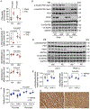
A. TAC increased cardiac PDH activity in control hearts 3 days after TAC, which was significantly inhibited by PKM1 deficiency. Pint<0.05. n=6 to 8. B. Mouse heart tissues were subjected to Western blotting 3 days after either sham or TAC. PDH phosphorylation and mitochondrial oxidative phosphorylation (OXPHOS) proteins were detected. C. Quantification of B showed that PDH phosphorylation (inhibitory) remained high in PKM1 cKO hearts after TAC, whereas NDUFB8 (complex I) and SDHB (complex II) were reduced. For NDUFB8, Pint<0.05; for all others, Pint>0.05. n=5 to 6. D. Immunoblotting analysis of PDH phosphorylation and OXPHOS proteins in NRVMs stimulated with either vehicle or PE after PKM1 knockdown. E. Quantification of D showed that PDH phosphorylation (inhibitory) was decreased by PE treatment, and PKM1 silencing prevented the decline. NDUFB8 (complex I) expression was reduced by PKM1 knockdown. All Pint<0.05. n=6.F. ATP level was decreased by PKM1 silencing in NRVMs, which was restored by supplementation of pyruvate. n=6 to 15. G. PKM1 protein expression was reduced in the failing human heart compared to the normal heart. Human heart tissue samples were obtained from patients undergoing heart transplantation who had been previously diagnosed with heart failure. Immunohistochemistry for PKM1 was conducted. Scale: 50 μm. Two-way ANOVA was conducted, followed by Tukey’s multiple comparisons test (A, C, E). Three-way ANOVA was conducted, followed by Tukey’s multiple comparisons test (F). Data are represented as mean±SEM.

References

    1. Virani SS, Alonso A, Benjamin EJ, Bittencourt MS, Callaway CW, Carson AP, Chamberlain AM, Chang AR, Cheng S, Delling FN, et al.Heart disease and stroke statistics-2020 update: a report from the American Heart Association. Circulation. 2020;141:e139–e596. - PubMed
    1. Drazner MH. The progression of hypertensive heart disease. Circulation. 2011;123:327–334. - PubMed
    1. Nakamura M and Sadoshima J. Mechanisms of physiological and pathological cardiac hypertrophy. Nat Rev Cardiol. 2018;15:387–407. - PubMed
    1. Hill JA and Olson EN. Cardiac plasticity. New Engl J Med. 2008;358:1370–1380. - PubMed
    1. Doenst T, Nguyen TD and Abel ED. Cardiac metabolism in heart failure: implications beyond ATP production. Circ Res. 2013;113:709–724. - PMC - PubMed

Publication types

MeSH terms