. 2021 Jul 16;373(6552):eabe4832.
doi: 10.1126/science.abe4832. Epub 2021 Jun 8.

Senolytics reduce coronavirus-related mortality in old mice

Affiliations

Senolytics reduce coronavirus-related mortality in old mice

Christina D Camell et al. Science. .

Abstract

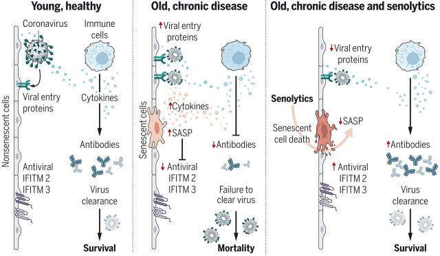
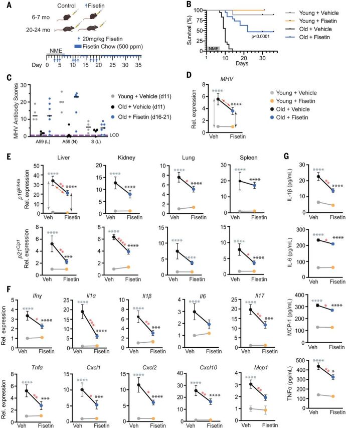
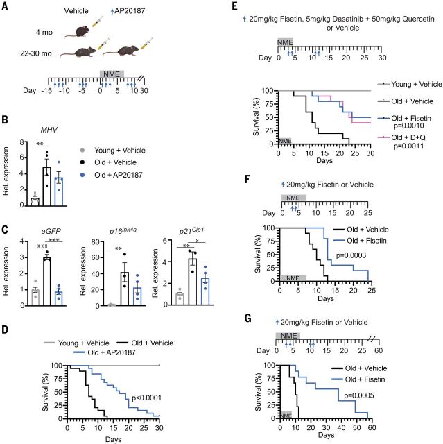
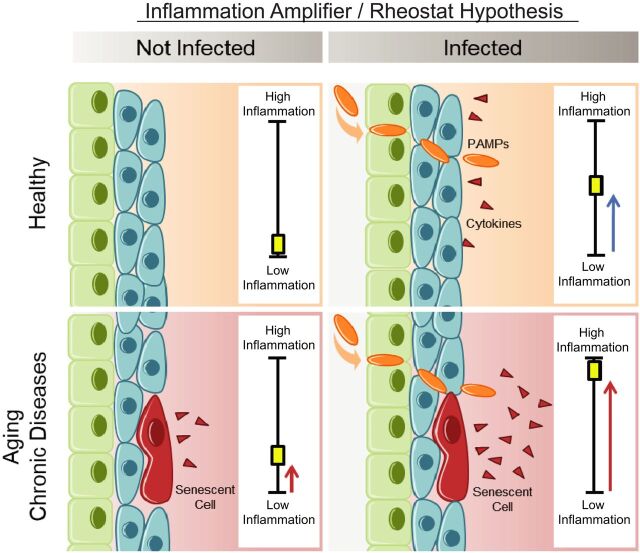
The COVID-19 pandemic has revealed the pronounced vulnerability of the elderly and chronically ill to severe acute respiratory syndrome coronavirus 2 (SARS-CoV-2)-induced morbidity and mortality. Cellular senescence contributes to inflammation, multiple chronic diseases, and age-related dysfunction, but effects on responses to viral infection are unclear. Here, we demonstrate that senescent cells (SnCs) become hyper-inflammatory in response to pathogen-associated molecular patterns (PAMPs), including SARS-CoV-2 spike protein-1, increasing expression of viral entry proteins and reducing antiviral gene expression in non-SnCs through a paracrine mechanism. Old mice acutely infected with pathogens that included a SARS-CoV-2-related mouse β-coronavirus experienced increased senescence and inflammation, with nearly 100% mortality. Targeting SnCs by using senolytic drugs before or after pathogen exposure significantly reduced mortality, cellular senescence, and inflammatory markers and increased antiviral antibodies. Thus, reducing the SnC burden in diseased or aged individuals should enhance resilience and reduce mortality after viral infection, including that of SARS-CoV-2.


Figures

None
SnCs that accumulate with age or chronic disease react to PAMPs such as SARS-CoV-2 S1 by amplifying the SASP, which increases viral entry protein expression and decreases viral defense IFITMs in normal cells.
Old mice exposed to pathogens such as the β-coronavirus MHV have increased inflammation and higher mortality. Treatment with a senolytic decreased SnCs, inflammation, and mortality and increased the antiviral antibody response.
Fig. 1
Fig. 1. The SASP is amplified by PAMP factors.
(A) Human adipocyte progenitors isolated from subcutaneous fat biopsies were induced to undergo senescence with 10 gray (Gy) of ionizing radiation (SnC) or not (non-SnC) (n = 5 subjects). Cells were treated with 10 ng of the prototype PAMP LPS for 3 hours before RNA isolation. Gene expression was measured with quantitative PCR, and the expression in LPS-treated cells was normalized to vehicle-treated samples. Means ± SEM. Statistical significance was calculated by using a mixed effect model for the effect of LPS on SnCs and its differential effects on SnCs compared with non-SnCs. Details are available in table S1. Arrows and asterisks: gray, vehicle-treated SnCs versus non-SnCs; black, LPS-treated SnCs versus non-SnCs; red, SnCs ± LPS. *P < 0.05, **P < 0.01, ***P < 0.001, ****P < 0.0001. (B) Young (2-month-old) and old (26-month-old) mice were treated with phosphate-buffered saline (PBS) (n = 5 young and 5 old) or LPS (n = 4 young and 3 old), and tissues were collected 24 hours later. RNA was isolated from liver, and gene expression measured by means of quamtitative PCR. Expression in LPS-treated mice was normalized to vehicle-treated animals. Means ± SEM, two-way analysis of variance (ANOVA) and post hoc comparison Tukey’s honestly significant difference used to compare the two animal cohorts within a treatment group. Arrows and asterisks: gray, vehicle-treated old versus young; black, LPS-treated old versus young; red, old ± LPS. **P < 0.01, ***P < 0.001, ****P < 0.0001. Kidney data are provided in fig. S2. (C) Serum protein from the same mice measured with enzyme-linked immunosorbent assay (ELISA). Statistics are as described in (B).
Fig. 2
Fig. 2. The SARS-CoV2 spike protein-1 (S1) exacerbates the secretory phenotype of senescent human endothelial cells, decreasing viral defenses and elevating viral entry/processing gene expression.
(A) Primary human kidney endothelial cells (n = 9 biological replicates) were induced to undergo senescence with 10 Gy of ionizing radiation (SnC) or not (non-SnC) then treated with 500 ng recombinant S1 or PBS vehicle for 24 hours. Thirty SASP-related proteins were measured in the conditioned media (CM) by means of Luminex xMAP technology. Relative abundance induced by S1, normalized to vehicle treated non-SnCs (non-SnC + Veh), is illustrated in the heat map. A mixed effects model was used to test the effect of S1, senescence, and their interaction, taking into account duplicate measures within a subject for each protein as well as the composite score. Margin effects of SnCs in the treatment group also were tested under the mixed-effects model framework. Overall, the effect of S1 on SnCs was significantly more pronounced than on non-SnCs (composite score change P < 0.0089; mean values and P values for each cytokine are in table S2). (B) Schematic of experiments in (C), (E), and (F). Primary human cells were induced to undergo senescence with 10 Gy of ionizing radiation (SnC) or not (non-SnC). Twenty days later, CM was collected (n = 4 biological replicates) and used to treat non-SnCs (n = 4 biological replicates) either with or without neutralizing antibodies to IL-1α, IL-18, and PAI-1 (alone or in combination) for 48 hours, then RNA was isolated to measure expression of genes related to SARS-CoV-2 pathogenesis by means of quantitative PCR. Expression in cells treated with SnC CM was normalized to cells treated with non-SnC CM. Data are displayed as mean ± SEM, mixed-effects model. *P < 0.05 **P < 0.01, ***P < 0.001, ****P < 0.0001. (C) IFITM expression in human kidney endothelial cells treated with CM from SnC versus non-SnC human kidney endothelial cells. (D) IFITM expression in human kidney endothelial cells exposed to two concentrations of IL-1α (n = 4 biological replicates). Expression was normalized to vehicle-treated samples. (E) Gene expression in human lung epithelial cells treated with CM from SnC versus non-SnC preadipocytes, HUVECs, or kidney endothelial cells. (F) TMPRSS2 expression in human kidney endothelial cells treated with CM from SnC versus non-SnC kidney endothelial cells with or without neutralizing antibodies or human kidney endothelial cells with recombinant IL-1α for 48 hours. (G) Human lung biopsies acquired for clinical indications of focal, noninfectious causes from elderly patients were stained for TMPRSS2, p16INK4a, and 4′,6-diamidino-2-phenylindole (DAPI) to detect nuclei (n = 5 subjects). Representative images are shown. Scale bar, 20 μm. (H) TMPRSS2+, p16INK4a+, and total nuclei were counted and expressed as a function of total nuclei in each field. TMPRSS2+ and p16INK4a+ cells/field were tightly linked (P < 0.0001; partial Pearson correlation). Each color series of dots indicates replicates from a single subject.
Fig. 3
Fig. 3. Old mice are vulnerable to a NME that includes acute mouse β-coronavirus infection.
(A) Young (3-month-old) and old (20- to 24-month-old) WT mice were exposed to NME bedding produced from pet store mice for 7 days. Survival was monitored for 35 days after initiation of NME (n = 10 young; n = 18 old). Log-rank (Mantel Cox) test. (B) Gene expression in three tissues of SPF or NME (6- to 7-day exposure) young and old mice (n = 3 young SPF; n = 5 old SPF; n = 14 young NME; n = 13 old NME) measured with quantitative PCR. Expression was normalized to young SPF mice. Means ± SEM, two-way ANOVA and post hoc comparison Tukey’s honestly significant difference were used to compare the two animal cohorts within a treatment group. *P < 0.05, **P < 0.01, ***P < 0.001, ****P < 0.0001. Arrows and asterisks: gray, SPF old versus young; black, NME old versus young; red, old SPF versus old NME. (C) Serum cytokine levels in young and old mice (n = 3 young SPF; n = 5 old SPF; n = 19 young NME; n = 17 old NME) measured with ELISA at day 5 after NME. Statistics are as described in (B). (D) Serology to detect antibodies against microbes in NME bedding. (Right) The mouse pathogens commonly tested for by Charles River Laboratory to define SPF housing. The pie charts illustrate the exposures detected in individual young and old mice (n = 24 young; n = 21 to 23 old) day 11 after initiation of NME. Serology of pet store mice is illustrated below. (E) Representative images of hematoxylin and eosin (H&E) staining or MHV immunohistochemistry in liver sections from young and old mice exposed to NME. (F) (Top) Schematic to illustrate the experimental design. Young (6-month-old) or old (22-month-old) female mice were inoculated with a sublethal dose of MHV. Thirty days later, naïve and inoculated mice were exposed to NME bedding for 3 weeks. (Bottom) Serum antibodies against three different MHV antigens measured 21 days after MHV inoculation and reported as relative scores. The dotted line indicates the limit of detection (LOD). Means ± SEM, unpaired two-tailed Student’s t test. **P < 0.01, ****P < 0.0001. (G) Survival of MHV-inoculated and naïve mice measured for 42 days after initiation of NME. Log-rank (Mantel Cox) test.
Fig. 4
Fig. 4. Treatment with the senolytic fisetin decreases mortality in NME-exposed old mice.
(A) Schematic of the experiment. Young (6 to 7 months) and old (20 to 24 months) mice were exposed to NME bedding containing mouse β-coronavirus MHV for 7 days. Mice were treated with 20 mg/kg/day Fisetin or vehicle only by means of oral gavage daily for 3 consecutive days starting on day 3 after initiation of NME. The 3 days of treatment were repeated (3 days on, 4 days off) for 3 weeks. Animals were also fed standard chow with Fisetin added (500 ppm) ad libitum after initiation of treatment. (B) Survival was measured for 36 days after initiation of NME (n = 9 young + vehicle; n = 5 young + Fisetin; n = 18 old + vehicle; n = 19 old + Fisetin). Log-rank (Mantel Cox) test. P < 0.0001 for old mice ± Fisetin. (C) Relative MHV antibody score in young and old mice in (B) on the indicated day after initiation of NME. (D to G) Young (2-month-old) and old (20-month-old) mice were exposed to NME bedding ± treatment with Fisetin as described in (A). On days 8 to 9 after initiation of NME, animals were euthanized, and tissues collected for measuring gene expression (n = 10 young + vehicle; n = 8 to 10 young + Fisetin; n = 10 to 11 old + vehicle; n = 13 old + Fisetin). All expression data were normalized to young mice treated with vehicle. Data are displayed as means ± SEM, two-way ANOVA and post-hoc comparison Tukey’s honestly significant difference used to compare the two animal cohorts within a treatment group. Arrows and asterisks: gray, vehicle-treated old versus young; black, Fisetin-treated old versus young; red, old ± Fisetin. *P < 0.05, **P < 0.01, ***P < 0.001, ****P < 0.0001. (D) MHV mRNA was quantified by means of quantitative PCR in fecal pellets collected from individual animals in (C). (E) Quantification of p16Ink4a and p21Cip1 mRNA in four tissues. (F) Quantification of SASP factor mRNA in liver. Data on other genes and tissues are available in fig. S9. (G) SASP protein levels in the liver measured with ELISA.
Fig. 5
Fig. 5. Pharmacologic and genetic ablation of SnCs reduces mortality in old mice exposed to NME.
(A) Schematic diagram of the experimental design for (B) to (D). Young (4-month-old) and old (22- to 30-month-old) male and female INK-ATTAC mice were treated with vehicle or AP20187 (n = 10 young; n = 19 old + vehicle; n = 19 old + AP20187) to dimerize FKBP-caspase-8 fusion protein expressed in p16Ink4a+ cells to kill SnC selectively. AP20187 (10 mg/kg) or vehicle was administered intraperitoneally daily for 3 days starting 2 weeks before initiating NME and ending 1 week after (3 days on and 4 days). NME was started on day 0 and lasted 1 week. Mice housed in SPF conditions were used as controls. Tissues were collected 7 days after initiation of NME in another cohort of male animals for molecular analysis (n = 5 young ; n = 3 or 4 old + vehicle; n = 4 old + AP20187). (B) Quantification of MHV mRNA in fecal pellets isolated from individual mice. Means ± SEM, one-way ANOVA with Tukey’s test. **P < 0.01. (C) Quantification of eGFP (a reporter of p16Ink4a expression in the INK-ATTAC construct), p16Ink4a, and p21Cip1 mRNA in the kidney of mice in (B). All expression data were normalized to young mice treated with vehicle. Means ± SEM, one-way ANOVA. *P < 0.05, **P < 0.01, ***P < 0.001. (D) Survival of male and female mice measured for 30 days after initiation of NME. Log-rank (Mantel Cox) test. (E) Young (2-month-old, n = 5) and old (22-month-old, n = 10/group) female mice were exposed to NME bedding for 4 days. Beginning on day 3, mice were treated with 20 mg/kg Fisetin or 5 mg/kg Dasatinib plus 50 mg/kg Quercetin at days 3, 4, 11, and 12 by means of oral gavage, or with vehicle only. Survival was measured for 30 days after initiation of NME. Log-rank (Mantel Cox) test. (F) Survival curves for 20-month-old WT female mice (n = 10/treatment group) treated with 20 mg/kg Fisetin or vehicle by oral gavage on days 3 and 4 after initiation of NME exposure. Log-rank (Mantel Cox) test. (G) Survival of 22-month-old WT female mice (n = 9/treatment group) treated with 20 mg/kg Fisetin or vehicle only by oral gavage at days 3, 4, 10, and 11 after NME exposure monitored out to 60 days after exposure. Log-rank (Mantel Cox) test.
Fig. 6
Fig. 6. SASP Amplifier/Rheostat hypothesis.
Schematic of the hypothesis generated from these data and tested herein. SnC amplified the response to PAMPs in vitro and in vivo, resulting in increased production of pro-inflammatory cytokines and chemokines. This could exacerbate acute systemic inflammatory responses and cytokine release by innate immune cells and amplify the spread of senescence. This model could explain the increased risk of cytokine storm during COVID-19 or other infections and adverse outcomes observed in the elderly or those with chronic conditions associated with an increased burden of SnC (obesity, diabetes, chronic lung or kidney disease, or cardiovascular disease).

Comment in

Similar articles

  • Fisetin for COVID-19 in skilled nursing facilities: Senolytic trials in the COVID era.
    Verdoorn BP, Evans TK, Hanson GJ, Zhu Y, Langhi Prata LGP, Pignolo RJ, Atkinson EJ, Wissler-Gerdes EO, Kuchel GA, Mannick JB, Kritchevsky SB, Khosla S, Rizza SA, Walston JD, Musi N, Lipsitz LA, Kiel DP, Yung R, LeBrasseur NK, Singh RJ, McCarthy T, Puskarich MA, Niedernhofer LJ, Robbins PD, Sorenson M, Tchkonia T, Kirkland JL. Verdoorn BP, et al. J Am Geriatr Soc. 2021 Nov;69(11):3023-3033. doi: 10.1111/jgs.17416. Epub 2021 Aug 20. J Am Geriatr Soc. 2021. PMID: 34375437 Free PMC article.
  • Treatment with the senolytics dasatinib/quercetin reduces SARS-CoV-2-related mortality in mice.
    Pastor-Fernández A, Bertos AR, Sierra-Ramírez A, Del Moral-Salmoral J, Merino J, de Ávila AI, Olagüe C, Villares R, González-Aseguinolaza G, Rodríguez MÁ, Fresno M, Gironés N, Bustos M, Smerdou C, Fernandez-Marcos PJ, von Kobbe C. Pastor-Fernández A, et al. Aging Cell. 2023 Mar;22(3):e13771. doi: 10.1111/acel.13771. Epub 2023 Jan 26. Aging Cell. 2023. PMID: 36704839 Free PMC article.
  • Virus-induced senescence is a driver and therapeutic target in COVID-19.
    Lee S, Yu Y, Trimpert J, Benthani F, Mairhofer M, Richter-Pechanska P, Wyler E, Belenki D, Kaltenbrunner S, Pammer M, Kausche L, Firsching TC, Dietert K, Schotsaert M, Martínez-Romero C, Singh G, Kunz S, Niemeyer D, Ghanem R, Salzer HJF, Paar C, Mülleder M, Uccellini M, Michaelis EG, Khan A, Lau A, Schönlein M, Habringer A, Tomasits J, Adler JM, Kimeswenger S, Gruber AD, Hoetzenecker W, Steinkellner H, Purfürst B, Motz R, Di Pierro F, Lamprecht B, Osterrieder N, Landthaler M, Drosten C, García-Sastre A, Langer R, Ralser M, Eils R, Reimann M, Fan DNY, Schmitt CA. Lee S, et al. Nature. 2021 Nov;599(7884):283-289. doi: 10.1038/s41586-021-03995-1. Epub 2021 Sep 13. Nature. 2021. PMID: 34517409
  • Senolytic drugs: from discovery to translation.
    Kirkland JL, Tchkonia T. Kirkland JL, et al. J Intern Med. 2020 Nov;288(5):518-536. doi: 10.1111/joim.13141. Epub 2020 Aug 4. J Intern Med. 2020. PMID: 32686219 Free PMC article. Review.
  • Insights into coronavirus immunity taught by the murine coronavirus.
    Grabherr S, Ludewig B, Pikor NB. Grabherr S, et al. Eur J Immunol. 2021 May;51(5):1062-1070. doi: 10.1002/eji.202048984. Epub 2021 Mar 22. Eur J Immunol. 2021. PMID: 33687066 Free PMC article. Review.

Cited by

References

    1. Jin J. M., Bai P., He W., Wu F., Liu X.-F., Han D.-M., Liu S., Yang J.-K., Gender differences in patients with COVID-19: Focus on severity and mortality. Front. Public Health 8, 152 (2020). 10.3389/fpubh.2020.00152 - DOI - PMC - PubMed
    1. Gebhard C., Regitz-Zagrosek V., Neuhauser H. K., Morgan R., Klein S. L., Impact of sex and gender on COVID-19 outcomes in Europe. Biol. Sex Differ. 11, 29 (2020). 10.1186/s13293-020-00304-9 - DOI - PMC - PubMed
    1. Palaiodimos L., Kokkinidis D. G., Li W., Karamanis D., Ognibene J., Arora S., Southern W. N., Mantzoros C. S., Severe obesity, increasing age and male sex are independently associated with worse in-hospital outcomes, and higher in-hospital mortality, in a cohort of patients with COVID-19 in the Bronx, New York. Metabolism 108, 154262 (2020). 10.1016/j.metabol.2020.154262 - DOI - PMC - PubMed
    1. Ruan Q., Yang K., Wang W., Jiang L., Song J., Clinical predictors of mortality due to COVID-19 based on an analysis of data of 150 patients from Wuhan, China. Intensive Care Med. 46, 846–848 (2020). 10.1007/s00134-020-05991-x - DOI - PMC - PubMed
    1. Huang C., Wang Y., Li X., Ren L., Zhao J., Hu Y., Zhang L., Fan G., Xu J., Gu X., Cheng Z., Yu T., Xia J., Wei Y., Wu W., Xie X., Yin W., Li H., Liu M., Xiao Y., Gao H., Guo L., Xie J., Wang G., Jiang R., Gao Z., Jin Q., Wang J., Cao B., Clinical features of patients infected with 2019 novel coronavirus in Wuhan, China. Lancet 395, 497–506 (2020). 10.1016/S0140-6736(20)30183-5 - DOI - PMC - PubMed

Publication types

MeSH terms