Review
. 2021 Sep;70(9):1782-1794.
doi: 10.1136/gutjnl-2020-323888. Epub 2021 Jun 8.

Hepatitis D virus in 2021: virology, immunology and new treatment approaches for a difficult-to-treat disease

Affiliations
Review

Hepatitis D virus in 2021: virology, immunology and new treatment approaches for a difficult-to-treat disease

Stephan Urban et al. Gut. 2021 Sep.

Abstract

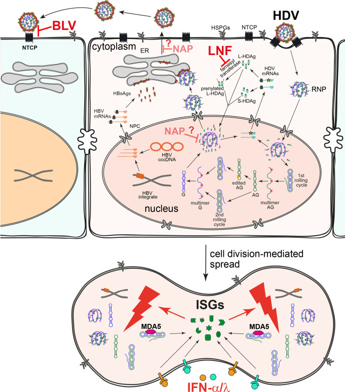
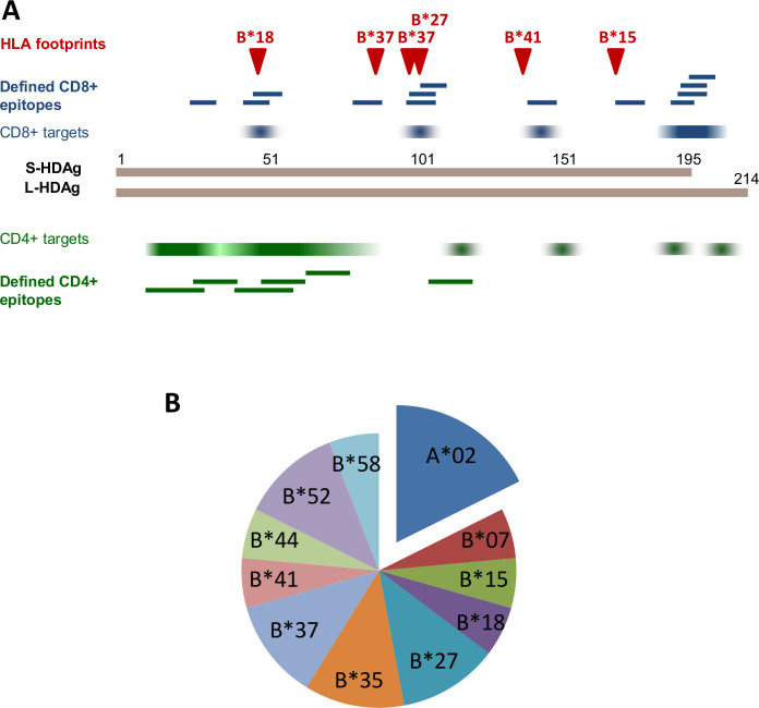
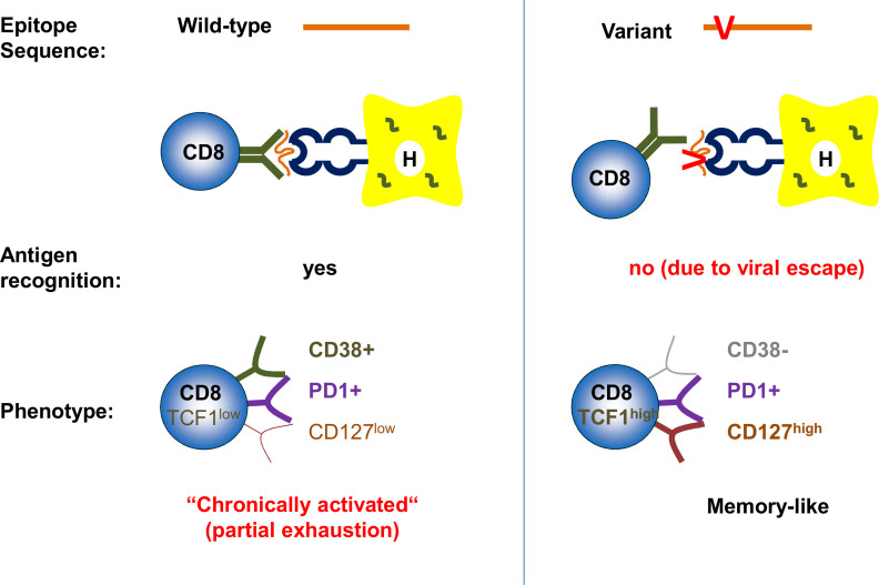
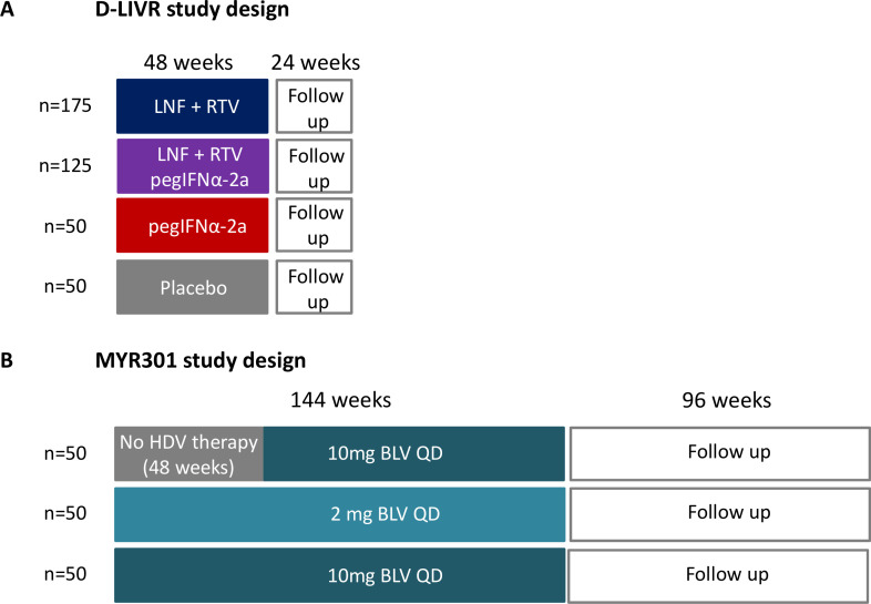
Approximately 5% of individuals infected with hepatitis B virus (HBV) are coinfected with hepatitis D virus (HDV). Chronic HBV/HDV coinfection is associated with an unfavourable outcome, with many patients developing liver cirrhosis, liver failure and eventually hepatocellular carcinoma within 5-10 years. The identification of the HBV/HDV receptor and the development of novel in vitro and animal infection models allowed a more detailed study of the HDV life cycle in recent years, facilitating the development of specific antiviral drugs. The characterisation of HDV-specific CD4+ and CD8+T cell epitopes in untreated and treated patients also permitted a more precise understanding of HDV immunobiology and possibly paves the way for immunotherapeutic strategies to support upcoming specific therapies targeting viral or host factors. Pegylated interferon-α has been used for treating HDV patients for the last 30 years with only limited sustained responses. Here we describe novel treatment options with regard to their mode of action and their clinical effectiveness. Of those, the entry-inhibitor bulevirtide (formerly known as myrcludex B) received conditional marketing authorisation in the European Union (EU) in 2020 (Hepcludex). One additional drug, the prenylation inhibitor lonafarnib, is currently under investigation in phase III clinical trials. Other treatment strategies aim at targeting hepatitis B surface antigen, including the nucleic acid polymer REP2139Ca. These recent advances in HDV virology, immunology and treatment are important steps to make HDV a less difficult-to-treat virus and will be discussed.

Keywords: antiviral therapy; chronic viral hepatitis; hepatitis D; immunology in hepatology.


Conflict of interest statement

Competing interests: SU: Advisory Board/Speaker Bureau for: GILEAD SCIENCES, MYR, VIRBIO, ASSEMBLY, JANSSEN, ENYO, PEPPERPRINT, ALIGOS. CN-H: Speaker Bureau for: ABBVIE, Falk Foundation, Novartis, MSD. PL: Advisory Board/Speaker Bureau for: BMS, ROCHE, GILEAD SCIENCES, GSK, ABBVIE, MSD, ARROWHEAD, ALNYLAM, JANSSEN, SBRING BANK, MYR, EIGER.

Figures

Figure 1
Figure 1
Structures of HDV virion and genome. (A) Schematic representation of HDV virion (left) and envelope proteins (right). HDV virion has a ribonucleoprotein (RNP) complex inside and an HBV derived envelope outside. The RNP consists of the HDV genome and two isoforms of hepatitis D antigen (HDAg), L-HDAg and S-HDAg. Prenylation of L-HDAg is essential for envelope acquisition. The envelope contains three HBV envelope proteins: small-HBsAg (S-HBsAg), medium-HBsAg (M-HBsAg) and large-HBsAg (L-HBsAg). M and L share the same sequence with S, however, contain N-terminal extensions: preS2 for M and preS1 plus preS2 for L. The preS1 domain of L is critical for binding of the receptor sodium taurocholate cotransporting polypeptide (NTCP), while the cytosolic loops (CLs) are important for encapsulation of HDV RNP through interaction with HDAg. (B) HDV genome structure and key elements. As a single-strand circular RNA, HDV genome forms an unbranched rod-like structure through high rate of intramolecular base-pairing. A representative region consisting of short stems and bulges is depicted on top. S-HDAg and L-HDAg are encoded by unedited and adenosine deaminases acting on RNA 1 (ADAR1)-edited (Amber stop codon to TGG (W)) genomic RNAs, respectively. The C terminal prenylation motif (CXXQ) is indicated. The numbering of nucleotide and protein sequences is based on a HDV genotype one strain (GenBank: M21012.1). HBsAg, hepatitis B surface antigen; HBV, hepatitis B virus; HDV, hepatitis D virus; L-HDAg, large HDAg; S-HDAg, small HDAg.
Figure 2
Figure 2
HDV life cycle, spreading pathways and drug targets. HDV virions first attach to heparan sulfate proteoglycans (HSPGs) and then to the viral receptor NTCP to enter host cells. After membrane fusion, the ribonucleoprotein (RNP) is released and further transported to the nucleus to initiate RNA replication. The incoming genome (G) serves as the template for the first rolling circle amplification. The resulting antigenome (AG) multimers are cleaved in cis by the intrinsic ribozyme and ligated into circular monomers. After a second rolling cycle using the AG as the template, HDV G multimers are synthesised and further cleaved to produce monomers. The HDV AG might be edited by ADAR1, yielding an extended HDAg ORF that produces L-HDAg, some of that is further prenylated. S-HDAg and L-HDAg (intact and prenylated) are transported into the nucleus to regulate virus replication or bind to the HDV RNA to form RNP. The G-containing RNP can be exported to the cytoplasm and encapsulated into HBV envelope through the interaction between L-HDAg and S-HBsAg. HDV virions are released through the ER-Golgi secretory pathway. besides the HBV envelope-dependent de novo infection, HDV can also spread through division of infected cells in an HBV-independent manner (below). Bulevirtide (BLV) blocks de novo infection by efficient binding of the viral receptor NTCP. Lonafarnib (LNF) prevents the prenylation of L-HDAg by inhibiting the farnesyl transferase and consequently impairs HDV assembly and secretion. The target(s) of nucleic acid polymer (NAP) is unclear. It may inhibit assembly/release of HDV virions and/or HDV ribonucleoprotein assembly via direct interaction with the HDAg. IFNs, including MDA5-mediated HDV-induced IFNs and therapeutic IFNα and IFNλ, induce IFN stimulated genes (ISGs) which profoundly suppress HDV amplification during cell division. HBV, hepatitis B virus; HDV, hepatitis D virus; IFN, interferon; L-HDAg, large hepatitis D antigen; NTCP, sodium taurocholate cotransporting polypeptide; S-HBsAg, small hepatitis B surface antigen.
Figure 3
Figure 3
HDV regions targeted by HDV-specific CD4+ and CD8+ T cell epitopes. (A) Dominantly targeted CD8+ and CD4+ epitope regions (identified in olp studies) are indicated in blue and green colour, respectively, with intensity representing frequency of recognition. Fine-mapped CD8+ and CD4+ T cell epitopes are indicated by blue and green bars, respectively. HLA class I associated HDV polymorphisms (‘HLA footprints’) that correlate to fine-mapped epitopes are depicted by red arrowheads. (B) HLA restriction of fine-mapped HDV-specific CD8+ T cell epitopes, demonstrating a clear dominance of HLA-B restriction. HDV, hepatitis D virus; L-HDAg, large hepatitis D antigen; S-HDAg, small HDAg.
Figure 4
Figure 4
Mechanisms of HDV-specific CD8+ T cell failure. HDV-specific CD8+ T cells targeting viral epitopes with wild-type sequence display a chronically activated phenotype and are functionally partially exhausted (left), HDV-specific CD8+ T cells targeting viral epitopes with sequence variations (viral escape) do not recognise the antigen anymore and display a memory-like phenotype (right). HDV, hepatitis D virus.
Figure 5
Figure 5
Study design of the two ongoing phase III studies assessing the efficacy and safety of new therapeutic regimens against HDV. (A) D-LIVR study. LNF +RTV: LNF 50 mg two times per day+RTV 100 mg two times per day. Primary endpoint: ≥2 log10 IU/mL decline in HDV RNA and ALT normalisation in week 48. All patients will be maintained on background HBV nucleoside/nucleotide analogue therapy. (B) MYR301 study. primary endpoint: undetectable HDV RNA or decrease by ≥2log10 IU/mL and ALT normalisation in week 48. If indicated treatment with nucleoside/nucleotide analogues according to European Association for the Study of the Liver (EASL)/American Association for the Study of the Liver (AASLD) guidelines. ALT, alanine aminotransferase; BLV, bulevirtide; HBV, hepatitis B virus; HDV, hepatitis D virus; LNF, lonafarnib; pegIFNα, pegylated interferon-α; RTV, ritonavir;

References

    1. Lempp FA, Ni Y, Urban S. Hepatitis delta virus: insights into a peculiar pathogen and novel treatment options. Nat Rev Gastroenterol Hepatol 2016;13:580–9. 10.1038/nrgastro.2016.126 - DOI - PubMed
    1. Lucifora J, Delphin M. Current knowledge on hepatitis delta virus replication. Antiviral Res 2020;179:104812. 10.1016/j.antiviral.2020.104812 - DOI - PubMed
    1. Perez-Vargas J, Amirache F, Boson B, et al. . Enveloped viruses distinct from HBV induce dissemination of hepatitis D virus in vivo. Nat Commun 2019;10:10. 10.1038/s41467-019-10117-z - DOI - PMC - PubMed
    1. Chemin I, Pujol FH, Scholtès C, et al. . Preliminary evidence for hepatitis delta virus exposure in patients who are apparently not infected with hepatitis B virus. Hepatology 2021;73:861–4. 10.1002/hep.31453 - DOI - PMC - PubMed
    1. Pflüger LS, Schulze Zur Wiesch J, Polywka S, et al. . Hepatitis delta virus propagation enabled by hepatitis C virus-Scientifically intriguing, but is it relevant to clinical practice? J Viral Hepat 2021;28:213–6. 10.1111/jvh.13385 - DOI - PubMed

Publication types