. 2021 Jun 8;12(6):593.
doi: 10.1038/s41419-021-03865-8.

RIP3 impedes transcription factor EB to suppress autophagic degradation in septic acute kidney injury

Affiliations

RIP3 impedes transcription factor EB to suppress autophagic degradation in septic acute kidney injury

Ruizhao Li et al. Cell Death Dis. .

Abstract

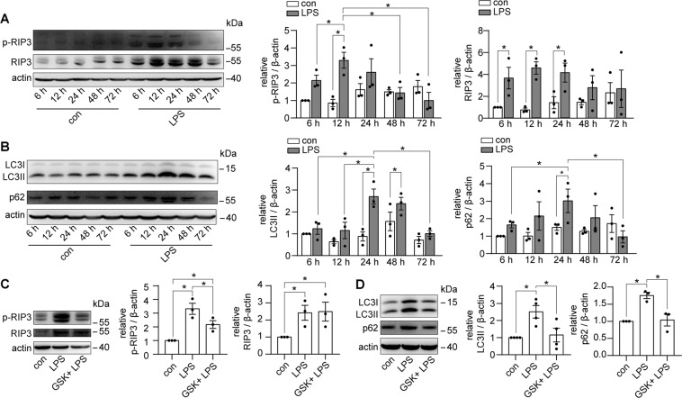
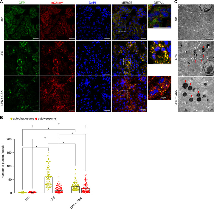
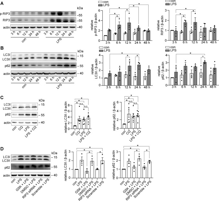
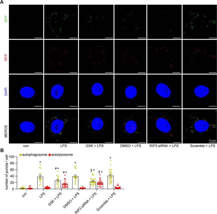
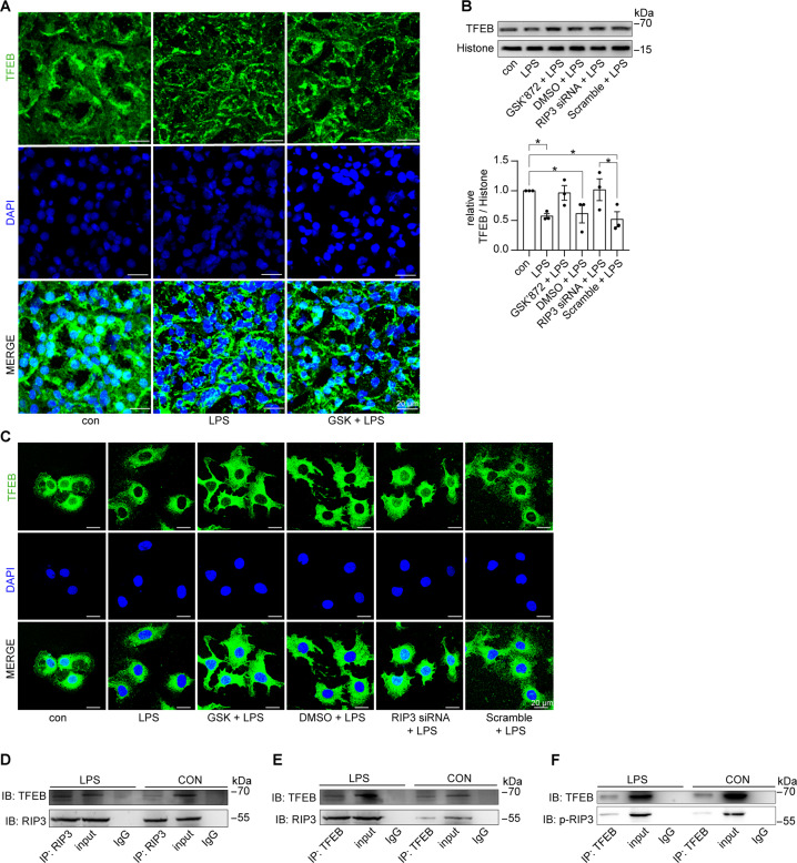
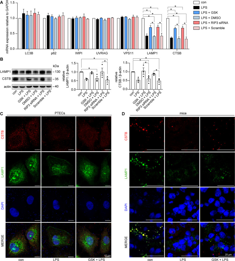
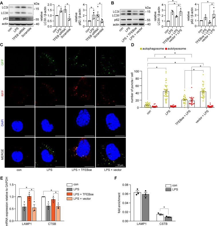
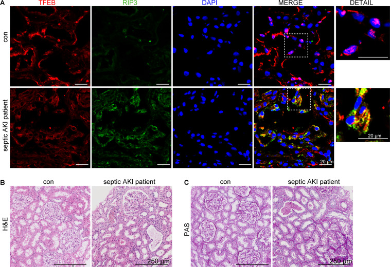
Autophagy is an important renal-protective mechanism in septic acute kidney injury (AKI). Receptor interacting protein kinase 3 (RIP3) has been implicated in the renal tubular injury and renal dysfunction during septic AKI. Here we investigated the role and mechanism of RIP3 on autophagy in septic AKI. We showed an activation of RIP3, accompanied by an accumulation of the autophagosome marker LC3II and the autophagic substrate p62, in the kidneys of lipopolysaccharide (LPS)-induced septic AKI mice and LPS-treated cultured renal proximal tubular epithelial cells (PTECs). The lysosome inhibitor did not further increase the levels of LCII or p62 in LPS-treated PTECs. Moreover, inhibition of RIP3 attenuated the aberrant accumulation of LC3II and p62 under LPS treatment in vivo and in vitro. By utilizing mCherry-GFP-LC3 autophagy reporter mice in vivo and PTECs overexpression mRFP-GFP-LC3 in vitro, we observed that inhibition of RIP3 restored the formation of autolysosomes and eliminated the accumulated autophagosomes under LPS treatment. These results indicated that RIP3 impaired autophagic degradation, contributing to the accumulation of autophagosomes. Mechanistically, the nuclear translocation of transcription factor EB (TFEB), a master regulator of the lysosome and autophagy pathway, was inhibited in LPS-induced mice and LPS-treated PTECs. Inhibition of RIP3 restored the nuclear translocation of TFEB in vivo and in vitro. Co-immunoprecipitation further showed an interaction of RIP3 and TFEB in LPS-treated PTECs. Also, the expression of LAMP1 and cathepsin B, two potential target genes of TFEB involved in lysosome function, were decreased under LPS treatment in vivo and in vitro, and this decrease was rescued by inhibiting RIP3. Finally, overexpression of TFEB restored the autophagic degradation in LPS-treated PTECs. Together, the present study has identified a pivotal role of RIP3 in suppressing autophagic degradation through impeding the TFEB-lysosome pathway in septic AKI, providing potential therapeutic targets for the prevention and treatment of septic AKI.


Conflict of interest statement

The authors declare no competing interests.

Figures

Fig. 1
Fig. 1. RIP3 was activated to inhibit autophagy in septic AKI mice.
A Kidney tissues were collected from C57BL/6 mice treated with LPS or sterilized saline (con) for immunoblot of phosphorylated RIP3 (p-RIP3, a marker of RIP3 activity) and total RIP3. p-RIP3 and RIP3 expression, against β-actin expression, were increased in the kidneys of LPS-induced mice (n = 3). B Immunoblot showed an accumulation of the expression of LC3II (marker for autophagosome) and p62 (substrate for autophagy) in the kidneys of LPS-induced mice (n = 3). C C57BL/6 mice were treated with sterilized saline (con), LPS, or LPS plus GSK’872 (GSK) for 12 h. Immunoblotting showed that GSK attenuated the increased p-RIP3 in the kidneys of LPS-induced mice (n = 3). D C57BL/6 mice were treated with sterilized saline (con), LPS, or LPS plus GSK for 24 h. Immunoblot showed that GSK attenuated the accumulated LC3II and p62 in the kidneys of LPS-induced mice (n = 3–4). *P < 0.05.
Fig. 2
Fig. 2. RIP3 was activated to suppress autophagic flux in septic AKI mice.
A Confocal laser scanning microscopy images of kidney tissues from renal tubular cell-specific mCherry-GFP-LC3 reporter mice treated as indicated. Scale bar = 50 μm for the left four lanes, 25 μm for the detailed images. B Quantitative analysis showed few autophagosomes (yellow puncta) and autolysosomes (red-only puncta) were detected in the renal tubular cells of the control group. The number of autophagosomes were largely increased, with a slight increase in the number of autolysosomes, in the LPS group. GSK induced the formation of autolysosomes and decreased the LPS-induced accumulation of autophagosomes (n = 3–4, 32–86 tubules from each group). C TEM showed that few autophagic vacuoles were detected in the renal tubular cells of the control mice. A lot of initial autophagic vacuoles (arrows) but few late autophagic vehicles (arrowheads) were detected in the renal tubular cells of LPS-induced mice. GSK induced the formation of late autophagic vehicles and decreased the number of initial autophagic vacuoles. Scale bar = 2 μm. *P < 0.05.
Fig. 3
Fig. 3. RIP3 activation inhibited autophagic degradation in LPS-induced cultured PTECs.
A, B Cultured PTECs were treated with DMSO or LPS for different time points as indicated. A Immunoblotting showed that the expression of p-RIP3 and RIP3 was increased at 6 and 12 h LPS treatment (n = 3). B Immunoblotting showed an increase in the expression of LC3II and p62 at 24 h LPS treatment (n = 3). C Cultured PTECs were treated with the lysosome inhibitor chloroquine (CQ), LPS, or LPS plus CQ for 24 h. Immunoblotting showed that the addition of CQ did not further increase the levels of LC3II and p62 in LPS-treated PTECs (n = 6). D Immunoblot of LC3 and p62 in cultured PTECs treated with LPS, LPS plus GSK’872 (GSK), LPS plus DMSO, LPS plus RIP3 siRNA, or LPS plus scrambled siRNA (scramble) for 24 h. GSK or RIP3 siRNA downregulated the LPS-induced increase of LC3II and p62 (n = 3–4). *P < 0.05. NS for nonsignificant.
Fig. 4
Fig. 4. RIP3 activation suppressed autophagic flux in LPS-induced cultured PTECs.
A, B Cultured PTECs transfected with mRFP-GFP-LC3 adenovirus and treated as indicated for 24 h. Representative confocal laser scanning microscopy images (A) and quantitative analysis (B) of autophagosomes (yellow puncta) and autolysosomes (red-only puncta). Few autophagosomes and autolysosomes were observed in the control group. The number of autophagosomes but not autolysosomes was increased in the LPS-treated PTECs (LPS, LPS + DMSO, and LPS + Scramble group). GSK or RIP3 siRNA induced the formation of autolysosomes, whereas it decreased the accumulation of autophagosomes in LPS-induced PTECs (n = 3–4, 21–26 cells from each group). *P < 0.05, significantly different from the control group. #P < 0.05, significantly different from the DMSO + LPS group. P < 0.05, significantly different from the scramble + LPS group.
Fig. 5
Fig. 5. RIP3 impeded the nuclear translocation of TFEB in septic AKI mice and LPS-treated cultured PTECs.
A Immunofluorescence staining for TFEB (green) and DAPI (blue) in the kidneys from C57BL/6 mice treated with sterilized saline (con), LPS, or LPS plus GSK’872 (GSK) for 24 h. GSK partially restored the decreased nuclear staining of TFEB in the renal tubular cells of LPS-induced mice. Scale bar = 20 μm. B Cultured PTECs treated with LPS, LPS plus GSK, LPS plus DMSO, LPS plus RIP3 siRNA, or LPS plus scrambled siRNA (Scramble) for 12 h. The nuclear protein fractions were immunoblotted for TFEB. Histone was used as the nuclear marker. TFEB expression against Histone was decreased by LPS treatment, which was restored by GSK or RIP3 siRNA (n = 3). C Immunofluorescence staining for TFEB (green) and DAPI (blue) in cultured PTECs treated as indicated. The nuclear translocation of TFEB was inhibited by LPS, which was rescued by GSK or RIP3 siRNA. Scale bar = 20 μm. D, E Lysates from cultured PTECs treated with LPS or DMSO (con) for 12 h were subjected to immunoprecipitation using an anti-RIP3 antibody (D) or anti-TFEB antibody (E), and IgG antibody followed by immunoblot for TFEB and RIP3. Input proteins were detected with anti-RIP3 and anti-TFEB antibodies. RIP3 interacted with TFEB in LPS-treated PTECs compared to the control group. F Lysates from cultured PTECs treated with LPS or DMSO (con) for 12 h were subjected to immunoprecipitation using anti-TFEB antibody and IgG antibody followed by immunoblot for p-RIP3. Input proteins were detected with anti-p-RIP3 and anti-TFEB antibodies. p-RIP3 interacted with TFEB in LPS-treated PTECs compared to the control group. *P < 0.05.
Fig. 6
Fig. 6. The expression of LAMP1 and CTSB was suppressed by RIP3 in LPS-induced cultured PTECs and septic AKI mice.
A RT-qPCR analyses of the expression of potential TFEB target genes in cultured PTECs treated with LPS, LPS plus GSK’872 (GSK), LPS plus DMSO, LPS plus RIP3 siRNA, or LPS plus scrambled siRNA (Scramble) for 12 h. The mRNA levels of LAMP1 and CTSB were inhibited by LPS, which was restored by GSK or RIP3 siRNA (n = 3). B Immunoblotting showed that the protein levels of LAMP1 and CTSB was decreased by LPS, which was restored by GSK’872 or RIP3 siRNA in cultured PTECs treated as indicated for 24 h (n = 3). C Immunofluorescence staining for CTSB (red), LAMP1 (green), and DAPI (blue) in cultured PTECs treated as indicated for 24 h. GSK restored the decreased expression of CTSB and LAMP1 in LPS-treated PTECs. Scale bar = 10 μm. D Immunofluorescence staining for CTSB (red), LAMP1 (green), and DAPI (blue) in the kidneys from C57BL/6 mice treated with vehicle (con), LPS, or LPS plus GSK for 24 h. The expression of CTSB and LAMP1 were suppressed in the renal tubular cells of LPS-induced mice, which were partially restored by GSK. Scale bar = 25 μm. *P < 0.05.
Fig. 7
Fig. 7. TFEB regulated the autophagic degradation in LPS-treated cultured PTECs.
A Immunoblot of LC3 and p62 in cultured PTECs treated with LPS, TFEB siRNA, or Scramble siRNA (scramble) for 24 h. Similar to LPS treatment, TFEB siRNA led to an accumulation of LC3II and p62 (n = 3–4). B Immunoblotting of LC3 and p62 in cultured PTECs treated with LPS, LPS plus TFEB-overexpression adenovirus (TFEBoe), or LPS plus vector adenovirus (vector) for 24 h. Overexpression of TFEB decreased the accumulation of LC3II and p62 in LPS-treated PTECs (n = 3). C, D Cultured PTECs transfected with mRFP-GFP-LC3 adenovirus and treated as indicated for 24 h. Representative confocal laser scanning microscopy images (C) and quantitative analysis (D) of autophagosomes (yellow puncta) and autolysosomes (red-only puncta). Overexpression of TFEB induced the formation of autolysosomes, whereas it decreased the accumulation of autophagosomes in LPS-induced PTECs (n = 3–4, 25–39 cells from each group). Scale bar = 10 μm. *P < 0.05. E RT-qPCR analyses of the expression of LAMP1 and CTSB in cultured PTECs treated as indicated for 12 h. Overexpression of TFEB rescued the decreased mRNA levels of LAMP1 and CTSB in LPS-treated PTECs (n = 3). F ChIP analyses of the binding of TFEB to the promoters of LAMP1 and CTSB in cultured PTECs treated as indicated for 24 h, using an antibody to TFEB. IgG was used as a negative control. Quantitative PCR was conducted to measure the immunoprecipitated DNA using a promoter-specific primer. TFEB binding to the promoter of CTSB but not LAMP1 was inhibited in the LPS-treated cultured PTECs (n = 3). Data were expressed as faction of input. *P < 0.05.
Fig. 8
Fig. 8. RIP3-TFEB pathway was involved in the patient with septic AKI.
A Immunofluorescence staining for TFEB (red), RIP3 (green), and DAPI (blue) of kidney specimens from one control subject and one patient with septic AKI. The RIP3 expression was enhanced, whereas the TFEB nuclear translocation was inhibited in the renal tubular cells in the septic AKI patient. TFEB colocalized with RIP3 in the cytosol in the renal tubular cells in the septic AKI patient. Scale bar = 20 μm. B, C Representative histology images of hematoxylin–eosin (H&E) staining and periodic acid–Schiff (PAS) staining of renal tissues from the septic AKI patient and the control subject. Scale bar = 250 μm.

Similar articles

Cited by

References

    1. Uchino S, et al. Acute renal failure in critically ill patients: a multinational, multicenter study. JAMA. 2005;294:813–818. doi: 10.1001/jama.294.7.813. - DOI - PubMed
    1. Ali T, et al. Incidence and outcomes in acute kidney injury: a comprehensive population-based study. J. Am. Soc. Nephrol. 2007;18:1292–1298. doi: 10.1681/ASN.2006070756. - DOI - PubMed
    1. Shum HP, Kong HH, Chan KC, Yan WW, Chan TM. Septic acute kidney injury in critically ill patients - a single-center study on its incidence, clinical characteristics, and outcome predictors. Ren. Fail. 2016;38:706–716. doi: 10.3109/0886022X.2016.1157749. - DOI - PubMed
    1. Yasuda H, Kato A, Fujigaki Y, Hishida A. Incidence and clinical outcomes of acute kidney injury requiring renal replacement therapy in Japan. Ther. Apher. Dial. 2010;14:541–546. doi: 10.1111/j.1744-9987.2010.00826.x. - DOI - PubMed
    1. Bagshaw SM, et al. Septic acute kidney injury in critically ill patients: clinical characteristics and outcomes. Clin. J. Am. Soc. Nephrol. 2007;2:431–439. doi: 10.2215/CJN.03681106. - DOI - PubMed

Publication types

MeSH terms

Substances