. 2021 Jun 8;12(1):3424.
doi: 10.1038/s41467-021-23832-3.

Synergistic immunotherapy of glioblastoma by dual targeting of IL-6 and CD40

Affiliations

Synergistic immunotherapy of glioblastoma by dual targeting of IL-6 and CD40

Fan Yang et al. Nat Commun. .

Abstract

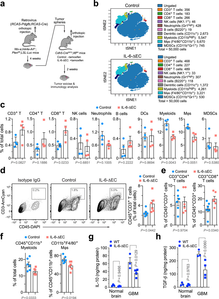
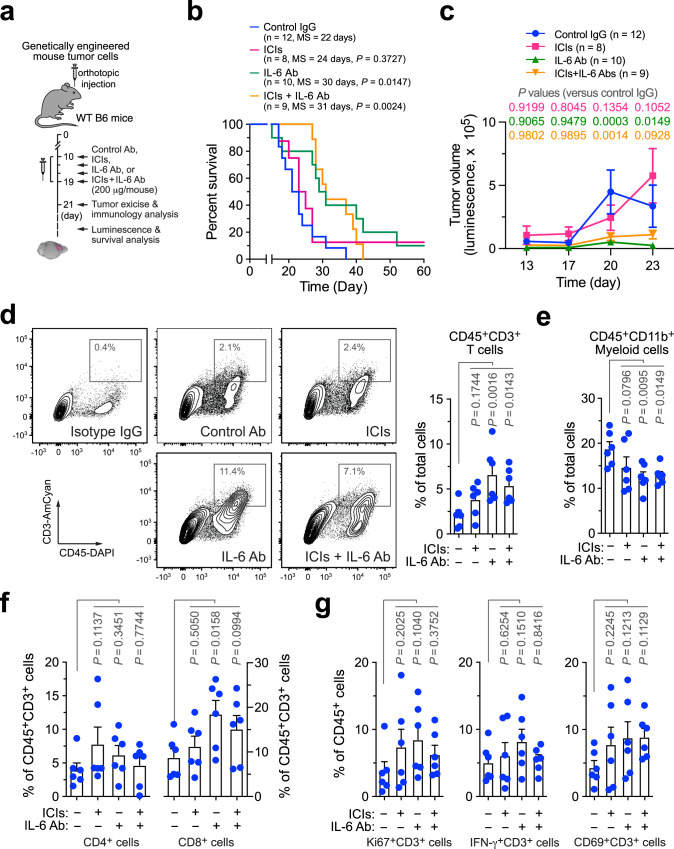
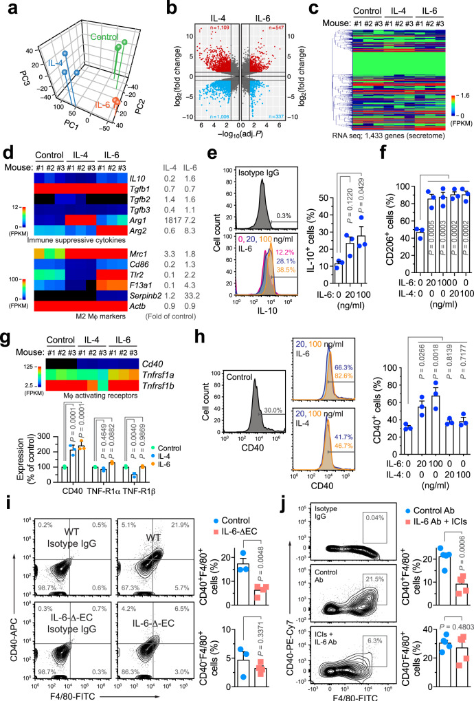
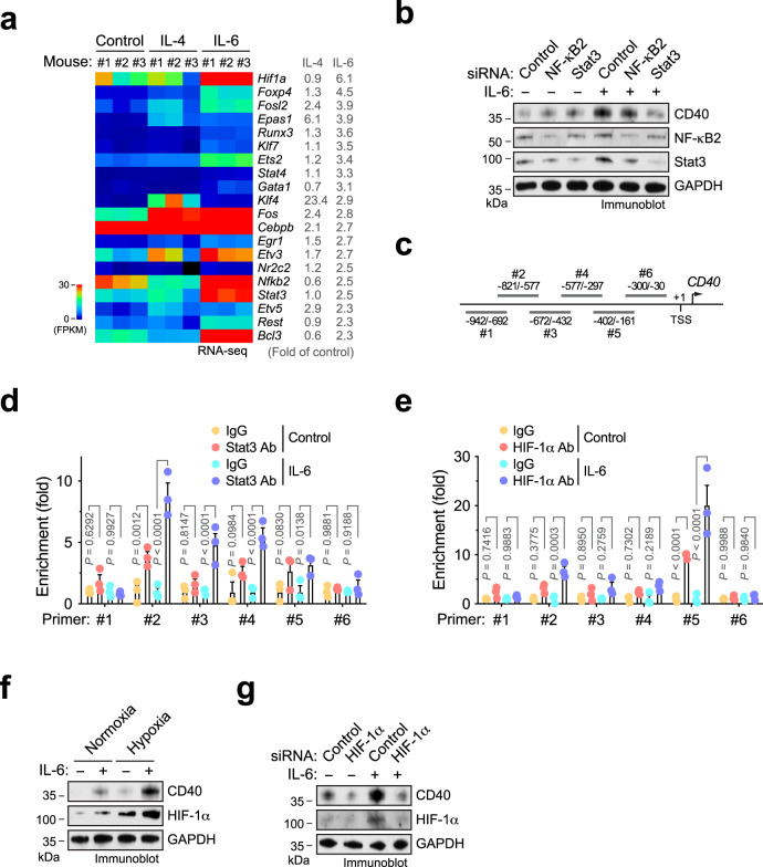
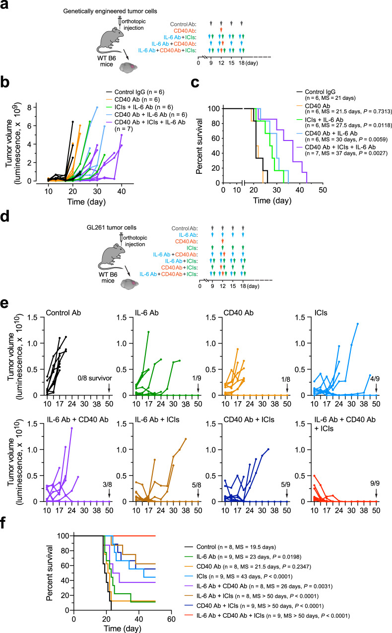
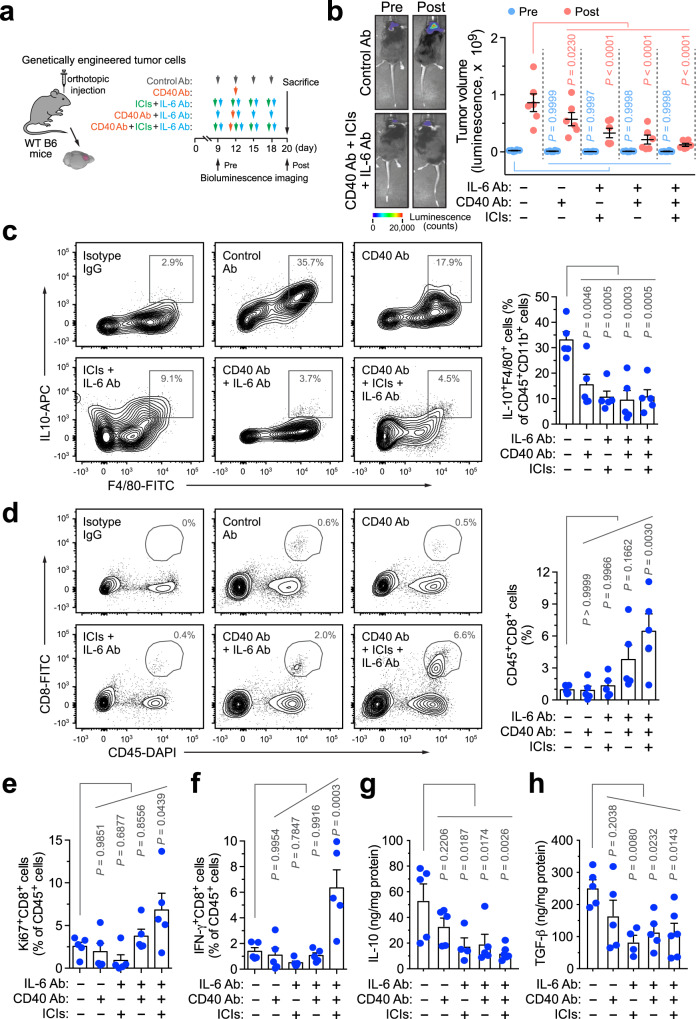
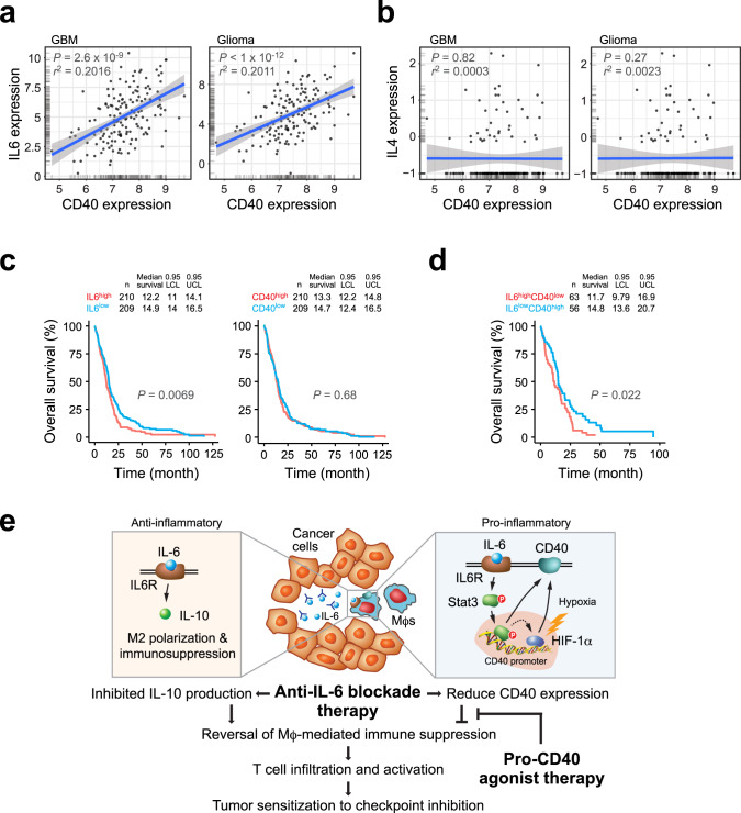
Immunologically-cold tumors including glioblastoma (GBM) are refractory to checkpoint blockade therapy, largely due to extensive infiltration of immunosuppressive macrophages (Mϕs). Consistent with a pro-tumor role of IL-6 in alternative Mϕs polarization, we here show that targeting IL-6 by genetic ablation or pharmacological inhibition moderately improves T-cell infiltration into GBM and enhances mouse survival; however, IL-6 inhibition does not synergize PD-1 and CTLA-4 checkpoint blockade. Interestingly, anti-IL-6 therapy reduces CD40 expression in GBM-associated Mϕs. We identify a Stat3/HIF-1α-mediated axis, through which IL-6 executes an anti-tumor role to induce CD40 expression in Mϕs. Combination of IL-6 inhibition with CD40 stimulation reverses Mϕ-mediated tumor immunosuppression, sensitizes tumors to checkpoint blockade, and extends animal survival in two syngeneic GBM models, particularly inducing complete regression of GL261 tumors after checkpoint blockade. Thus, antibody cocktail-based immunotherapy that combines checkpoint blockade with dual-targeting of IL-6 and CD40 may offer exciting opportunities for GBM and other solid tumors.


Conflict of interest statement

S.J.B. is an advisor to Bayer, Novocure, and Sumitomo Dainippon. S.J.B. has received research funding support from Incyte, GSK, Novocure, and Eli Lilly. L.Z. reports having received research funding from AstraZeneca, Bristol-Myers Squibb/Celgene, and Prelude Therapeutics. X.X. owns stocks in CureBiotech and Exio Bioscience. R.H.V. reports being an inventor on a licensed patent application for cellular immunotherapy. R.H.V. reports receiving royalties for the license of a research grade monoclonal antibody. Y.F. is a co-founder of Radix Therapeutics. The other authors have no competing interests.

Figures

Fig. 1
Fig. 1. Genetic ablation of IL-6 reverses GBM immunosuppression.
GBM was induced by RCAS-mediated genetic engineering in Ntv-a;Ink4a-Arf−/−;Ptenfl/fl;LSL-Luc donor mice, followed by orthotopic tumor implantation into Cdh5-CreERT2;Il6fl/fl recipient mice that were pretreated with (IL-6-ΔEC) or without (Control) tamoxifen. Two weeks after tumor implantation, tumors were excised. a Schematic approach. b, c Tumor-derived single-cell suspensions were analyzed by CyTOF. b Representative CyTOF sorting. c Quantitative results (mean ± SEM, n = 4 mice). Statistical analysis by two-tailed Student’s t-test. df Tumor-derived single-cell suspensions were analyzed by flow cytometry. d Analysis for CD3+ T cells. Left, representative cell sortings. Right, quantified results (n = 6 mice, mean ± SEM). Statistical analysis by two-tailed Student’s t-test. e, f Analysis for e CD4+/CD8+ T cells or f myeloid cells (n = 6 mice, mean ± SEM). Statistical analysis by two-tailed Student’s t-test. g, h Tissue lysates from normal brains and tumors were subjected to ELISA analysis for g IL-10 and h TGF-β expression (mean ± SEM, n = 4 mice for IL-6-ΔEC GBM group and n = 3 mice for other groups). Statistical analysis by two-way ANOVA with Sidak’s test. Source data are provided as a Source data file.
Fig. 2
Fig. 2. IL-6 neutralization enhances T-cell infiltration into GBM tumors and improves animal survival but does not sensitize tumor to immune checkpoint blockade.
GBM was induced in WT B6 mice, followed by injection with control IgG, anti-IL-6 antibody (Ab), immune checkpoint inhibitors (ICIs), or ICIs plus anti-IL-6 Ab. a Schematic approach. b, c Survival and tumor growth analyses (n = 8–12 mice, specific n numbers are shown in the figure). b Mouse survival was monitored for 60 days and subjected to two-sided log-rank Mantel–Cox analysis. MS, median survival. c Tumor volume was analyzed by bioluminescence imaging during days 13–23 (mean ± SEM). Statistical analysis by two-way ANOVA with Dunnett’s test. dg Tumors were excised 2 days after treatment. Tumor-derived single-cell suspensions were stained with antibodies against CD45, d CD3, e CD11b, f CD4, CD8, CD3, and g Ki67, IFN-γ, and CD69, followed by flow cytometry analyses. d Analysis for CD3+ T cells. Left, representative cell sortings. Right, quantified results (n = 6 mice, mean ± SEM). Statistical analysis by one-way ANOVA with Fisher’s LSD test. eg Quantified results for immune cells (n = 6 mice, mean ± SEM). Statistical analysis by one-way ANOVA with Fisher’s LSD test. Source data are provided as a Source data file.
Fig. 3
Fig. 3. IL-6 induces Mϕ-mediated immunosuppression but stimulates CD40 expression.
ae Bone marrow (BM)-derived Mϕs were isolated from mice and treated with 50 ng/ml IL-4 and IL-6 for 2 days, followed by RNA-seq analysis (n = 3 mice). Genes were mapped and subjected to a principal component and b volcano plot analyses. c Heatmap of secretome genes. d Expression of immunosuppressive cytokines (top) and M2 Mϕ activation-associated genes. Left, heatmap. Right, means of fold expression of control. e, f BM-derived mouse Mϕs were treated with IL-4 and IL-6 for 2 days, and analyzed by flow cytometry. e IL-10 expression. Left, representative sortings. Right, quantitative results (n = 3 mice, mean ± SEM). Statistical analysis by one-way ANOVA with Dunnett’s test. f CD206 expression (n = 3 mice, mean ± SEM). Statistical analysis by one-way ANOVA with Dunnett’s test. g Expression of Mϕ activation-associated receptor genes. Left, heatmap. Right, quantitative results (n = 3 mice, mean ± SEM). Statistical analysis by two-way ANOVA with Dunnett’s test. h BM-derived mouse Mϕs were treated with IL-4 and IL-6, and analyzed by flow cytometry. Left, representative sortings. Right, quantitative results (n = 3 mice, mean ± SEM). Statistical analysis by one-way ANOVA with Dunnett’s test. i GBM was induced in control WT or IL-6-ΔEC mice. Two weeks after tumor implantation, tumor-derived single-cell suspensions were analyzed by flow cytometry (mean ± SEM, n = 3 mice for control group and n = 4 mice for IL-6-ΔEC group). Statistical analysis by two-tailed Student’s t-test. j GBM was induced in mice. Two days after treatment with IL-6 Ab and ICIs or with control Ab, tumors were analyzed by flow cytometry (n = 5 mice, mean ± SEM). Statistical analysis by two-tailed Student’s t-test. Source data are provided as a Source data file.
Fig. 4
Fig. 4. IL-6 induces CD40 expression through Stat3 and HIF-1α.
a BM-derived Mϕs were isolated from mice and treated with 50 ng/ml IL-4 and IL-6 for 2 days, followed by RNA-seq analysis (n = 3 mice). Shown are top upregulated transcriptional factors induced by IL-6. Left, heatmap. Right, means of fold expression of control. b Human monocytes were transfected with siRNA targeting NF-κB2, Stat3, or control sequence and treated with IL-6 or control medium. Cell lysates were immunoblotted. This experiment was repeated independently twice with similar results. ce Human monocytes were treated with IL-6 or control medium under d normoxia or e hypoxia. Nuclei protein was immunoprecipitated with d anti-Stat3 or e anti-HIF-1α antibody, or IgG, and subjected to ChIP analysis with different primers. c Results shown are from quantitative real-time polymerase chain reaction (RT-PCR) analysis (n = 3 human samples, means ± SEM). Statistical analysis by two-way ANOVA with Tukey’s test. f Human monocytes were treated with IL-6 or control medium under normoxia or hypoxia, followed by immunoblot analysis. This experiment was repeated independently twice with similar results. g Human monocytes were pretreated with siRNA targeting HIF-1α or control sequence, and treated with IL-6 or control medium under hypoxia. Cell lysates were immunoblotted. This experiment was repeated independently twice with similar results. Source data are provided as a Source data file.
Fig. 5
Fig. 5. IL-6 neutralization and CD40 stimulation sensitizes GBM to immune checkpoint blockade treatment.
GBM was induced in mice by transplantation with ac, tumor cells derived from RCAS-genetically engineered model (n = 6–7 mice, specific n numbers are shown in the figure) or df GL261 tumor cells (n = 8–9 mice, specific n numbers are shown in the figure), followed by different treatment and survival analyses. a, d Experimental procedure. b, e Tumor volume was analyzed by bioluminescence imaging. c, f Mouse survival was monitored and analyzed by two-sided Log-rank Mantel–Cox analysis. MS, median survival. Source data are provided as a Source data file.
Fig. 6
Fig. 6. IL-6 neutralization and CD40 stimulation plus immune checkpoint blockade synergistically reverses Mϕ-mediated immune suppression and activates GBM-associated T cells.
GBM was induced in mice, followed by different treatment and endpoint analyses. a Experimental procedure. b Tumor volume was analyzed pre- and post treatment by bioluminescence imaging. Left, representative images. Right, quantified results (n = 6 mice, mean ± SEM). Statistical analysis by two-way ANOVA with Dunnett’s test. cf Tumor-derived single-cell suspensions were analyzed by flow cytometry. c, d Cells were probed with c, anti-F4/80 and anti-IL-10, or d anti-CD45 and anti-CD8 antibodies. Left, representative sortings. Right, quantified results (n = 5 mice, mean ± SEM). Statistical analysis by one-way ANOVA with Dunnett’s test. e, f Cells were probed with e anti-CD8 and anti-Ki67, or f anti-CD8 and anti-IFN-γ antibodies. Quantified results are shown (n = 5 mice, mean ± SEM). Statistical analysis by one-way ANOVA with Dunnett’s test. g, h Tumor lysates were subjected to g IL-10 and h TGF-β ELISA analysis (mean ± SEM, n = 4 mice for ICI plus IL-6 Ab treatment group, n = 6 mice for ICIs, CD40 Ab, plus IL-6 Ab treatment group, and n = 5 mice for other groups). Statistical analysis by one-way ANOVA with Dunnett’s test. Source data are provided as a Source data file.
Fig. 7
Fig. 7. High IL-6 expression and low CD40 expression correlate with poor survival in human GBM patients.
a, b Correlation of CD40 expression with a IL-6 and b IL-4 expression was subjected to linear regression analyses using GlioVis/TCGA GBM-RNA-seq (n = 160 patients) and low-grade glioma (n = 513 patients) data sets. Statistical analysis by linear regression analysis. c, d Correlation of IL-6 and CD40 expression (high/low cutoff of 40%) with overall survival was analyzed using TCGA-Firehose data set. Statistical analysis by two-sided log-rank test. e A schematic model. IL-6 induces anti-inflammatory and pro-inflammatory functions in Mϕs, through IL-10 and CD40 expression, respectively. Combination therapy by anti-IL-6 neutralization and CD40 activation reverses Mϕ-mediated tumor immunosuppression and promotes T-cell infiltration and activation, sensitizing tumor to checkpoint inhibition treatment.

References

    1. Bovenberg MS, Degeling MH, Tannous BA. Cell-based immunotherapy against gliomas: from bench to bedside. Mol. Ther. 2013;21:1297–1305. doi: 10.1038/mt.2013.80. - DOI - PMC - PubMed
    1. Datta M, Coussens LM, Nishikawa H, Hodi FS, Jain RK. Reprogramming the tumor microenvironment to improve immunotherapy: emerging strategies and combination therapies. Am. Soc. Clin. Oncol. Educ. Book. 2019;39:165–174. doi: 10.1200/EDBK_237987. - DOI - PMC - PubMed
    1. Finocchiaro G, Pellegatta S. Immunotherapy for glioma: getting closer to the clinical arena? Curr. Opin. Neurol. 2011;24:641–647. doi: 10.1097/WCO.0b013e32834cbb17. - DOI - PubMed
    1. Jackson CM, Choi J, Lim M. Mechanisms of immunotherapy resistance: lessons from glioblastoma. Nat. Immunol. 2019;20:1100–1109. doi: 10.1038/s41590-019-0433-y. - DOI - PubMed
    1. Riley RS, June CH, Langer R, Mitchell MJ. Delivery technologies for cancer immunotherapy. Nat. Rev. Drug Discov. 2019;18:175–196. doi: 10.1038/s41573-018-0006-z. - DOI - PMC - PubMed

Publication types

MeSH terms