. 2021 Aug;42(8):1235-1247.
doi: 10.1038/s41401-021-00682-9. Epub 2021 Jun 8.

Inhibition of Nav1.7 channel by a novel blocker QLS-81 for alleviation of neuropathic pain

Affiliations

Inhibition of Nav1.7 channel by a novel blocker QLS-81 for alleviation of neuropathic pain

He-Ling Niu et al. Acta Pharmacol Sin. 2021 Aug.

Abstract

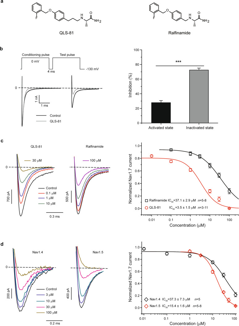
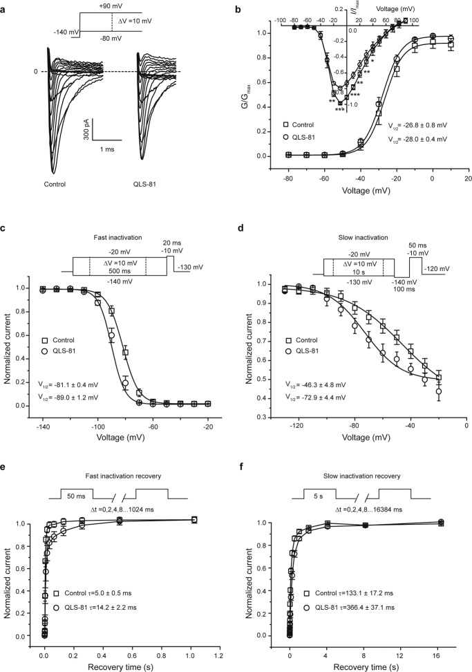
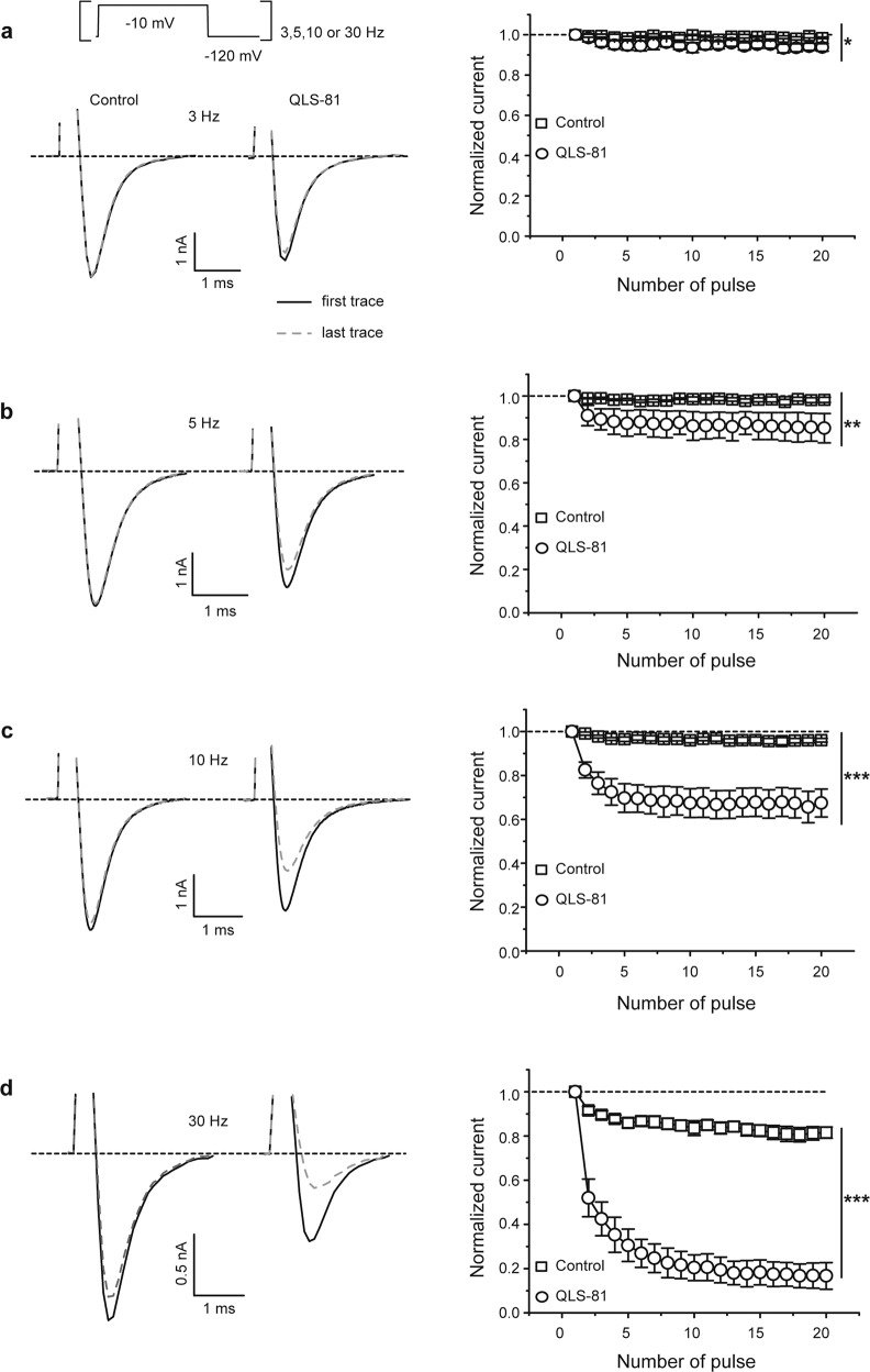
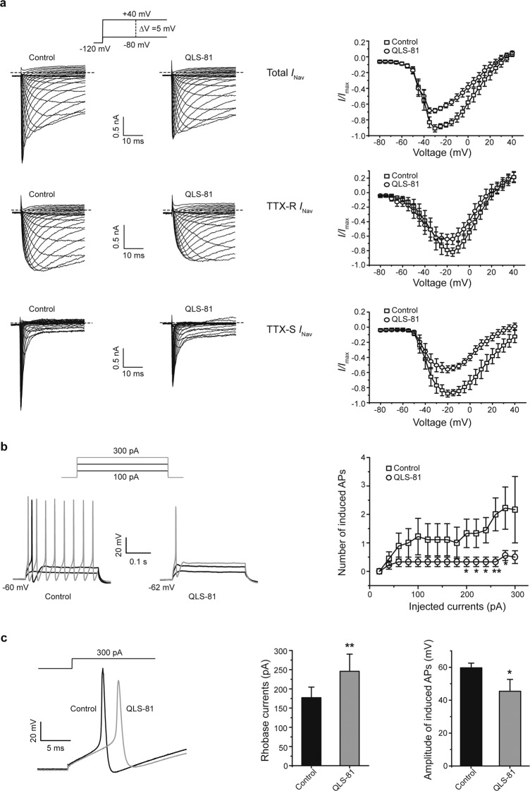
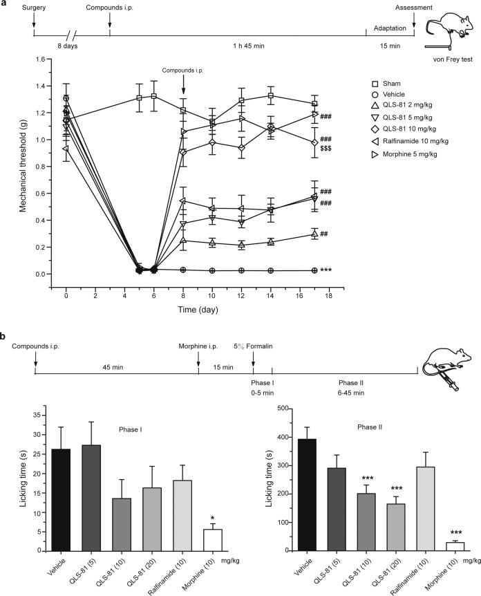
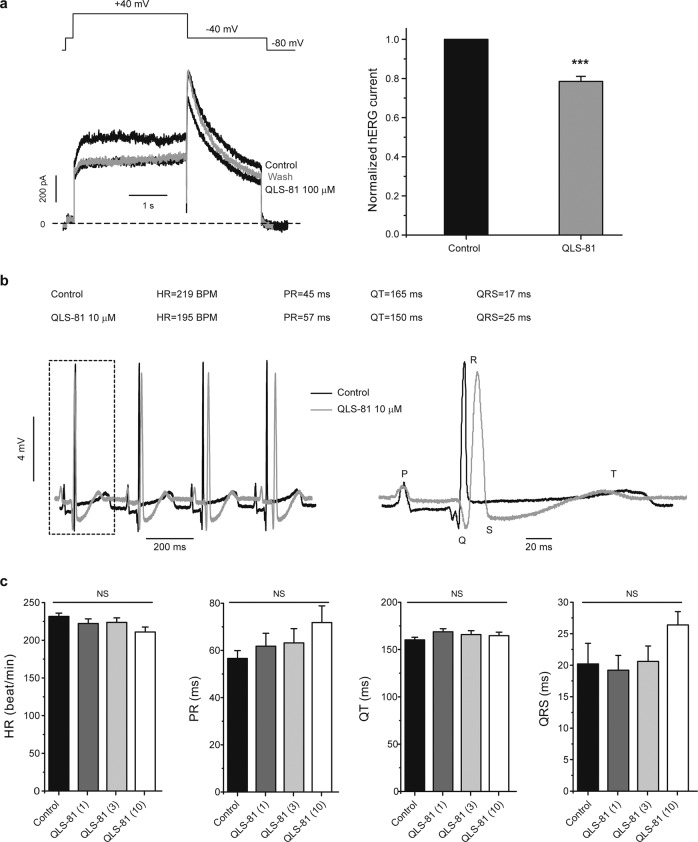
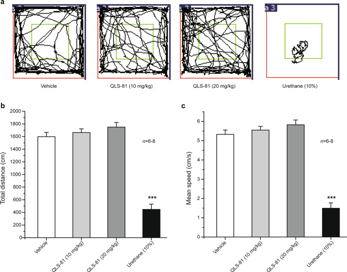
Voltage-gated sodium channel Nav1.7 robustly expressed in peripheral nociceptive neurons has been considered as a therapeutic target for chronic pain, but there is no selective Nav1.7 inhibitor available for therapy of chronic pain. Ralfinamide has shown anti-nociceptive activity in animal models of inflammatory and neuropathic pain and is currently under phase III clinical trial for neuropathic pain. Based on ralfinamide, a novel small molecule (S)-2-((3-(4-((2-fluorobenzyl) oxy) phenyl) propyl) amino) propanamide (QLS-81) was synthesized. Here, we report the electrophysiological and pharmacodynamic characterization of QLS-81 as a Nav1.7 channel inhibitor with promising anti-nociceptive activity. In whole-cell recordings of HEK293 cells stably expressing Nav1.7, QLS-81 (IC50 at 3.5 ± 1.5 μM) was ten-fold more potent than its parent compound ralfinamide (37.1 ± 2.9 μM) in inhibiting Nav1.7 current. QLS-81 inhibition on Nav1.7 current was use-dependent. Application of QLS-81 (10 μM) caused a hyperpolarizing shift of the fast and slow inactivation of Nav1.7 channel about 7.9 mV and 26.6 mV, respectively, and also slowed down the channel fast and slow inactivation recovery. In dissociated mouse DRG neurons, QLS-81 (10 μM) inhibited native Nav current and suppressed depolarizing current pulse-elicited neuronal firing. Administration of QLS-81 (2, 5, 10 mg· kg-1· d-1, i.p.) in mice for 10 days dose-dependently alleviated spinal nerve injury-induced neuropathic pain and formalin-induced inflammatory pain. In addition, QLS-81 (10 μM) did not significantly affect ECG in guinea pig heart ex vivo; and administration of QLS-81 (10, 20 mg/kg, i.p.) in mice had no significant effect on spontaneous locomotor activity. Taken together, our results demonstrate that QLS-81, as a novel Nav1.7 inhibitor, is efficacious on chronic pain in mice, and it may hold developmental potential for pain therapy.

Keywords: Nav1.7; QLS-81; anti-nociception; inflammatory pain; neuropathic pain; ralfinamide.


Conflict of interest statement

The authors declare no competing interests.

Figures

Fig. 1
Fig. 1. Concentration-dependent inhibition of Nav1.7 currents expressed in HEK293 cells by compound QLS-81.
a Chemical structure of QLS-81: (S)-2-((3-(4-((2-fluorobenzyl) oxy) phenyl) propyl) amino) propanamide and ralfinamide. b Left panel, top, Nav1.7 currents stably expressed in HEK293 cells held at −130 mV were elicited by a double-pulse protocol consisting of a conditional pulse at 0 mV (15 ms) for channel activation and a test pulse at 0 mV (15 ms) for 50% channel inactivation. Left panel, bottom, representative current traces elicited by the second test pulse before and after application of QLS-81. The dashed line represents zero-current levels. Right panel, summary for normalized inhibition of activated and inactivated Nav1.7 currents by QLS-81. Paired t-test, ***P < 0.001, n = 10. c Inhibition of representative Nav1.7 current traces elicited by the test pulse (panel (b)) before and after QLS-81 (left panel) and ralfinamide (middle panel) at different concentrations. The dashed line represents zero-current levels. Right panel, curves were fitted using a logistic function for concentration-dependent inhibition of Nav1.7 by QLS-81 (black) with IC50 of 3.5 ± 1.5 μM (n = 3–11) and ralfinamide (red) with IC50 of 37.1 ± 2.9 μM (n = 5–8). d Representative current traces of Nav1.4 (left panel) and Nav1.5 (middle panel) elicited by the depolarizing potential at 0 mV (test pulse of a panel (b)) before and after different concentrations of QLS-81. The dashed line represents zero-current levels. Right panel, curves were fitted using the logistic function for concentration-dependent inhibition of Nav1.4 with IC50 of 37.3 ± 7.3 μM (n = 5) and Nav1.5 with 15.4 ± 1.6 μM (n = 5–8) by QLS-81. All data were expressed as the mean ± SEM.
Fig. 2
Fig. 2. Leftward shift of Nav1.7 inactivation and prolongation of recovery from inaction by QLS-81.
a Top panel, a family of potentials from −80 mV to +90 mV in a 10-mV increment were applied from the holding potential at −140 mV for 14 ms with 1 s intervals. Bottom panels, representative Nav1.7 current traces without (left) or with (right) 10 μM QLS-81. b Normalized conductance versus voltage was plotted for Nav1.7 channels. Curves were fitted by Boltzmann function of half activation voltage (V1/2) for Nav1.7 channels with −26.8 ± 0.8 mV (control) and −28.0 ± 0.4 mV (QLS-81), n = 8. Inset, current–voltage (IV) relationships with or without QLS-81 (10 μM) normalized with the maximum peak currents obtained from panel a. Paired t-test, *P < 0.05, **P < 0.01, and ***P < 0.001 vs. Control, n = 8. c Top panel, fast inactivation currents of Nav1.7 were elicited with starting by a holding voltage of −130 mV, a family of conditional pulses ranging from −160 mV to −20 mV (500 ms) in a 10-mV increment to cause channel inactivation before a 20 ms test pulse at −10 mV to assess the available non-inactivated channels. Bottom panel, a steady-state fast-inactivation curves of Nav1.7 channels were obtained by Boltzmann function with V1/2 of −89.0 ± 1.2 mV (QLS-81, circles) and the control value of −81.1 ± 0.4 mV (squares), n = 4. d Top panel, Voltage-dependent steady-state slow inactivation of Nav1.7 currents was measured using a series of 10 s pre-pulses, ranging from −130 to −20 mV in a 10-mV increment, followed by a 100 ms step to −140 mV to remove fast inactivation, and a 50 ms test pulse at −10 mV to assess the available non-inactivated channels. Bottom, steady-state slow-inactivation curves of Nav1.7 channels were obtained by Boltzmann function with V1/2 of −72.9 ± 4.4 mV (QLS-81, circles) and the control value of −46.3 ± 4.8 mV (squares), n = 6. e, f Top panels, a standard two-pulse protocol consisting of two depolarizing pulses to −10 mV lasting 50 ms for fast inactivation or 5 s for slow inactivation, and a variable duration step between two depolarizing steps at −120 mV (1–1024 ms for fast inactivation or 1–16384 ms for slow inactivation) for recovery. Bottom panels, channel recovery was determined by normalizing the current elicited from the second test pulse to the first conditioning pulse and plotted versus recovery time. The curves were fitted with a one-exponential function. QLS-81 (circles) delayed the Nav1.7 recovery from fast (e) and slow (f) inactivation with time constants of 14.2 ± 2.2 ms (n = 8) and 366.4 ± 37.1 ms (n = 5) as compared with control value of 5.0 ± 0.5 ms (n = 8) and 133.1 ± 17.2 ms (n = 5). All data were expressed as the mean ± SEM.
Fig. 3
Fig. 3. Use-dependent block of Nav1.7 currents by QLS-81.
ad HEK293 cells expressing Nav1.7 channels were held at −120 mV and depolarized to −10 mV for four different frequencies (3, 5, 10, and 30 Hz), with interval pulse potential set at −120 mV. Left panels, representative first (solid line) and last (dashed line) traces of Nav1.7 currents evoked by different frequencies in the absence (left) or presence (right) of QLS-81 (10 μM). Right panels, currents were normalized to the amplitude of the first pulse in the absence (open squares) or presence (open circles) of QLS-81. All data were expressed as the mean ± SEM. Paired t-test, *P < 0.05, **P < 0.01, and ***P < 0.001 vs. Control, n = 4–6.
Fig. 4
Fig. 4. Inhibition of native Nav currents and firing in DRG neurons by QLS-81.
a Left and middle panels, representative total (top panel) currents, TTX-R (middle panel), and TTX-S (bottom panel) currents in DRG neurons of small or medium size diameter were elicited by a family of depolarizing voltage steps in a 5-mV increment from −80 mV to +40 mV with holding potential at −120 mV in the absence (open squares) or presence (open circles) of 10 μM QLS-81. Right panel, current–voltage (IV) relationships of peak currents normalized with the maximum amplitude of Nav current obtained from left panels n = 7–8. b Representative traces of action potential (AP) induced by injection of different depolarizing currents without (left panel) or with (middle panel) QLS-81. Right panel, summary for the number of action potentials induced by increasing depolarizing currents with or without QLS-81 (10 μM). Paired t-test, *P < 0.05, and **P < 0.01 vs. Control, n = 9. c Left panel, representative traces of AP induced by injection of 300 pA depolarizing currents with (gray) or without (black) QLS-81 (10 μM). Middle panel, a summary of the depolarization current threshold for eliciting the first AP with or without QLS-81 (10 μM). Right panel, summary for the amplitude of induced APs by injection of 300 pA current with or without QLS-81. Paired t-test, *P < 0.05, and **P < 0.01 vs. Control, n = 9. All data were expressed as the mean ± SEM.
Fig. 5
Fig. 5. Antinociceptive effects of QLS-81 on pain induced by spinal nerve injury (SNI) and inflammation in mice.
a Top panel, a schematic time-course of paw mechanical withdrawal threshold (PMWT) measured by von Frey hair stimulation in a mouse model of SNI. Bottom panel, dose-dependent antinociception by QLS-81 (2, 5, and 10 mg/kg, i.p.), morphine (5 mg/kg), and ralfinamide (10 mg/kg) in SNI model. Two-way ANOVA followed by Bonferroni post hoc tests revealed a significant decrease of PMWT between-group sham and group vehicle, ***P < 0.001; a significant increase of PMWT in QLS-81 (2, 5, 10 mg/kg, i.p.) injected groups, compared with group vehicle, ##P < 0.01, ###P < 0.001; and also a significant difference between group QLS-81 (10 mg/kg) and group ralfinamide (10 mg/kg), $$$P < 0.001. All data were expressed as the mean ± SEM, n = 10–12. b Top panel, experimental design, and schedule for formalin test in a mouse model of inflammatory pain. Bottom panels, formalin-induced spontaneous pain behavior was divided into two phases and analyzed following subcutaneous (sc.) injection of formalin. QLS-81 (5, 10, and 20 mg/kg) dose-dependently attenuated the nocifensive behaviors induced by formalin at phase II (right) but not at phase I. One-way ANOVA followed by Tukey test, *P < 0.01 and ***P < 0.001 vs. vehicle, n = 10–12. All data were expressed as the mean ± SEM.
Fig. 6
Fig. 6. Lack of hERG inhibition and induction of cardiac toxicity by QLS-81 in whole-cell patch-clamp recording and electrical mapping of a guinea pig heart.
a Left panel, representative hERG currents elicited by depolarization steps of +40 mV before repolarization to −40 mV. The dashed line represents zero-current levels. Right panel, a summary for hERG current inhibition of about 21.5% ± 2.6% by 100 μM QLS-81. Paired t-test, ***P < 0.001 vs. Control, n = 4. b Representative electrocardiograms (ECG) of ex vivo guinea pig heart in electrical mapping assay in the absence (black) or presence (gray) of QLS-81 (10 μM). c Summary for parameters of heart rate (HR), QT interval (the time from the start of the Q wave to the end of the T wave), PR interval (the time from the beginning of the P wave to the beginning of QRS complex) and QRS duration obtained from panel (b). One-way ANOVA, n = 5.
Fig. 7
Fig. 7. Lack of QLS-81 effect on locomotion.
a Representative traces of mouse travel in the open field test after injections of saline, QLS-81 (10 and 20 mg/kg, i.p.), and urethane (10%, i.p.). b The total distance traveled in the open field for 5 min after injections of saline, QLS-81 (10 and 20 mg/kg, i.p.), and urethane (10%, i.p.). c The mean travel speed in open field for 5 min after injection of saline, QLS-81 (10 and 20 mg/kg, i.p.) and urethane (10%, i.p.). One-way ANOVA followed by Tukey test, n = 6–8. All data were expressed as the mean ± SEM. ***P < 0.001 vs. Vehicle.

Similar articles

Cited by

References

    1. Xu L, Ding X, Wang T, Mou S, Sun H, Hou T. Voltage-gated sodium channels: structures, functions, and molecular modeling. Drug Discov Today. 2019;24:1389–97. doi: 10.1016/j.drudis.2019.05.014. - DOI - PubMed
    1. Minett MS, Nassar MA, Clark AK, Passmore G, Dickenson AH, Wang F, et al. Distinct Nav1.7-dependent pain sensations require different sets of sensory and sympathetic neurons. Nat Commun. 2012;3:791. doi: 10.1038/ncomms1795. - DOI - PMC - PubMed
    1. Choi JS, Boralevi F, Brissaud O, Sanchez-Martin J, Te Morsche RH, Dib-Hajj SD, et al. Paroxysmal extreme pain disorder: a molecular lesion of peripheral neurons. Nat Rev Neurol. 2011;7:51–5. doi: 10.1038/nrneurol.2010.162. - DOI - PubMed
    1. Claes L, Del-Favero J, Ceulemans B, Lagae L, Van Broeckhoven C, De Jonghe P. De novo mutations in the sodium-channel gene SCN1A cause severe myoclonic epilepsy of infancy. Am J Hum Genet. 2001;68:1327–32. doi: 10.1086/320609. - DOI - PMC - PubMed
    1. Gardella E, Becker F, Moller RS, Schubert J, Lemke JR, Larsen LH, et al. Benign infantile seizures and paroxysmal dyskinesia caused by an SCN8A mutation. Ann Neurol. 2016;79:428–36. doi: 10.1002/ana.24580. - DOI - PubMed

MeSH terms