. 2021 May 19:2021:6677955.
doi: 10.1155/2021/6677955. eCollection 2021.

PGC-1 α Protects against Hepatic Ischemia Reperfusion Injury by Activating PPAR α and PPAR γ and Regulating ROS Production

Affiliations

PGC-1 α Protects against Hepatic Ischemia Reperfusion Injury by Activating PPAR α and PPAR γ and Regulating ROS Production

Chaoqun Wang et al. Oxid Med Cell Longev. .

Abstract

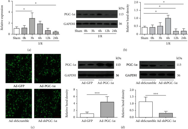
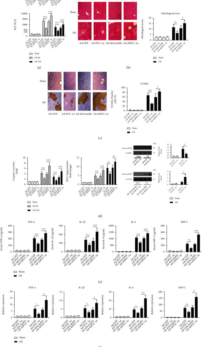
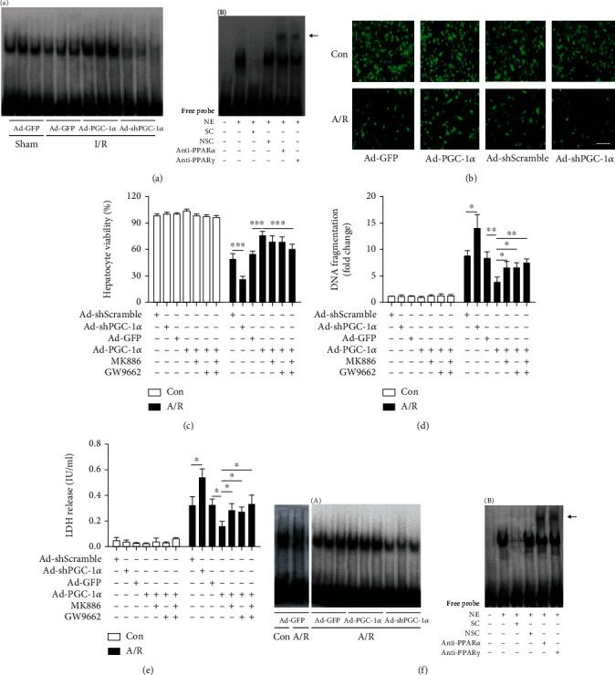
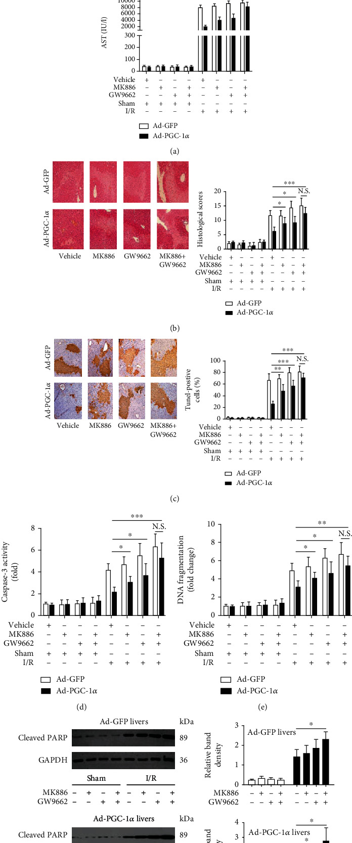
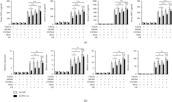
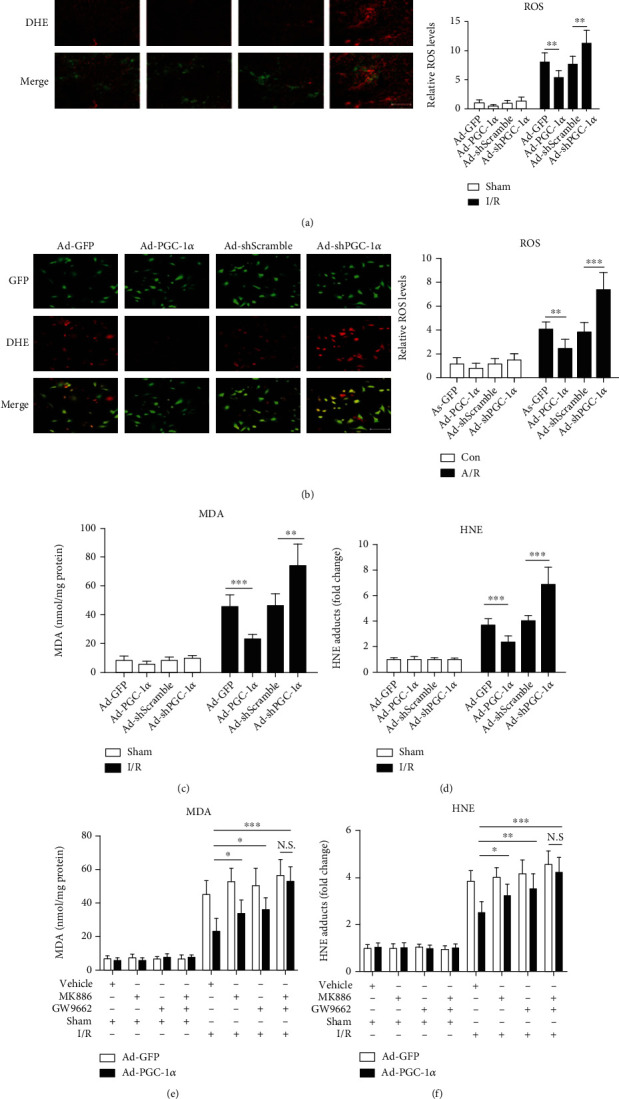
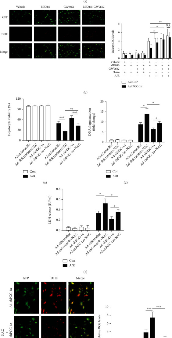
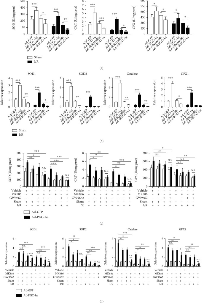
Peroxisome proliferator-activated receptors (PPARs) α and γ have been shown to be protective in hepatic ischemia/reperfusion (I/R) injury. However, the precise role of PPARγ coactivator-1α (PGC-1α), which can coactivate both of these receptors, in hepatic I/R injury, remains largely unknown. This study was designed to test our hypothesis that PGC-1α is protective during hepatic I/R injury in vitro and in vivo. Our results show that endogenous PGC-1α is basally expressed in normal livers and is moderately increased by I/R. Ectopic PGC-1α protects against hepatic I/R and hepatocyte anoxia/reoxygenation (A/R) injuries, whereas knockdown of endogenous PGC-1α aggravates such injuries, as evidenced by assessment of the levels of serum aminotransferases and inflammatory cytokines, necrosis, apoptosis, cell viability, and histological examination. The EMSA assay shows that the activation of PPARα and PPARγ is increased or decreased by the overexpression or knockdown of PGC-1α, respectively, during hepatic I/R and hepatocyte A/R injuries. In addition, the administration of specific antagonists of either PPARα (MK886) or PPARγ (GW9662) can effectively decrease the protective effect of PGC-1α against hepatic I/R and hepatocyte A/R injuries. We also demonstrate an important regulatory role of PGC-1α in reactive oxygen species (ROS) metabolism during hepatic I/R, which is correlated with the induction of ROS-detoxifying enzymes and is also dependent on the activations of PPARα and PPARγ. These data demonstrate that PGC-1α protects against hepatic I/R injury, mainly by regulating the activation of PPARα and PPARγ. Thus, PGC-1α may be a promising therapeutic target for the protection of the liver against I/R injury.


Conflict of interest statement

The authors declare no competing financial interests.

Figures

Figure 1
Figure 1
Hepatic I/R enhances the in situ expression of PGC-1α, and transduction with adenoviral (Ad) vectors is effective for inducing intrahepatic PGC-1α overexpression or knockdown. (a) The mRNA expression of PGC-1α in the mouse liver of each group was assessed by real-time RT-PCR (n = 3). (b) The protein expression of PGC-1α was detected by western blot. (c) Fluorescent microscopy showed the efficient transduction of hepatocytes, as indicated by GFP expression, 72 h following administration of the Ad vectors. (d) PGC-1α protein expression in the mouse liver of each group was assessed by western blot analysis. Bar: 100 μm. P < 0.05, ∗∗P < 0.01, and ∗∗∗P < 0.001. The results are representative of three independent experiments.
Figure 2
Figure 2
PGC-1α protects the liver against I/R injury. (a) Serum levels of aminotransferases (ALT and AST) were detected in the mice subjected to Ad-GFP, Ad-PGC-1α, Ad-shScramble, and Ad-shPGC-1α at 6 h and 24 h after liver I/R (n = 6). (b) Representative images (200x magnification) of H&E-stained liver sections (6 h after I/R) were taken, and histopathological scoring of hepatic injury was performed. (c) Representative images (200x magnification) of liver sections (6 h after I/R) stained by TUNEL were taken, and TUNEL-positive cells were counted as described in Materials and Methods. (d) Caspase-3 activity, DNA fragmentation, and cleaved PARP expression in the mouse livers were assessed by ELISA and western blot (n = 3-6). (e) Systemic TNF-α, IL-1β, IL-6, and MIP-2 levels at 6 h after liver I/R were measured by ELISA (n = 6). (f) The relative mRNA expression levels of TNF-α, IL-1β, IL-6, and MIP-2 in the mouse liver tissues at 6 h after I/R were determined by quantitative RT-PCR (n = 3). P < 0.05, ∗∗P < 0.01, and ∗∗∗P < 0.001.
Figure 3
Figure 3
PGC-1α regulates the activation of PPARα and PPARγ in vivo and in vitro and protects A/R-induced hepatocyte injury. (a) The activation of PPARα and PPARγ in liver tissues was assessed in the mice subjected to Ad-PGC-1α, Ad-shPGC-1α, and the related control at 6 h after liver I/R by EMSA (A), and the specificity of the PPARα and PPARγ bands were confirmed with competition and supershift assays; the supershift bands are indicated by the arrow (B). (b) Representative fluorescence microscopy images of the hepatocytes from certain groups were taken at 24 h after A/R injury. (c) Cell viability was determined at 24 h after reoxygenation by a CCK-8 assay. (d) DNA fragmentation was determined at 24 h after reoxygenation. (e) LDH release was measured in each group. (f) The activation of PPARα and PPARγ in the hepatocytes was assessed by EMSA (A), and the specificity of the PPAR α and γ bands was also confirmed (B). The data was presented as the means ± SD of three independent experiments. Bar: 200 μm, P < 0.05, ∗∗P < 0.01, and ∗∗∗P < 0.001. NE: nuclear extracts; SC: specific competition; NSC: nonspecific competition; anti-PPARα: PPARα antibody; anti-PPARγ: PPARγ antibody.
Figure 4
Figure 4
Inhibition of either PPARα or PPARγ alleviates the hepatoprotective effect of PGC-1α in vivo. (a) The serum levels of aminotransferases were measured in the mice subjected to Ad-PGC-1α, Ad-PGC-1α+MK886, Ad-PGC-1α+GW9662, and Ad-PGC-1α+MK886+GW9662 at 6 h after liver I/R (n = 6). (b) Representative images (200x magnification) of the H&E-stained liver sections (6 h after I/R) were taken, and histopathological scoring of hepatic injury was performed (n = 6). (c) Representative images (200x magnification) of the liver sections (6 h after I/R) stained by TUNEL were taken, and TUNEL-positive cells were counted as described in Materials and Methods (n = 6). (d–f) Caspase-3 activity, DNA fragmentation, and cleaved PARP expression in the mouse livers of each group were assessed by ELISA and western blot analysis at 6 h after I/R (n = 3-6). P < 0.05, ∗∗P < 0.01, and ∗∗∗P < 0.001.
Figure 5
Figure 5
Inhibition of either PPARα or PPARγ alleviates the effect of PGC-1α on inflammatory mediator production induced by liver I/R injury. (a) Systemic TNF-α, IL-1β, IL-6, and MIP-2 levels were assessed at 6 h after liver I/R by ELISA. (b) the relative mRNA expression levels of TNF-α, IL-1β, IL-6, and MIP-2 in the mouse liver tissues were determined by qRT-PCR at 6 h after liver I/R. The data are expressed as the mean ± SD of 6 animals per group. P < 0.05, ∗∗P < 0.01, and ∗∗∗P < 0.001.
Figure 6
Figure 6
PGC-1α regulates the metabolism of ROS in hepatocytes in vivo and in vitro and decreases liver I/R-induced damage of oxidative stress. (a) The representative images of DHE-stained liver cryosections from mice subjected to Ad-GFP, Ad-PGC-1α, Ad-shScramble, and Ad-shPGC-1α at 1 h after liver I/R and the relative ROS levels. Bar: 200 μm (n = 6). (b) Representative images of hepatocytes stained by DHE, and the relative ROS levels. Bar: 200 μm (n = 3). (c) The hepatic content of MDA at 6 h after liver I/R (n = 6). (d) The hepatic content of HNE at 6 h after liver I/R (n = 6). (e) The hepatic content of MDA from mice subjected to Ad-PGC-1α, Ad-PGC-1α+MK886, Ad-PGC-1α+GW9662, and Ad-PGC-1α+MK886+GW9662 at 6 h after liver I/R (n = 6). (f) The hepatic content of HNE in the groups. P < 0.05, ∗∗P < 0.01, and ∗∗∗P < 0.001.
Figure 7
Figure 7
Inhibition of either PPARα or PPARγ alleviates the effect of PGC-1α on ROS production induced by liver I/R injury, and NAC pretreatment significantly reduces the aggravating effects of PGC-1α knockdown on A/R-induced hepatocyte injury. (a) The representative images of DHE-stained liver cryosections from mice subjected to Ad-PGC-1α, Ad-PGC-1α+MK886, Ad-PGC-1α+GW9662, and Ad-PGC-1α+MK886+GW9662, relative to the Ad-GFP control at 1 h after I/R, and the relative ROS levels. Bar: 200 μm (n = 6). (b) The representative photographs of the DHE-stained hepatocytes subjected to Ad-PGC-1α, Ad-PGC-1α+MK886, Ad-PGC-1α+GW9662, and Ad-PGC-1α+MK886+GW9662, relative to the Ad-GFP control at 1 h after A/R, and the relative ROS levels in the hepatocytes of each group; bar: 200 μm (n = 3). (c) The Ad-shScramble- and Ad-shPGC-1α-transduced hepatocytes were pretreated with NAC for 1 h before the onset of A/R, respectively. At 24 h after A/R, the cell viability was determined by a CCK-8 assay (n = 3). (d) DNA fragmentation was determined at 24 h after A/R by the Cell Death Detection ELISA assay (n = 3). (e) LDH release was measured in each group (n = 3). (f) The DHE staining of the Ad-shScramble- and Ad-shPGC-1α-transduced hepatocytes at 1 h after A/R, with or without NAC pretreatment, and the relative ROS levels. Bar: 200 μm (n = 3). P < 0.05, ∗∗P < 0.01, and ∗∗∗P < 0.001.
Figure 8
Figure 8
PGC-1α induces the gene expression of ROS-detoxifying enzymes and increase the activities of SOD, CAT, and GPX in the liver, which is associated with the activities of PPARα and PPARγ. (a) The activities of SOD, CAT, and GPX in the liver tissues from mice subjected to Ad-GFP, Ad-PGC-1α, Ad-shScramble, and Ad-shPGC-1α at 6 h after liver I/R (n = 6). (b) The relative gene expression of SOD1, SOD2, catalase, and GPX1 in the liver tissues from the mice were assessed by qRT-PCR (n = 6). (c) The activities of SOD, CAT, and GPX in the liver tissues from mice subjected to Ad-PGC-1α, Ad-PGC-1α+MK886, Ad-PGC-1α+GW9662, and Ad-PGC-1α+MK886+GW9662, relative to the Ad-GFP control at 6 h after liver I/R (n = 6). (d) The relative gene expression levels of SOD1, SOD2, catalase, and GPX1 in the liver tissues (n = 6). P < 0.05, ∗∗P < 0.01, and ∗∗∗P < 0.001.

Similar articles

Cited by

References

    1. Bamboat Z. M., Balachandran V. P., Ocuin L. M., Obaid H., Plitas G., DeMatteo R. P. Toll-like receptor 9 inhibition confers protection from liver ischemia–reperfusion injury. Hepatology. 2010;51(2):621–632. doi: 10.1002/hep.23365. - DOI - PMC - PubMed
    1. Yang L., Wang W., Wang X., et al. Creg in hepatocytes ameliorates liver ischemia/reperfusion injury in a TAK1-dependent manner in mice. Hepatology. 2019;69(1):294–313. doi: 10.1002/hep.30203. - DOI - PubMed
    1. Akahori T., Sho M., Hamada K., et al. Importance of peroxisome proliferator-activated receptor-γ in hepatic ischemia/reperfusion injury in mice. Journal of Hepatology. 2007;47(6):784–792. doi: 10.1016/j.jhep.2007.07.030. - DOI - PubMed
    1. Kuboki S., Shin T., Huber N., et al. Peroxisome proliferator-activated receptor-gamma protects against hepatic ischemia/reperfusion injury in mice. Hepatology. 2008;47(1):215–224. doi: 10.1002/hep.21963. - DOI - PubMed
    1. Xiang S., Chen K., Xu L., Wang T., Guo C. Bergenin exerts hepatoprotective effects by inhibiting the release of inflammatory factors, apoptosis and autophagy via the PPAR-γ pathway. Drug Design, Development and Therapy. 2020;Volume 14:129–143. doi: 10.2147/DDDT.S229063. - DOI - PMC - PubMed

Substances