. 2021 Jun 8;116(1):40.
doi: 10.1007/s00395-021-00881-9.

Small extracellular vesicles obtained from hypoxic mesenchymal stromal cells have unique characteristics that promote cerebral angiogenesis, brain remodeling and neurological recovery after focal cerebral ischemia in mice

Affiliations

Small extracellular vesicles obtained from hypoxic mesenchymal stromal cells have unique characteristics that promote cerebral angiogenesis, brain remodeling and neurological recovery after focal cerebral ischemia in mice

Jonas Gregorius et al. Basic Res Cardiol. .

Abstract

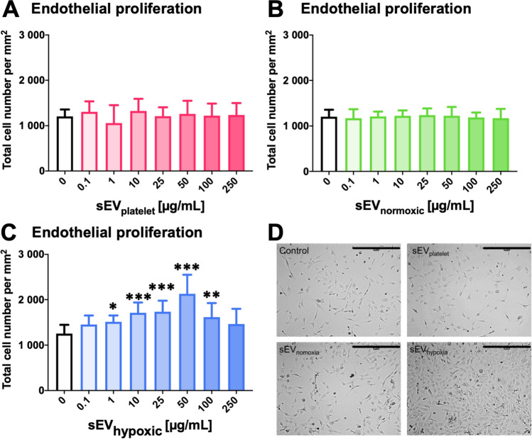
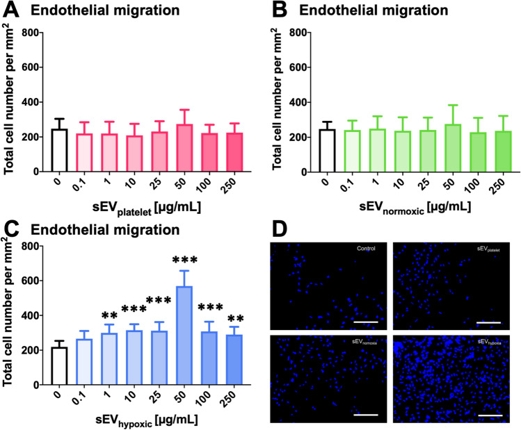
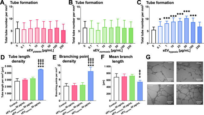
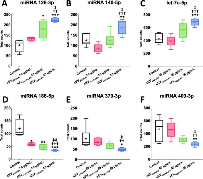
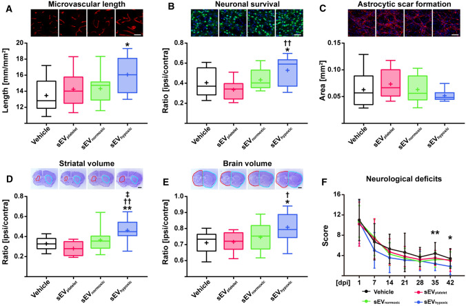
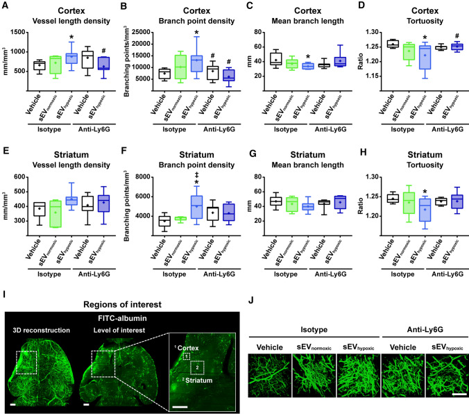
Obtained from the right cell-type, mesenchymal stromal cell (MSC)-derived small extracellular vesicles (sEVs) promote stroke recovery. Within this process, microvascular remodeling plays a central role. Herein, we evaluated the effects of MSC-sEVs on the proliferation, migration, and tube formation of human cerebral microvascular endothelial cells (hCMEC/D3) in vitro and on post-ischemic angiogenesis, brain remodeling and neurological recovery after middle cerebral artery occlusion (MCAO) in mice. In vitro, sEVs obtained from hypoxic (1% O2), but not 'normoxic' (21% O2) MSCs dose-dependently promoted endothelial proliferation, migration, and tube formation and increased post-ischemic endothelial survival. sEVs from hypoxic MSCs regulated a distinct set of miRNAs in hCMEC/D3 cells previously linked to angiogenesis, three being upregulated (miR-126-3p, miR-140-5p, let-7c-5p) and three downregulated (miR-186-5p, miR-370-3p, miR-409-3p). LC/MS-MS revealed 52 proteins differentially abundant in sEVs from hypoxic and 'normoxic' MSCs. 19 proteins were enriched (among them proteins involved in extracellular matrix-receptor interaction, focal adhesion, leukocyte transendothelial migration, protein digestion, and absorption), and 33 proteins reduced (among them proteins associated with metabolic pathways, extracellular matrix-receptor interaction, focal adhesion, and actin cytoskeleton) in hypoxic MSC-sEVs. Post-MCAO, sEVs from hypoxic MSCs increased microvascular length and branching point density in previously ischemic tissue assessed by 3D light sheet microscopy over up to 56 days, reduced delayed neuronal degeneration and brain atrophy, and enhanced neurological recovery. sEV-induced angiogenesis in vivo depended on the presence of polymorphonuclear neutrophils. In neutrophil-depleted mice, MSC-sEVs did not influence microvascular remodeling. sEVs from hypoxic MSCs have distinct angiogenic properties. Hypoxic preconditioning enhances the restorative effects of MSC-sEVs.

Keywords: Endothelial migration; Microvascular network characteristics; Microvascular remodeling; Neuronal survival; Polymorphonuclear neutrophil; Tube formation.


Conflict of interest statement

B.G. and D.M.H. hold patents for the application of extracellular vesicles for the treatment of inflammatory conditions (EP2687219A1 pending; US9877989B2 issued). B.G. is a Scientific Board member of Evox Therapeutics and Innovex Therapeutics SL. Besides, the authors report no conflicts of interest.

Figures

Fig. 1
Fig. 1
Small extracellular vesicles (sEVs) obtained from mesenchymal stromal cells (MSCs) cultured under hypoxic conditions increase cerebral microvascular endothelial cell proliferation. Relative number of human microvascular endothelial cells (hCMEC/D3) after exposure to different concentrations of A sEVs obtained from MSC culture media that contain platelet lysates (sEVplatelet), B sEVs obtained from MSCs ( source 41.5) cultured under regular ‘normoxic’ conditions (21% O2; sEVnormoxic) or C sEVs obtained from MSCs (source 41.5) cultured under hypoxic conditions (1% O2; sEVhypoxic). In D, representative microphotographs for hCMEC/D3 cells exposed to control conditions or sEVs at a concentration of 50 µg/mL are shown. Data are mean ± SD values (n = 9 independent experiments). *p < 0.05, **p < 0.01, ***p < 0.001 compared with control. Scale bar: 400 µm in D
Fig. 2
Fig. 2
sEVs from hypoxic MSCs increase cerebral microvascular endothelial cell migration. Relative number of migrating hCMEC/D3 cells, determined in a modified Boyden chamber transwell migration assay, after exposure to different concentrations of A sEVs obtained from MSC culture media that contain platelet lysates (sEVplatelet), B sEVs obtained from MSCs ( source 41.5) cultured under ‘normoxic’ conditions (21% O2; sEVnormoxic) or C sEVs obtained from MSCs (source 41.5) cultured under hypoxic conditions (1% O2; sEVhypoxic). In D, representative microphotographs for hCMEC/D3 cells exposed to control conditions or sEVs at a 50 µg/mL concentration are shown. Data are mean ± SD values (n = 9 independent experiments). *p < 0.05, **p < 0.01, ***p < 0.001 compared with control. Scale bars: 125 µm in D
Fig. 3
Fig. 3
sEVs from hypoxic MSCs increase the tube formation of cerebral microvascular endothelial cells. Relative tube number, evaluated in a Matrigel-based tube formation assay, of hCMEC/D3 cells exposed to different concentrations of A sEVs obtained from MSC culture media that contain platelet lysates (sEVplatelet), B sEVs obtained from MSCs ( source 41.5) cultured under regular ‘normoxic’ conditions (21% O2; sEVnormoxic) or C sEVs obtained from MSCs (source 41.5) cultured under hypoxic conditions (1% O2; sEVhypoxic). D Microvascular tube length density, E microvascular branching point density and F mean branch length between two branching points of hCMEC/D3 cells exposed to control conditions or sEVplatelet, sEVnormoxic (MSC source 41.5) or sEVhypoxic (MSC source 41.5) at a concentration of 50 µg/mL (n = 3–9 independent experiments). In G, representative microphotographs for hCMEC/D3 cells exposed to control conditions or 50 µg/mL sEVs are shown. Data are mean ± SD values (n = 9 independent experiments [in AC], 3–9 independent experiments [in DF]). *p < 0.05, ***p < 0.001 compared with control/p < 0.05, †††p < 0.001 compared with sEVplatelet/p < 0.05, ‡‡‡p < 0.001 compared with sEVnormoxic. Scale bars: 500 µm
Fig. 4
Fig. 4
sEVs from hypoxic MSCs increase the survival of cerebral microvascular endothelial cells exposed to oxygen–glucose deprivation (OGD), but do not influence the viability of cells cultured under regular ‘normoxic’ conditions. Relative absorbance of hCMEC/D3 cells cultured under (A–C) regular ‘normoxic’ conditions (21% O2) or (D–F) 24 h OGD (1% O2, glucose deprivation) followed by 6 h reoxygenation (21% O2)/glucose recultivation, determined in a 3-(4,5-dimethylthiazol-2-yl)-2,5-diphenyltetrazolium bromide (MTT) assay after exposure to different concentrations of A, D sEVs obtained from MSC culture media that contain platelet lysates (sEVplatelet), B, E sEVs obtained from MSCs ( source 41.5) cultured under regular ‘normoxic’ conditions (21% O2; sEVnormoxic) or C, F sEVs obtained from MSCs (source 41.5) cultured under hypoxic conditions (1% O2; sEVhypoxic). Data are mean ± SD values (n = 3 independent experiments [in AE], 8 independent experiments [in F]). **p < 0.01, ***p < 0.001 compared with control
Fig. 5
Fig. 5
Cerebral microvascular endothelial cells exposed to sEVs obtained from hypoxic MSCs exhibit a distinct microRNA signature associated with angiogenesis. Total counts of A miR-126-3p, B miR-140-5p, C let-7c-5p, D miR-186-5p, E miR-370-3p and F miR-409-3p, evaluated by NanoString analysis, in hCMEC/D3 cells exposed to control conditions, sEVs obtained from MSC culture media that contain platelet lysates (50 µg/mL; sEVplatelet), sEVs obtained from MSCs ( source 41.5) cultured under regular ‘normoxic’ conditions (21% O2; 50 µg/mL; sEVnormoxic) or sEVs obtained from MSCs (source 41.5) cultured under hypoxic conditions (1% O2; 50 µg/mL; sEVhypoxic). Data are box plots with medians (lines inside boxes)/means (crosses inside boxes) ± interquartile ranges (IQR; boxes) with minimum/maximum values as whiskers (n = 4–6 samples per group). *p < 0.05, **p < 0.01, ***p < 0.001 compared with control/††p < 0.01, †††p < 0.001 compared with sEVplatelet/p < 0.05, ‡‡p < 0.01 compared with sEVnormoxic
Fig. 6
Fig. 6
sEVs obtained from hypoxic MSCs induce post-ischemic angiogenesis, brain remodeling and neurological recovery in a mouse model of ischemic stroke. A Density of CD31+ cerebral microvessels in the previously ischemic striatum, B number of NeuN+ surviving neurons in the previously ischemic striatum and C area of GFAP+ astrocytic scar in the brain infarct at the rostrocaudal level of the bregma, which is the core of the middle cerebral artery territory, as well as D striatum volume, E whole-brain volume and F neurological deficits evaluated using the Clark score of mice exposed to 40 min middle cerebral artery occlusion (MCAO), which were intravenously treated after 24 h, 72 h and 120 h with vehicle (normal saline), sEVs obtained from MSC culture media that contain platelet lysate (sEVplatelet), sEVs released by MSCs ( source 41.5) cultured under regular ‘normoxic’ conditions (21% O2; sEVnormoxic; equivalent released by 2 × 106 cells) or sEVs released by MSCs (source 41.5) cultured under hypoxic conditions (1% O2; sEVhypoxic; equivalent released by 2 × 106 cells) followed by animal sacrifice after 56 days. Representative microphotographs are also shown. Data are box plots with medians (lines inside boxes)/means (crosses inside boxes) ± IQR (boxes) with minimum/maximum values as whiskers (in AE) or mean ± SD values (in F) (n = 10 animals vehicle, 6 animals sEVplatelet, 9 animals sEVnormoxic, 9 animals sEVhypoxic). *p < 0.05, **p < 0.01 compared with control/p < 0.05, ††p < 0.01 compared with sEVplatelet/p < 0.05 compared with sEVnormoxic. Scale bars: 50 µm (in AC)/1 mm (in D, E)
Fig. 7
Fig. 7
sEVs obtained from hypoxic MSCs increase microvascular remodeling following ischemic stroke, which is abolished in the absence of polymorphonuclear neutrophils (PMNs) in vivo. Microvascular network characteristics, that is, A, E microvascular length density, B, F branch point density, C, G mean branch length, and D, H tortuosity in the previously ischemic A–D cerebral cortex and E–H striatum evaluated by 3D light sheet microscopy in mice exposed to 40 min MCAO followed by 14 days survival, in which vehicle (normal saline), sEVs released by MSCs ( source 41.5) cultured under ‘normoxic’ conditions (21% O2; sEVnormoxic; equivalent released by 2 × 106 cells) or sEVs released by MSCs (source 41.5) cultured under hypoxic conditions (1% O2; sEVhypoxic; equivalent released by 2 × 106 cells) were intravenously administered at 1, 3, and 5 days post-MCAO, while control (isotype) IgG or anti-Ly6G (clone 1A8; Ly6G indicates lymphocyte antigen-6, locus G) antibody was intraperitoneally applied at 1, 3, 5, and 7 days post-MCAO. I Representative axial overview images of a vehicle-treated mouse brain with magnifications depicting the regions of interest in the ischemic striatum and cortex, in which microvascular network characteristics were determined. J Representative maximum intensity projection (MIP) images in the ischemic cortex for the five experimental groups. Data are box plots with medians (lines inside boxes)/means (crosses inside boxes) ± IQR (boxes) with minimum/maximum values as whiskers (n = 6 animals isotype/vehicle, 5 animals isotype/sEVnormoxic, 8 animals isotype/sEVhypoxic, 7 animals anti-Ly6G/vehicle, 7 animals anti-Ly6G/sEVhypoxic). *p < 0.05 compared with isotype/vehicle/p < 0.05 compared with isotype/sEVnormoxic/#p < 0.05 compared with isotype/sEVhypoxic. Scale bars: 500 µm (in I)/100 µm (in J)

References

    1. Almeria C, Weiss R, Roy M, Tripisciano C, Kasper C, Weber V, Egger D. Hypoxia conditioned mesenchymal stem cell-derived extracellular vesicles induce increased vascular tube formation in vitro. Front Bioeng Biotechnol. 2019;7:292. doi: 10.3389/fbioe.2019.00292. - DOI - PMC - PubMed
    1. Anderson JD, Johansson HJ, Graham CS, Vesterlund M, Pham MT, Bramlett CS, Montgomery EN, Mellema MS, Bardini RL, Contreras Z, Hoon M, Bauer G, Fink KD, Fury B, Hendrix KJ, Chedin F, El-Andaloussi S, Hwang B, Mulligan MS, Lehtiö J, Nolta JA. Comprehensive proteomic analysis of mesenchymal stem cell exosomes reveals modulation of angiogenesis via nuclear factor-kappaB signaling. Stem Cells. 2016;34:601–613. doi: 10.1002/stem.2298. - DOI - PMC - PubMed
    1. Börger V, Staubach S, Dittrich R, Stambouli O, Giebel B. Scaled isolation of mesenchymal stem/stromal cell-derived extracellular vesicles. Curr Protoc Stem Cell Biol. 2020;55:e128. doi: 10.1002/cpsc.128. - DOI - PubMed
    1. Burkhart JM, Schumbrutzki C, Wortelkamp S, Sickmann A, Zahedi RP. Systematic and quantitative comparison of digest efficiency and specificity reveals the impact of trypsin quality on MS-based proteomics. J Proteom. 2012;75:1454–1462. doi: 10.1016/j.jprot.2011.11.016. - DOI - PubMed
    1. Chen Z, Lai TC, Jan YH, Lin FM, Wang WC, Xiao H, Wang YT, Sun W, Cui X, Li YS, Fang T, Zhao H, Padmanabhan C, Sun R, Wang DL, Jin H, Chau GY, Huang HD, Hsiao M, Shyy JY. Hypoxia-responsive miRNAs target argonaute 1 to promote angiogenesis. J Clin Invest. 2013;123:1057–1067. doi: 10.1172/JCI65344. - DOI - PMC - PubMed

Publication types

MeSH terms

LinkOut - more resources